


ABSTRACT
Background Posterior instrumentation of the spine presents a challenge, especially in conditions with low bone quality. Pedicle screw insertion with cortical bone trajectory is designed to add interface between the screw and the bone through engagement between pedicles and the cortex when compared to conventional screw course. Pedicle screw insertion trajectory from cortical infero-superior and the proposed cortical supero-inferior should obtain better pull-out performance than conventional pedicle trajectory. We aim to evaluate the pull-out strength differences among conventional pedicle screw, cortical infero-superior, and cortical supero-inferior trajectories.
Methods Samples from Yorkshire porcine lumbar spine (L1–L5; n = 30) were relieved of soft tissue attachments and dried. Morphometric measurements were conducted, and the samples were randomly assigned to 3 groups. The screws were inserted into the vertebrae by drilling with the 3 trajectories: conventional, cortical infero-superior, and cortical supero-inferior. The trajectories of the screws were examined using x-rays. Pull-out tests were conducted by applying uniaxial traction in line with the screw trajectory with a translational speed of 5 mm/min. The results of the pull-out are measured in Newtons.
Results We obtained a mean value of pull-out force in conventional trajectory of 491.72 ± 187.2 N, cortical infero-superior of 822.16 ± 295.73 N, and cortical supero-inferior of 644.14 ± 201.97 N. Cortical infero-superior and cortical supero-inferior trajectories attained 67% and 30% higher pull-out mean, respectively. Using 1-way analysis of variance and a post hoc Tukey test revealed a significant difference between cortical infero-superior and conventional trajectories (P < .01). Differing pull-out strengths between cortical infero-superior and supero-inferior trajectories showed no statistical significance. Results of our study showed a 30% higher pull-out strength in our proposed trajectory than the conventional one, although not statistically significant.
Conclusions The trajectory of the screws within the lumbar spine seemed to have an impact in pull-out strength. Cortical bone engagement using the novel trajectories may increase pull-out strength of pedicle screws.
Level of Evidence Level 5.
INTRODUCTION
The increase in life expectancy of the general population will lead to an increase in problems of the spine due to aging or degeneration process. In 2010, the number of people over the age of 65 was 524 million, with the number increasing to 1.5 billion in 2015, or 16% of the world's population.1
Problems arising from the spine may be due to the degenerative process of bone quality or deterioration of spinal disc and facet joints. Some of these problems may need posterior instrumentation of the spine.2–4
Posterior instrumentation is aimed at achieving spinal fusion, which is helped by maintaining stable screw insertion within the pedicle. However, posterior instrumentation in porotic pedicle lumbal spine may cause loosening and failure of fixation. Pedicle screw insertion with cortical bone trajectory (CBT) is designed to add interface between the screw and the bone through engagement between pedicles and the cortex when compared to conventional pedicle screw insertion. Pedicle screw insertion from cortical infero-superior and the proposed cortical supero-inferior trajectories should obtain better pull-out performance than conventional pedicle trajectory. We aim to evaluate the pull-out strength differences among conventional pedicle screw, CBT infero-superior, and proposed CBT supero-inferior trajectories.5–8
METHODS
For our experimental study, we obtained 6 female Yorkshire pigs. The age of the pigs were between 6 and 8 months; all were healthy with no deformities. We separated lumbar vertebrae 1–5. Then we fixated the specimens in formaldehyde 10% for 2 weeks. We then removed the surrounding tissue from the lumbar spine and dried our samples in a dry box with 30% humidity at 26°C for 3 weeks until only the bone remained. We measured the vertebral body height, body diameter, superior pedicle diameter, pedicle anterior corpus width, and spinal canal width by using a digital caliper (Mitutoyo 500-182-30; Mitutoyo, Kawasaki, Japan).
We then randomly assigned 10 lumbar vertebrae into 3 groups (conventional, cortical supero-inferior, and cortical infero-superior trajectories) and performed pedicle screw insertions using the methods described by Weinstein et al8 for our conventional trajectory, Santoni et al6 for the cortical infero-superior trajectory and our proposed trajectory (Figure 1). Insertional holes were made using an awl. We then prepared the trajectory using a Lenke probe and a 3-mm diameter drill bit with a power tool.

Trajectory proposed by the authors. (a) The screw insertional point. (b) Sagittal view of the screw after insertion for superior to inferior. (c) An axial cut of the screw trajectory to slightly lateral.
We used traditional polyaxial screws (length 35 mm and outer diameter of 4.5 mm; Changzhou Waston Medical Appliance Co, Changzhou, China) that complied with the standards of GB/T13810 and ISO5832-3: 1996. After screw insertion, we confirmed their positions using a radiograph and to ensure correct trajectories of the screws.
Screw pull-out testing was performed using a universal testing machine (Gotech AI-7000S; Gotech, Taichung, Taiwan). We positioned our samples on a platform that we designed to ensure an axial placement between the screw and the testing machine. The screws were then connected to an insertion screwdriver and pulled out at a speed of 5 mm/min. The point of deflection in the force profile was noted as the pull-out strength of the screws (Figures 2–5).

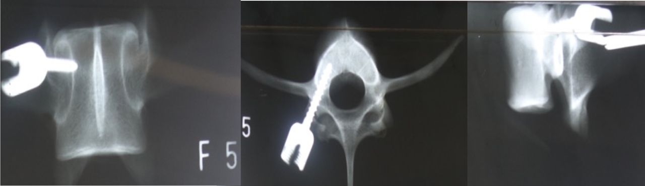
Pedicle screw insertion with conventional trajectory.

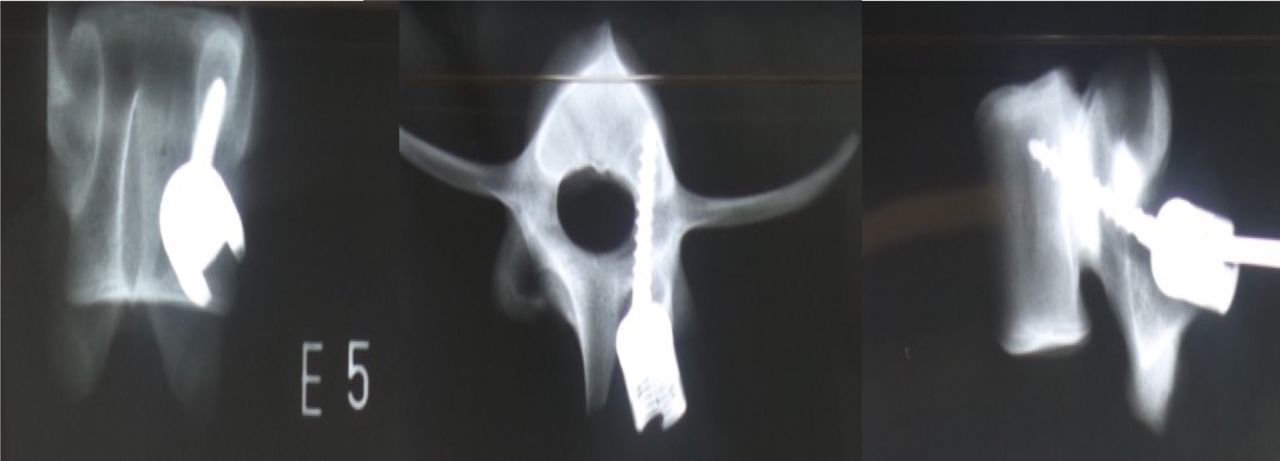
Pedicle screw insertion with cortical bone infero-superior trajectory.

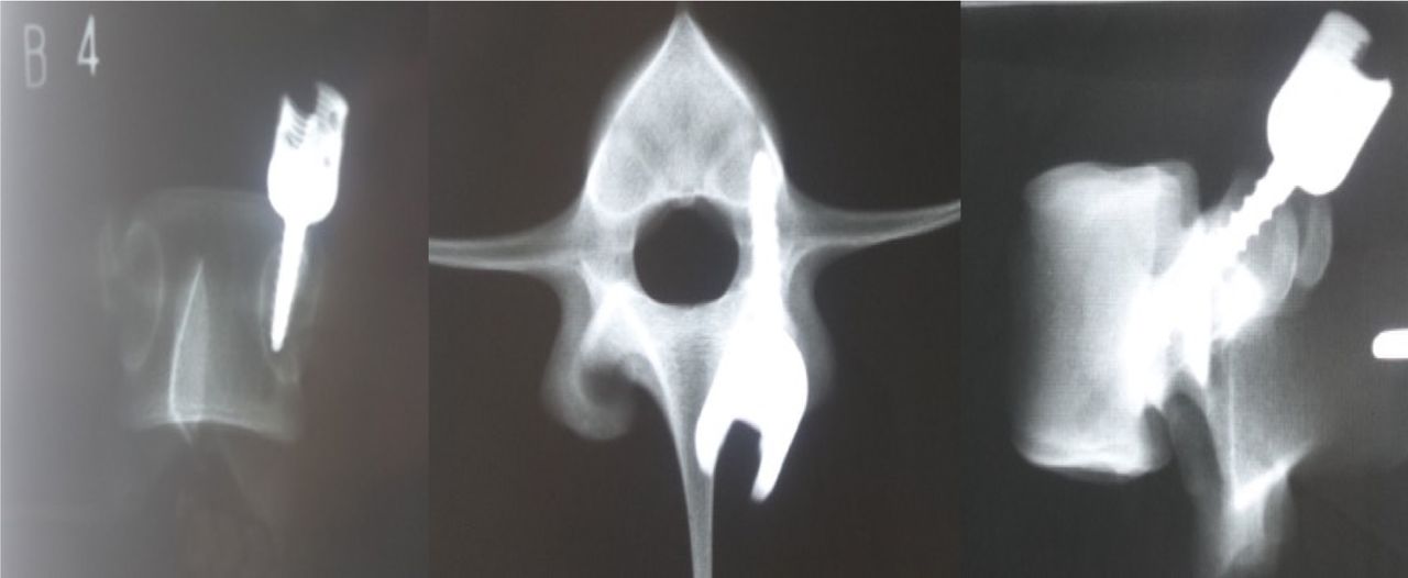
Pedicle screw insertion with cortical bone supero-inferior trajectory.
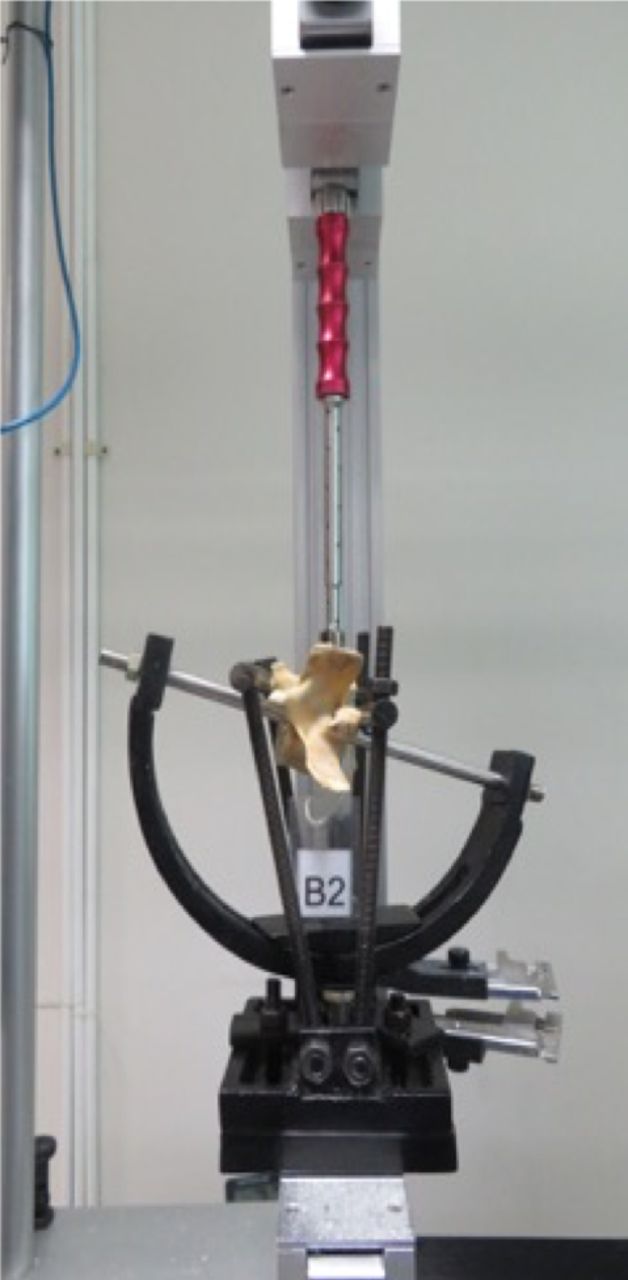
Pull-out testing. The screw is connected to the insertion screwdriver in the apparatus that was designed to hold the vertebrae and the screw in line with the machine. The screw was then pulled out in the direction of the insertion.
We recorded the results and performed statistical analysis using SPSS Statistics version 25 (SPSS Inc, Chicago, IL). The means of the pull-out value were analyzed using 1-way analysis of variance (ANOVA). The P value < .05 was considered statistically significant.
RESULTS AND DISCUSSION
The measurements of the porcine lumbar vertebrae were comparable with the studies performed by Dath et al9 and Busscher et al11 We found no discernable morphologic differences between of the trajectory groups (Table). The porcine lumbar pedicle height and width were comparable to humans.9–11
Sample characteristics.
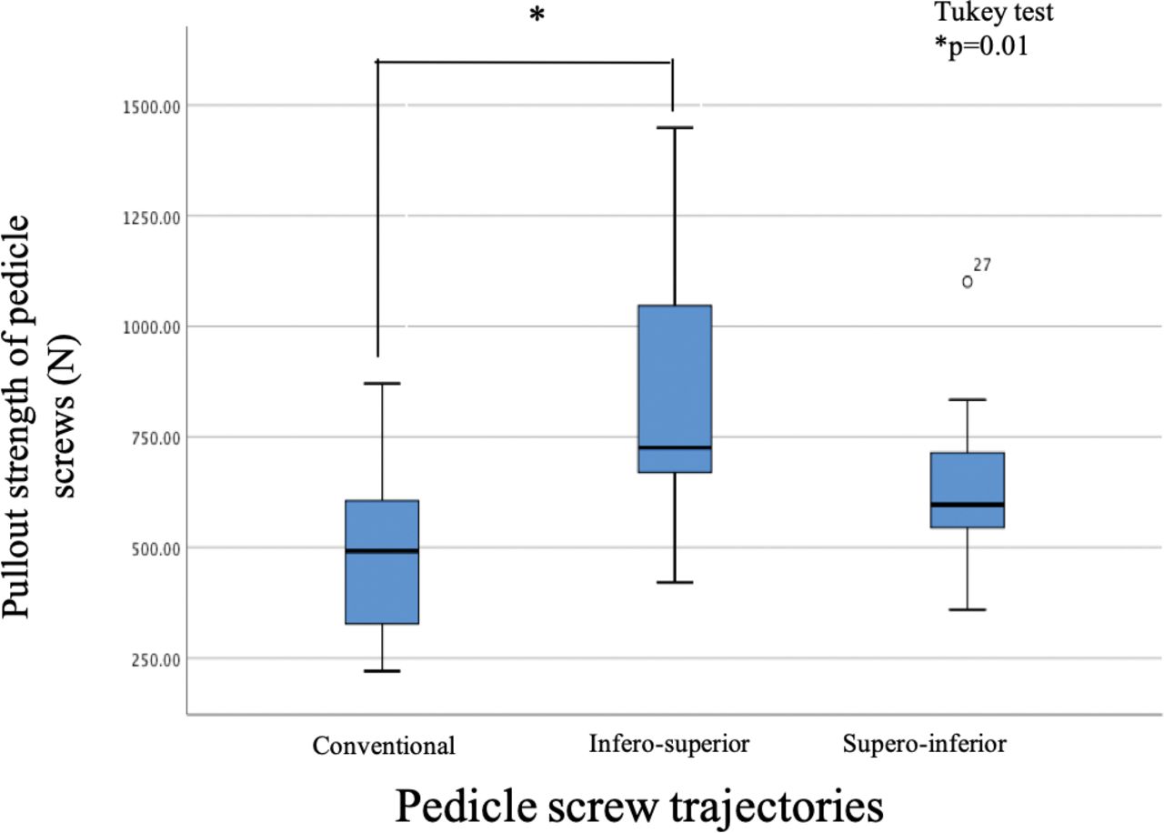
The mean resistance of pull-out strength for the conventional trajectory was 491.72 ± 187.2 N, CBT infero-superior 822.16 ± 295.73 N, and CBT supero-inferior 644.14 ± 201.97 N (Figure 6). The pull-out values of the screws were normally distributed. One-way ANOVA test showed a statistically significant difference between groups. The post hoc Tukey test showed significance only between the conventional and infero-superior trajectory groups.6

Maximum pull-out strength of the screws during pull-out testing. Pull-out strength was greater in the cortical bone infero-superior group. A statistically significant result was found only between the conventional and cortical bone infero-superior groups. Error bars indicate significant differences.
Different screw trajectory will lead to difference in pull-out strength. Santoni et al6 in 2009 showed a 30% increase in pull-out strength with screws with CBT compared with conventional trajectory. Ueno et al12 produced similar results: a 29.5% increase in pull-out strength compared with conventional screws. Matsukawa et al13 showed that controlling bone mineral denisty and screw type yields different pull-out strengths. Our results showed a 67% increase in the trajectory of Santoni et al6 and 30% increase in pull-out strength from our proposed trajectory compared with the conventional trajectory.6,12,13
Decreasing bone quality leads to decrease in screw-bone interface. The infero-superior trajectory start point may engage the inferior lamina cortex first, “force nucleus” (confluence area at the transverse process, lamina, facet inferior, and superior pedicle). To catch more cancellous bone and to engage the cortical area, the supero-inferior trajectory engages the inferior end plate, which is a denser area of the lumbar spine compared with the superior end plate.13,14
Pull-out strength is still considered an effective method of measuring screw strength within a bone. Pull-out test methodology differs between investigators, especially in the manner of fixation of the specimen to the measuring device. Some prefer fixation using embedded resin; others prefer mechanical fixture. No authors have showed superiority over these differing fixation methods.15
CONCLUSIONS
There is a difference between pull-out strengths in pedicle screws with different trajectories; the highest pull-out strength was obtained with the cortical infero-superior group, the lowest with the conventional trajectory. We found a statistically significant difference between pull-out strength of the conventional trajectory and the cortical infero-superior group. No statistical difference of pull-out strength was noted between our proposed trajectory, the conventional trajectory, and the cortical infero-superior group. This study may be used as a baseline for further studies using human cadaveric studies and other biomechanical outcomes.
ACKNOWLEDGMENTS
The authors would like to thank all the staff of Department of Orthopaedics and Traumatology at the Faculty of Medicine University of Indonesia for the suggestions and corrections in writing this article. We would like to express our gratitude to all contributors.
Footnotes
Disclosures and COI: The authors have no conflicts of interest to disclose. Ethical clearance was obtained from the Ethical Review Board of the Faculty of Medicine, University of Indonesia.
- This manuscript is generously published free of charge by ISASS, the International Society for the Advancement of Spine Surgery. Copyright © 2020 ISASS.
In this issue


Jump to section
Related Articles
Cited By...
- No citing articles found.