
Main menu

  • Home
  • Content
    • Current Issue
    • Advance Online Publication
    • Archive
  • About Us
    • About ISASS
    • About the Journal
    • Author Instructions
    • Editorial Board
    • Reviewer Guidelines & Publication Criteria
  • More
    • Advertise
    • Subscribe
    • Alerts
    • Feedback
  • Join Us
  • Reprints & Permissions
  • Sponsored Content
  • Other Publications
    • ijss

User menu

  • My alerts

Search

  • Advanced search
International Journal of Spine Surgery
  • My alerts
International Journal of Spine Surgery

Advanced Search

  • Home
  • Content
    • Current Issue
    • Advance Online Publication
    • Archive
  • About Us
    • About ISASS
    • About the Journal
    • Author Instructions
    • Editorial Board
    • Reviewer Guidelines & Publication Criteria
  • More
    • Advertise
    • Subscribe
    • Alerts
    • Feedback
  • Join Us
  • Reprints & Permissions
  • Sponsored Content
  • Follow ijss on Twitter
  • Visit ijss on Facebook
Research ArticleTumors

Metastatic Paraganglioma of the Spine With SDHB Mutation: Case Report and Review of the Literature

Rashad Jabarkheel, Arjun V. Pendharkar, Jonathan L. Lavezo, Justin Annes, Kaniksha Desai, Hannes Vogel and Atman M. Desai
International Journal of Spine Surgery February 2021, 14 (s4) S37-S45; DOI: https://doi.org/10.14444/7163
Rashad Jabarkheel
1Department of Neurosurgery, Division of Endocrinology and Endocrine Tumor Program, Stanford University School of Medicine, Stanford, California
MD
  • Find this author on Google Scholar
  • Find this author on PubMed
  • Search for this author on this site
Arjun V. Pendharkar
1Department of Neurosurgery, Division of Endocrinology and Endocrine Tumor Program, Stanford University School of Medicine, Stanford, California
MD
  • Find this author on Google Scholar
  • Find this author on PubMed
  • Search for this author on this site
Jonathan L. Lavezo
2Department of Pathology, Division of Endocrinology and Endocrine Tumor Program, Stanford University School of Medicine, Stanford, California
MD
  • Find this author on Google Scholar
  • Find this author on PubMed
  • Search for this author on this site
Justin Annes
3Department of Medicine, Division of Endocrinology and Endocrine Tumor Program, Stanford University School of Medicine, Stanford, California
MD, PHD
  • Find this author on Google Scholar
  • Find this author on PubMed
  • Search for this author on this site
Kaniksha Desai
3Department of Medicine, Division of Endocrinology and Endocrine Tumor Program, Stanford University School of Medicine, Stanford, California
MD
  • Find this author on Google Scholar
  • Find this author on PubMed
  • Search for this author on this site
Hannes Vogel
2Department of Pathology, Division of Endocrinology and Endocrine Tumor Program, Stanford University School of Medicine, Stanford, California
MD
  • Find this author on Google Scholar
  • Find this author on PubMed
  • Search for this author on this site
Atman M. Desai
1Department of Neurosurgery, Division of Endocrinology and Endocrine Tumor Program, Stanford University School of Medicine, Stanford, California
MD
  • Find this author on Google Scholar
  • Find this author on PubMed
  • Search for this author on this site
  • Article
  • Figures & Data
  • Info & Metrics
  • PDF
Loading

ABSTRACT

Background: Paragangliomas (PGLs) are rare neuroendocrine tumors that can arise from any autonomic ganglion of the body. Most PGLs do not metastasize. Here, we present a rare case of metastatic PGL of the spine in a patient with a germline pathogenic succinate dehydrogenase subunit B (SDHB) mutation.

Methods: In addition to a case report we provide a literature review of metastatic spinal PGL to highlight the importance of genetic testing and long-term surveillance of these patients.

Results: A 45-year-old woman with history of spinal nerve root PGL, 17 years prior, presented with back pain of several months' duration. Imaging revealed multilevel lytic lesions throughout the cervical, thoracic, and lumbar spine as well as involvement of the right mandibular condyle and clavicle. Percutaneous biopsy of the L1 spinal lesion confirmed metastatic PGL and the patient underwent posterior tumor resection and instrumented fusion of T7–T11. Postoperatively the patient was found to have a pathogenic SDHB deletion.

Conclusions: Patients with SDHx mutation, particularly SDHB, have increased risk of developing metastatic PGLs. Consequently, these individuals require long-term surveillance given the risk for developing new tumors or disease recurrence, even years to decades after primary tumor resection. Surgical management of spinal metastatic PGL involves correcting spinal instability, minimizing tumor burden, and alleviating epidural cord compression. In patients with metastatic PGL of the spine, genetic testing should be considered.

  • paraganglioma
  • SDHB
  • spine metastasis

INTRODUCTION

Paragangliomas (PGLs) are rare neuroendocrine tumors that can develop anywhere along the sympathetic and parasympathetic ganglia of the body with an estimated prevalence of 0.2 to 1 per 100,000.1,2 When PGLs arise from chromaffin cells they frequently overproduce catecholamines. PGLs arising from adrenal chromaffin cells are commonly known as pheochromocytomas and account for 80%–85% of chromaffin cell PGLs.3 Extra-adrenal PGLs are less frequent, are primarily found along the parasympathetic ganglia of the head and neck, and are more likely to be biochemically silent.4,5 Extra-adrenal PGLs generally present between ages 40 and 50 with symptoms of mass effect specific to their location of origin.5–7

Most PGLs do not metastasize, with only 10%–17% ultimately being metastatic, although extra-adrenal PGLs are thought to have greater metastatic potential.8,9 Diagnosis of metastatic PGL is difficult as there are no reliable cellular or molecular markers of metastatic disease, and thus progression is necessary for diagnosis.8,10 Given the rarity of metastatic disease, there is a relative paucity of literature on metastatic extra-adrenal PGL, especially for spinal metastasis.4,10–14 Here, we present a report of a patient with metastatic extra-adrenal PGL of the spine presenting 27 years after resection of primary spinal nerve root PGL who was found to have a germline succinate dehydrogenase subunit B (SDHB) mutation.

CASE PRESENTATION

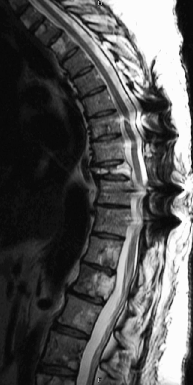
A 45-year-old woman with history of a large (> 8 cm) spinal nerve root PGL, status postresection 27 years prior at an outside hospital, with a recurrence in the right tibia, status postresection 6 years prior also at an outside hospital, presented for care at our institution with severe thoracic back pain radiating around the rib cage for several months' duration, which was worse with movement. She endorsed feeling weak, although she was able to ambulate normally. She denied lower extremity symptoms, saddle anesthesia, and changes in urination or defecation. Physical exam was normal except for tenderness to palpation along the lower thoracic spine. She reported no overt signs of catecholamine excess such as tremors, headaches, visual symptoms, palpitations, weight loss, and diaphoresis. Preoperatively, both her blood pressure and pulse were within normal limits and she was not on any antihypertensive medications. On imaging with computed tomography (CT), magnetic resonance imaging (MRI), and fluorodeoxyglucose positron emission tomography, she was found to have multilevel lytic lesions throughout the cervical, thoracic, and lumbar spine at C1, C4–C7, L1–L3, as well as involvement of the right mandibular condyle and clavicle. She had compression fractures of the C3 and T9 vertebral bodies both with greater than 80% loss of height, and retropulsion causing moderate-to-severe spinal canal stenosis. Spinal canal extension was present at T4, T5, T9, L1, and L4, and most severe at T9, which was considered to be the symptomatic level (Figure 1).

Figure 1
  • Download figure
  • Open in new tab
  • Download powerpoint
Figure 1

Preoperative T2 magnetic resonance imaging (MRI) complete spine images showing extensive metastatic disease of the spine. (Left) Sagittal T2 MRI of the upper spine showing pathologic compression fracture of the T9 vertebral body with retropulsion of the posterior vertebral body obliterating the cerebrospinal fluid space. (Middle) Sagittal T2 MRI of the lower spine showing extensive metastatic disease throughout the lumbar spine. (Right) Axial T2 MRI at the level of the T9 vertebral body.

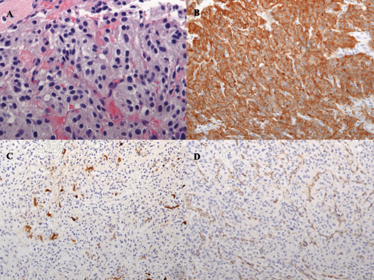
On the day after admission, percutaneous biopsy was performed of the L1 soft tissue lesion, and this confirmed metastatic PGL. Histologic sections of the biopsy specimen contained abundant groups of nested cells characterized by mild anisonucleosis, stippled nuclear chromatin, and abundant pale eosinophilic and slightly granular cytoplasm. Mitotic figures were seen at 7 in 10 high-power fields (Figure 2A). Immunohistochemical stains for S100, synaptophysin, chromogranin, and succinate dehydrogenase subunit B (SDHB) were performed. Tumor cells stained positive for synaptophysin (Figure 2B) and chromogranin. The S100 stain highlighted a fragmented network of sustenacular cells surrounding the neoplastic cells (Figure 2C). SDHB showed intracytoplasmic granular positivity within vascular endothelium but complete loss of staining in the tumor cells (Figure 2D), indicative of an underlying SDHx gene mutation.

Figure 2
  • Download figure
  • Open in new tab
  • Download powerpoint
Figure 2

Histologic and immunohistochemical sections of metastatic paraganglioma biopsy specimen. (A) Hematoxylin-eosin stain of metastatic paraganglioma (×60). Histologic sections of the biopsy specimen showing an epithelioid proliferation of cells with nested architecture (“Zellballen”) with a delicate tumor vascular network between nests. The epithelioid cells show round to ovoid nuclei, minimal nuclear pleomorphism, and stippled chromatin with abundant granular amphophilic cytoplasm. Mitotic figures are seen at 7 in 10 high-power fields. (B) Immunohistochemical stain for synaptophysin showing positive granular cytoplasmic staining in tumor cells (×20). (C) Immunohistochemical stain for S-100 highlighting fragmented network of sustenacular cells surrounding tumor cells (×20). (D) Immunohistochemical stain for succinate dehydrogenase subunit B (SDHB) showing absence of SDHB among tumor cells, while maintaining positive internal control within vascular endothelium (×20).

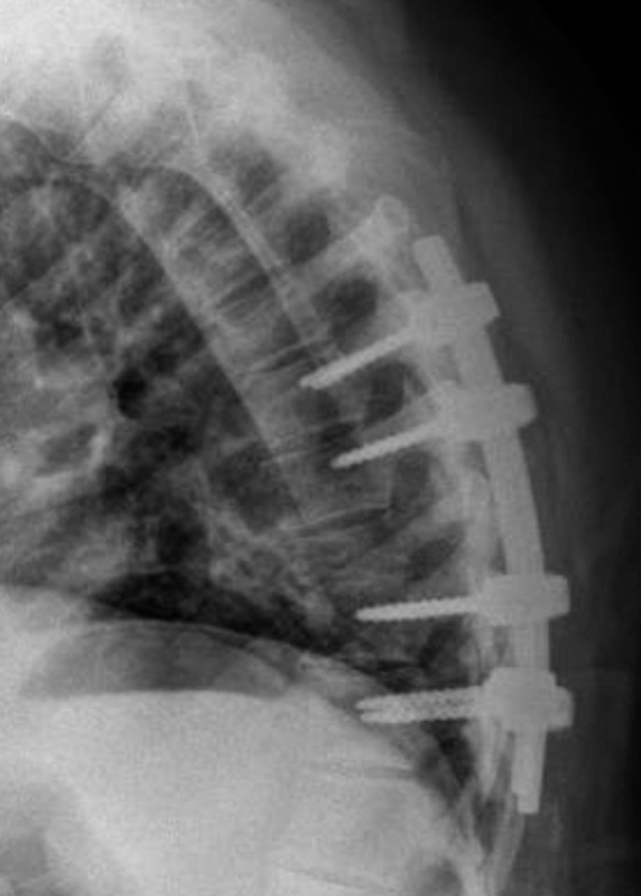
Given clinical concern for spinal instability and epidural cord compression, the patient underwent lateral extracavitary approach for partial corpectomy and laminectomy for tumor resection at T9 and posterior instrumented fusion of T7–T11. The patient tolerated the procedure well with 400 mL of blood loss and no hypertensive complications. In consultation with anesthesiology, preoperative alpha blockade was not performed.15 Grossly, the tumor appeared as fragments of red-brown soft tumor mixed with blood and trabecular bone fragments. Histologic and immunohistochemical analysis was consistent with recent biopsy. A postoperative x-ray was obtained to confirm adequate positioning of hardware (Figure 3). Patient was discharged on postoperative day 5 with a thoracic lumbosacral orthosis brace and cervical collar. Although no family history of pheochromocytoma or PGL was obtained on review, germline testing was recommended with particular concern for SDHB mutation given negative SDHB immunohistochemistry and the aggressive metastatic nature of the PGL. The patient was found to have a pathogenic SDHB deletion (c.166_170delCCTCA) which resulted in a frame shift and protein truncation (p.P56Yfs*5). Genetic testing was arranged for at-risk relatives, where several affected individuals were identified.

Figure 3
  • Download figure
  • Open in new tab
  • Download powerpoint
Figure 3

Postoperative sagittal x-ray of the spine showing instrumentation.

At 18 months after surgery the patient's metastatic disease is stable (Figure 4). She has received 10 sessions of radiation to the spine and 8 cycles of chemotherapy with cyclophosphamide, dacarbazine, and vincristine. Additionally, she has been receiving monthly octreotide and denosumab injections to promote disease stabilization and prevent further bone loss, respectively. Given the patient's advanced disease, additional screening beyond standard response surveillance was not pursued.

Figure 4
  • Download figure
  • Open in new tab
  • Download powerpoint
Figure 4

Post-operative T2 magnetic resonance imaging of thoracic spine at 1.5 years showing stable disease.

RESULTS AND DISCUSSION

Metastatic extra-adrenal PGL to the spine is a rare phenomenon with limited case reports and 2 small case series described in the English literature (Table 1).4,10,11 Here, we present what is, to our knowledge, the fourth case report of metastatic PGL to the spine found to have a mutation of an SDH subunit, (SDHA, SDHB, SDHC, SDHD) or assembly factor (SDH-AF2).4 While there are limited data available on the metastatic tendencies of extra-adrenal PGL, studies looking at both metastatic pheochromocytomas and extra-adrenal PGLs have found bone to be the most common site of metastasis followed by liver and lung.16–19 The spine is the most common site of bone metastasis.16 In this case our patient had an interval of 27 years between total resection of her primary tumor and spinal metastasis. This extended interval is consistent with previous case reports as shown in Table 1.10,12,20

View this table:
  • View inline
  • View popup
  • Download powerpoint
Table 1

Previous reports of metastatic extra-adrenal PGL to the spine.

Genetic testing is recommended for all patients with PGLs and their first-degree relatives.3,43 Over the past several years it has been shown that 20%–40% of patients with pheochromocytoma and extra-adrenal PGL have a germline mutation in SDHx, NF1, VHL, or RET.44–46 While mutations in NF1, VHL, and RET genes cause well-characterized hereditary syndromes, the association between SDHx mutations and PGLs was more recently elucidated.6,47–49 SDH is a mitochondrial enzyme complex that plays a role in both the tricarboxylic acid cycle and in the electron transport chain. SDH genes function as classical tumor suppressor genes where somatic loss of heterozygosity of the wild-type allele is observed in tumors.6,50 While the precise pathogenic mechanism of SDH-mutation-dependent tumor formation is incompletely understood, the leading theory revolves around the role of succinate as an oncometabolite.51 Specifically, it is thought that disruption of SDH complex function, which leads to an accumulation of succinate, increases the risk of cancer because succinate accumulation competitively inhibits α-ketoglutarate dependent enzymes such as prolyl hydroxylases and histone demethylases. Inhibition of hypoxia-inducible factor prolyl hydroxylases leads to the stabilization of hypoxia-inducible factors, which normally promote angiogenesis and cell survival in hypoxic conditions. Inhibition of histone demethylases causes a cell to adopt a hypermethylator phenotype that is thought to silence genes associated with neuroendocrine differentiation.52–54 SDHx mutations are the most frequent hereditary cause of extra-adrenal PGLs with autosomal-dominant mutations in SDHD and SDHB being the most common.6,49 Immunohistochemical and genetic testing all PGLs for SDHx mutation, and in particular for SDHB mutation, is critically important as 30%–70% of metastatic PGLs have been found to have an SDHB germline mutation.48,55,56 Thus, while there are no definite histological markers of metastatic PGL, SDHB mutation is a strong independent predictor in addition to primary tumor size > 5 cm and extra-adrenal location.8 Interestingly, although our patient's primary tumor was reportedly > 8 cm in size and extra-adrenal in location she was not screened for SDHB mutation prior to receiving care at our institution for her spinal metastasis.

Whole-body scanning for detection of metastatic PGL is recommended at the time of primary tumor detection.3 Metastatic disease can be detected through a combination of anatomical (CT, MRI) and scintigraphic imaging techniques (fluorodeoxyglucose positron emission tomography, metaiodobenzylguanidine scintigraphy, and more recently somatostatin receptor scintigraphy with 68Ga-DOTATATE).57,58 Fluorodeoxyglucose positron emission tomography is currently the preferred imaging technique for detecting metastatic disease although several recent studies suggest that 68Ga-DOTATATE may have greater sensitivity particularly in the context of SDH-related disease.3,59,60 Long-term imaging follow-up in addition to annual clinical evaluation and laboratory testing is required for all patients with PGLs as approximately 50% of metastatic PGLs present metachronously and in particular for patients with SDHx mutations, who have an increased risk for metastatic disease.19 At this time no clear guidelines exist regarding the optimal frequency of imaging for PGL patients.3 In terms of laboratory testing, the Endocrine Society recommends lifelong annual testing of plasma or urine metanephrine levels to assess for recurrent or persistent disease.3

Treatment of metastatic PGL of the spine involves a combination of surgery, radiation therapy, and chemotherapy. As with other metastatic tumors of the spine the primary goals of surgery are management of spinal instability caused by lytic lesions, and decompression of the spinal cord secondary to any epidural tumor.61 Surgical resection of the primary tumor has been shown to improve overall survival in cases of synchronous metastatic PGL.62 Radiation therapy is the primary of method of local control for metastatic disease that is unresectable. Chemotherapy is reserved for widely metastatic disease with cyclophosphamide, dacarbazine, and vincristine being the preferred regimen.63 Overall, metastatic PGL is difficult to treat with 5-year overall survival at approximately 60%.61

In conclusion, metastasis to the spine is a rare but important complication in patients with PGL that can lead to significant pain and disability. Genetic testing is recommended for all patients with PGLs. Patients with SDHx mutations are more likely to develop metastatic disease and SDHx mutation status is the current best predictor of metastatic PGL. SDHx mutation carriers must have frequent long-term imaging surveillance performed given the potential for metastasis several years to decades after primary resection. Neurosurgical management of metastatic PGL of the spine involves correcting spinal instability and alleviating epidural cord compression. When caring for patient with metastatic PGL of the spine, genetic testing for patients and their families should be considered.

Footnotes

  • Disclosures and COI: None declared. This research did not receive any specific grant from funding agencies in the public, commercial, or not-for-profit sectors. Patient's written informed consent for publication was obtained.

  • This manuscript is generously published free of charge by ISASS, the International Society for the Advancement of Spine Surgery. Copyright © 2020 ISASS

REFERENCES

  1. 1 .↵
    1. Simpson LN,
    2. Hughes BD,
    3. Karikari IO, et al.
    Catecholamine-secreting paraganglioma of the thoracic spinal column: report of an unusual case and review of the literature. Neurosurgery. 2012;70(4):E1049–1052; discussion E1052.
    OpenUrlPubMed
  2. 2 .↵
    1. Baysal BE.
    Hereditary paraganglioma targets diverse paraganglia. J Med Genet. 2002;39(9):617–622.
    OpenUrlAbstract/FREE Full Text
  3. 3 .↵
    1. Lenders JW,
    2. Duh QY,
    3. Eisenhofer G, et al.
    Pheochromocytoma and paraganglioma: an Endocrine Society clinical practice guideline. J Clin Endocrinol Metab. 2014;99(6):1915–1942.
    OpenUrlCrossRefPubMed
  4. 4 .↵
    1. Lau D,
    2. La Marca F,
    3. Camelo-Piragua S,
    4. Park P.
    Metastatic paraganglioma of the spine: case report and review of the literature. Clin Neurol Neurosurg. 2013;115(9):1571–1574.
    OpenUrl
  5. 5 .↵
    1. Erickson D,
    2. Kudva YC,
    3. Ebersold MJ, et al.
    Benign paragangliomas: clinical presentation and treatment outcomes in 236 patients. J Clin Endocrinol Metab. 2001;86(11):5210–5216.
    OpenUrlCrossRefPubMed
  6. 6 .↵
    1. Welander J,
    2. Soderkvist P,
    3. Gimm O.
    Genetics and clinical characteristics of hereditary pheochromocytomas and paragangliomas. Endocr Relat Cancer. 2011;18(6):R253–276.
    OpenUrlAbstract/FREE Full Text
  7. 7 .↵
    1. Al-Harthy M,
    2. Al-Harthy S,
    3. Al-Otieschan A,
    4. Velagapudi S,
    5. Alzahrani AS.
    Comparison of pheochromocytomas and abdominal and pelvic paragangliomas with head and neck paragangliomas. Endocr Pract. 2009;15(3):194–202.
    OpenUrlPubMed
  8. 8 .↵
    1. Plouin PF,
    2. Fitzgerald P,
    3. Rich T, et al.
    Metastatic pheochromocytoma and paraganglioma: focus on therapeutics. Horm Metab Res. 2012;44(5):390–399.
    OpenUrlCrossRefPubMed
  9. 9 .↵
    1. Laird AM,
    2. Gauger PG,
    3. Doherty GM,
    4. Miller BS.
    Paraganglioma: not just an extra-adrenal pheochromocytoma. Langenbecks Arch Surg. 2012;397(2):247–253.
    OpenUrlPubMed
  10. 10 .↵
    1. Jia Q,
    2. Yin H,
    3. Yang J, et al.
    Treatment and outcome of metastatic paraganglioma of the spine. Eur Spine J. 2018;27(4):859–867.
    OpenUrl
  11. 11 .↵
    1. Kapetanakis S,
    2. Chourmouzi D,
    3. Gkasdaris G,
    4. Katsaridis V,
    5. Eleftheriadis E,
    6. Givissis P.
    Functional extra-adrenal paraganglioma of the retroperitoneum giving thoracolumbar spine metastases after a five-year disease-free follow-up: a rare malignant condition with challenging management. Pan Afr Med J. 2017;28:94.
    OpenUrl
  12. 12 .↵
    1. Lehmen JA,
    2. Babbel DM,
    3. Mikhitarian K,
    4. Choma TJ.
    Paraganglioma presenting as metastatic lesion in a cervical vertebra: a case report and review of the literature. Spine (Phila Pa 1976). 2010;35(5):E152–154.
    OpenUrl
  13. 13 .
    1. Hamilton MA,
    2. Tait D.
    Metastatic paraganglioma causing spinal cord compression. Br J Radiol. 2000;73(872):901–904.
    OpenUrlPubMed
  14. 14 .↵
    1. Kwan RB,
    2. Erasmus AM,
    3. Hunn AW, et al.
    Pre-operative embolisation of metastatic paraganglioma of the thoracic spine. J Clin Neurosci. 2010;17(3):394–396.
    OpenUrlCrossRefPubMed
  15. 15 .↵
    1. Isaacs M,
    2. Lee P.
    Preoperative alpha-blockade in phaeochromocytoma and paraganglioma: is it always necessary? Clin Endocrinol (Oxf). 2017;86(3):309–314.
    OpenUrl
  16. 16 .↵
    1. Zelinka T,
    2. Timmers HJ,
    3. Kozupa A, et al.
    Role of positron emission tomography and bone scintigraphy in the evaluation of bone involvement in metastatic pheochromocytoma and paraganglioma: specific implications for succinate dehydrogenase enzyme subunit B gene mutations. Endocr Relat Cancer. 2008;15(1):311–323.
    OpenUrlAbstract/FREE Full Text
  17. 17 .
    1. Chrisoulidou A,
    2. Kaltsas G,
    3. Ilias I,
    4. Grossman AB.
    The diagnosis and management of malignant phaeochromocytoma and paraganglioma. Endocr Relat Cancer. 2007;14(3):569–585.
    OpenUrlAbstract/FREE Full Text
  18. 18 .
    1. Mediouni A,
    2. Ammari S,
    3. Wassef M, et al.
    Malignant head/neck paragangliomas. Comparative study. Eur Ann Otorhinolaryngol Head Neck Dis. 2014;131(3):159–166.
    OpenUrl
  19. 19 .↵
    1. Ayala-Ramirez M,
    2. Palmer JL,
    3. Hofmann MC, et al.
    Bone metastases and skeletal-related events in patients with malignant pheochromocytoma and sympathetic paraganglioma. J Clin Endocrinol Metab. 2013;98(4):1492–1497.
    OpenUrlCrossRefPubMed
  20. 20 .↵
    1. Ayala-Ramirez M,
    2. Feng L,
    3. Johnson MM, et al.
    Clinical risk factors for malignancy and overall survival in patients with pheochromocytomas and sympathetic paragangliomas: primary tumor size and primary tumor location as prognostic indicators. J Clin Endocrinol Metab. 2011;96(3):717–725.
    OpenUrlCrossRefPubMed
  21. 21 .
    1. Narechania S,
    2. Bath A,
    3. Ghassemi L, et al.
    Paraganglioma presenting as postpartum fever of unknown origin. Case Rep Endocrinol. 2015;2015:864719.
    OpenUrl
  22. 22 .
    1. Bickmann JK,
    2. Sollfrank S,
    3. Schad A, et al.
    Phenotypic variability and risk of malignancy in SDHC-linked paragangliomas: lessons from three unrelated cases with an identical germline mutation (p.Arg133*). J Clin Endocrinol Metab. 2014;99(3):E489–496.
    OpenUrlPubMed
  23. 23 .
    1. Feng N,
    2. Li X,
    3. Gao HD,
    4. Liu ZL,
    5. Shi LJ,
    6. Liu WZ.
    Urinary bladder malignant paraganglioma with vertebral metastasis: a case report with literature review. Chin J Cancer. 2013;32(11):624–628.
    OpenUrl
  24. 24 .
    1. Sasaki K,
    2. Inose H,
    3. Kawabata S, et al.
    Combined surgical and radiosurgical treatment for a symptomatic cervical metastasis in a case of malignant paraganglioma: a case report. BMC Res Notes. 2013;6:494.
    OpenUrl
  25. 25 .
    1. He J,
    2. Wang X,
    3. Zheng W,
    4. Zhao Y.
    Retroperitoneal paraganglioma with metastasis to the abdominal vertebra: a case report. Diagn Pathol. 2013;8:52.
    OpenUrl
  26. 26 .
    1. Richter A,
    2. Halm HF,
    3. Lerner T,
    4. Liljenqvist UR,
    5. Quante M.
    Long-term follow-up after en bloc resection and reconstruction of a solitary paraganglioma metastasis in the first lumbar vertebral body: a case report. J Med Case Rep. 2011;5:45.
    OpenUrl
  27. 27 .
    1. Persu A,
    2. Amyere M,
    3. Gutierrez-Roelens I, et al.
    Rare presentation of familial paraganglioma without evidence of mutation in the SDH, RET and VHL genes: towards further genetic heterogeneity. J Hypertens. 2009;27(1):76–82.
    OpenUrlPubMed
  28. 28 .
    1. Prabhu S,
    2. Jacob JJ,
    3. Thomas N,
    4. Oommen R.
    Visual vignette. Solitary sacral metastasis from a malignant paraganglioma. Endocr Pract. 2008;14(1):131.
    OpenUrlPubMed
  29. 29 .
    1. Yamaguchi S,
    2. Hida K,
    3. Nakamura N,
    4. Seki T,
    5. Iwasaki Y.
    Multiple vertebral metastases from malignant cardiac pheochromocytoma—case report. Neurol Med Chir (Tokyo). 2003;43(7):352–355.
    OpenUrlCrossRefPubMed
  30. 30 .
    1. U-King-Im JM,
    2. Carroll TA,
    3. Morris K.
    Vertebral metastatic chemodectoma: imaging and therapeutic octreotide. Case report. J Neurosurg. 2002;97(1 Suppl):106–109.
    OpenUrlPubMed
  31. 31 .
    1. Mori S,
    2. Okura T,
    3. Kitami Y, et al.
    A case of metastatic extra-adrenal pheochromocytoma 12 years after surgery. Hypertens Res. 2002;25(1):141–144.
    OpenUrlCrossRefPubMed
  32. 32 .
    1. Absher KJ,
    2. Witte DA,
    3. Truong LD,
    4. Ramzy I,
    5. Mody DR,
    6. Ostrowski ML.
    Aspiration biopsy of osseous metastasis of retroperitoneal paraganglioma. Report of a case with cytologic features and differential diagnostic considerations. Acta Cytol. 2001;45(2):249–253.
    OpenUrlPubMed
  33. 33 .
    1. Blasius S,
    2. Brinkschmidt C,
    3. Poremba C, et al.
    Metastatic retroperitoneal paraganglioma in a 16-year-old girl. Case report, molecular pathological and cytogenetic findings. Pathol Res Pract. 1998;194(6):439–444.
    OpenUrlPubMed
  34. 34 .
    1. Brodkey JA,
    2. Brodkey JS,
    3. Watridge CB.
    Metastatic paraganglioma causing spinal cord compression. Spine (Phila Pa 1976). 1995;20(3):367–372.
    OpenUrl
  35. 35 .
    1. Gabriel EM,
    2. Sampson JH,
    3. Dodd LG,
    4. Turner DA.
    Glomus jugulare tumor metastatic to the sacrum after high-dose radiation therapy: case report. Neurosurgery. 1995;37(5):1001–1005.
    OpenUrlPubMed
  36. 36 .
    1. North CA,
    2. Zinreich ES,
    3. Christensen WN,
    4. North RB.
    Multiple spinal metastases from paraganglioma. Cancer. 1990;66(10):2224–2228.
    OpenUrlCrossRefPubMed
  37. 37 .
    1. Siddiqui MZ,
    2. Von Eyben FE,
    3. Spanos G.
    High-voltage irradiation and combination chemotherapy for malignant pheochromocytoma. Cancer. 1988;62(4):686–690.
    OpenUrlCrossRefPubMed
  38. 38 .
    1. Osborn RE,
    2. Mojtahedi S.
    Paraganglioma metastatic to the cervical spine. Comput Radiol. 1986;10(4):167–170.
    OpenUrlCrossRefPubMed
  39. 39 .
    1. Kapetanakis S,
    2. Chourmouzi D,
    3. Gkasdaris G,
    4. Katsaridis V,
    5. Eleftheriadis E,
    6. Givissis P.
    A rare case of spinal cord compression due to cervical spine metastases from paraganglioma of the jugular foramen-how should it be treated? J Surg Case Rep. 2018;2018(2):rjy005.
  40. 40 .
    1. Lv G,
    2. Lu L,
    3. Dai Z.
    Paragangliomas of the spine. Turk Neurosurg. 2017;27(3):401–407.
    OpenUrl
  41. 41 .
    1. Jang Khan NA,
    2. Ullah S,
    3. Siddiqui HU,
    4. Karim A.
    spinal cord compression by metastatic thoracic spine paraganglioma. J Ayub Med Coll Abbottabad. 2016;28(3):617–619.
    OpenUrl
  42. 42 .
    1. Kitagawa R,
    2. Murakami H,
    3. Kato S,
    4. Nakada M,
    5. Demura S,
    6. Tsuchiya H.
    En bloc resection and reconstruction using a frozen tumor-bearing bone for metastases of the spine and cranium from retroperitoneal paraganglioma. World Neurosurg. 2016;90:698.e691–698.e695.
    OpenUrl
  43. 43 .↵
    1. Cavenagh T,
    2. Patel J,
    3. Nakhla N, et al.
    Succinate dehydrogenase mutations: paraganglioma imaging and at-risk population screening. Clin Radiol. 2019;74(3):169–177.
    OpenUrl
  44. 44 .↵
    1. Buffet A,
    2. Venisse A,
    3. Nau V, et al.
    A decade (2001–2010) of genetic testing for pheochromocytoma and paraganglioma. Horm Metab Res. 2012;44(5):359–366.
    OpenUrlCrossRefPubMed
  45. 45 .
    1. Neumann HP,
    2. Bausch B,
    3. McWhinney SR, et al.
    Germ-line mutations in nonsyndromic pheochromocytoma. N Engl J Med. 2002;346(19):1459–1466.
    OpenUrlCrossRefPubMed
  46. 46 .↵
    1. Fishbein L,
    2. Merrill S,
    3. Fraker DL,
    4. Cohen DL,
    5. Nathanson KL.
    Inherited mutations in pheochromocytoma and paraganglioma: why all patients should be offered genetic testing. Ann Surg Oncol. 2013;20(5):1444–1450.
    OpenUrlCrossRefPubMed
  47. 47 .↵
    1. Baysal BE,
    2. Ferrell RE,
    3. Willett-Brozick JE, et al.
    Mutations in SDHD, a mitochondrial complex II gene, in hereditary paraganglioma. Science. 2000;287(5454):848–851.
    OpenUrlAbstract/FREE Full Text
  48. 48 .↵
    1. Neumann HP,
    2. Pawlu C,
    3. Peczkowska M, et al.
    Distinct clinical features of paraganglioma syndromes associated with SDHB and SDHD gene mutations. JAMA. 2004;292(8):943–951.
    OpenUrlCrossRefPubMed
  49. 49 .↵
    1. van Nederveen FH,
    2. Gaal J,
    3. Favier J, et al.
    An immunohistochemical procedure to detect patients with paraganglioma and phaeochromocytoma with germline SDHB, SDHC, or SDHD gene mutations: a retrospective and prospective analysis. Lancet Oncol. 2009;10(8):764–771.
    OpenUrlCrossRefPubMed
  50. 50 .↵
    1. Fishbein L,
    2. Nathanson KL.
    Pheochromocytoma and paraganglioma: understanding the complexities of the genetic background. Cancer Genet. 2012;205(1–2):1–11.
    OpenUrlCrossRefPubMed
  51. 51 .↵
    1. Sajnani K,
    2. Islam F,
    3. Smith RA,
    4. Gopalan V,
    5. Lam AK.
    Genetic alterations in Krebs cycle and its impact on cancer pathogenesis. Biochimie. 2017;135:164–172.
    OpenUrlCrossRef
  52. 52 .↵
    1. Letouze E,
    2. Martinelli C,
    3. Loriot C, et al.
    SDH mutations establish a hypermethylator phenotype in paraganglioma. Cancer Cell. 2013;23(6):739–752.
    OpenUrlCrossRefPubMed
  53. 53 .
    1. Tretter L,
    2. Patocs A,
    3. Chinopoulos C.
    Succinate, an intermediate in metabolism, signal transduction, ROS, hypoxia, and tumorigenesis. Biochim Biophys Acta. 2016;1857(8):1086–1101.
    OpenUrlCrossRefPubMed
  54. 54 .↵
    1. Fliedner SM,
    2. Shankavaram U,
    3. Marzouca G, et al.
    Hypoxia-inducible factor 2alpha mutation-related paragangliomas classify as discrete pseudohypoxic subcluster. Neoplasia. 2016;18(9):567–576.
    OpenUrl
  55. 55 .↵
    1. King KS,
    2. Prodanov T,
    3. Kantorovich V, et al.
    Metastatic pheochromocytoma/paraganglioma related to primary tumor development in childhood or adolescence: significant link to SDHB mutations. J Clin Oncol. 2011;29(31):4137–4142.
    OpenUrlAbstract/FREE Full Text
  56. 56 .↵
    1. Gimenez-Roqueplo AP,
    2. Favier J,
    3. Rustin P, et al.
    Mutations in the SDHB gene are associated with extra-adrenal and/or malignant phaeochromocytomas. Cancer Res. 2003;63(17):5615–5621.
    OpenUrlAbstract/FREE Full Text
  57. 57 .↵
    1. Gimenez-Roqueplo AP,
    2. Caumont-Prim A,
    3. Houzard C, et al.
    Imaging work-up for screening of paraganglioma and pheochromocytoma in SDHx mutation carriers: a multicenter prospective study from the PGL.EVA Investigators. J Clin Endocrinol Metab. 2013;98(1):E162–173.
    OpenUrlCrossRefPubMed
  58. 58 .↵
    1. Lepoutre-Lussey C,
    2. Caramella C,
    3. Bidault F, et al.
    Screening in asymptomatic SDHx mutation carriers: added value of (1)(8)F-FDG PET/CT at initial diagnosis and 1-year follow-up. Eur J Nucl Med Mol Imaging. 2015;42(6):868–876.
    OpenUrlCrossRefPubMed
  59. 59 .↵
    1. Timmers HJ,
    2. Kozupa A,
    3. Chen CC, et al.
    Superiority of fluorodeoxyglucose positron emission tomography to other functional imaging techniques in the evaluation of metastatic SDHB-associated pheochromocytoma and paraganglioma. J Clin Oncol. 2007;25(16):2262–2269.
    OpenUrlAbstract/FREE Full Text
  60. 60 .↵
    1. Janssen I,
    2. Blanchet EM,
    3. Adams K, et al.
    Superiority of [68ga]-dotatate pet/ct to other Functional Imaging Modalities in the Localization of SDHB-associated metastatic pheochromocytoma and paraganglioma. Clin Cancer Res. 2015;21(17):3888–3895.
    OpenUrlAbstract/FREE Full Text
  61. 61 .↵
    1. Jimenez P,
    2. Tatsui C,
    3. Jessop A,
    4. Thosani S,
    5. Jimenez C.
    Treatment for malignant pheochromocytomas and paragangliomas: 5 years of progress. Curr Oncol Rep. 2017;19(12):83.
    OpenUrl
  62. 62 .↵
    1. Roman-Gonzalez A,
    2. Zhou S,
    3. Ayala-Ramirez M, et al.
    Impact of surgical resection of the primary tumor on overall survival in patients with metastatic pheochromocytoma or sympathetic paraganglioma. Ann Surg. 2018;268(1):172–178.
    OpenUrl
  63. 63 .↵
    1. Fliedner SM,
    2. Lehnert H,
    3. Pacak K.
    Metastatic paraganglioma. Semin Oncol. 2010;37(6):627–637.
    OpenUrlCrossRefPubMed
PreviousNext
Back to top

In this issue

International Journal of Spine Surgery
Vol. 14, Issue s4
1 Feb 2021
  • Table of Contents
  • Index by author

Print
Download PDF
Article Alerts
Sign In to Email Alerts with your Email Address
Email Article

Thank you for your interest in spreading the word on International Journal of Spine Surgery.

NOTE: We only request your email address so that the person you are recommending the page to knows that you wanted them to see it, and that it is not junk mail. We do not capture any email address.

Enter multiple addresses on separate lines or separate them with commas.
Metastatic Paraganglioma of the Spine With SDHB Mutation: Case Report and Review of the Literature
(Your Name) has sent you a message from International Journal of Spine Surgery
(Your Name) thought you would like to see the International Journal of Spine Surgery web site.
CAPTCHA
This question is for testing whether or not you are a human visitor and to prevent automated spam submissions.
Citation Tools
Metastatic Paraganglioma of the Spine With SDHB Mutation: Case Report and Review of the Literature
Rashad Jabarkheel, Arjun V. Pendharkar, Jonathan L. Lavezo, Justin Annes, Kaniksha Desai, Hannes Vogel, Atman M. Desai
International Journal of Spine Surgery Feb 2021, 14 (s4) S37-S45; DOI: 10.14444/7163

Citation Manager Formats

  • BibTeX
  • Bookends
  • EasyBib
  • EndNote (tagged)
  • EndNote 8 (xml)
  • Medlars
  • Mendeley
  • Papers
  • RefWorks Tagged
  • Ref Manager
  • RIS
  • Zotero
Share
Metastatic Paraganglioma of the Spine With SDHB Mutation: Case Report and Review of the Literature
Rashad Jabarkheel, Arjun V. Pendharkar, Jonathan L. Lavezo, Justin Annes, Kaniksha Desai, Hannes Vogel, Atman M. Desai
International Journal of Spine Surgery Feb 2021, 14 (s4) S37-S45; DOI: 10.14444/7163
Twitter logo Facebook logo Mendeley logo
  • Tweet Widget
  • Facebook Like
  • Google Plus One

Jump to section

  • Article
    • ABSTRACT
    • INTRODUCTION
    • CASE PRESENTATION
    • RESULTS AND DISCUSSION
    • Footnotes
    • REFERENCES
  • Figures & Data
  • Info & Metrics
  • PDF

Related Articles

  • No related articles found.
  • PubMed
  • Google Scholar

Cited By...

  • No citing articles found.
  • Google Scholar

More in this TOC Section

  • A Rare Entity in the Lumbar Epidural Region: T-Cell Lymphoblastic Lymphoma
  • Fibrous Dysplasia of the Spine—A Case Involving the Polyostotic Form Isolated to the Thoracolumbar Spine
Show more Tumors

Similar Articles

Keywords

  • paraganglioma
  • SDHB
  • spine metastasis

Content

  • Current Issue
  • Latest Content
  • Archive

More Information

  • About IJSS
  • About ISASS
  • Privacy Policy

More

  • Subscribe
  • Alerts
  • Feedback

Other Services

  • Author Instructions
  • Join ISASS
  • Reprints & Permissions

© 2025 International Journal of Spine Surgery

International Journal of Spine Surgery Online ISSN: 2211-4599

Powered by HighWire