
Main menu

  • Home
  • Content
    • Current Issue
    • Advance Online Publication
    • Archive
  • About Us
    • About ISASS
    • About the Journal
    • Author Instructions
    • Editorial Board
    • Reviewer Guidelines & Publication Criteria
  • More
    • Advertise
    • Subscribe
    • Alerts
    • Feedback
  • Join Us
  • Reprints & Permissions
  • Sponsored Content
  • Other Publications
    • ijss

User menu

  • My alerts

Search

  • Advanced search
International Journal of Spine Surgery
  • My alerts
International Journal of Spine Surgery

Advanced Search

  • Home
  • Content
    • Current Issue
    • Advance Online Publication
    • Archive
  • About Us
    • About ISASS
    • About the Journal
    • Author Instructions
    • Editorial Board
    • Reviewer Guidelines & Publication Criteria
  • More
    • Advertise
    • Subscribe
    • Alerts
    • Feedback
  • Join Us
  • Reprints & Permissions
  • Sponsored Content
  • Follow ijss on Twitter
  • Visit ijss on Facebook
Research ArticleOther & Special Categories

Symptomatic Vertebral Hemangioma of the Posterior Elements Sharing Blood Supply with a Radiculomedullary Artery

AMIT R. PERSAD, RICHARD J. FOX and JEREMY L. REMPEL
International Journal of Spine Surgery August 2018, 12 (4) 415-418; DOI: https://doi.org/10.14444/5049
AMIT R. PERSAD
1Faculty of Medicine and Dentistry, University of Alberta, Edmonton, Alberta, Canada
  • Find this author on Google Scholar
  • Find this author on PubMed
  • Search for this author on this site
RICHARD J. FOX
2Division of Neurosurgery, Department of Surgery, University of Alberta, Edmonton, Alberta, Canada
MD
  • Find this author on Google Scholar
  • Find this author on PubMed
  • Search for this author on this site
JEREMY L. REMPEL
MD
  • Find this author on Google Scholar
  • Find this author on PubMed
  • Search for this author on this site
  • Article
  • Figures & Data
  • Info & Metrics
  • PDF
Loading

ABSTRACT

Vertebral hemangiomas are common lesions usually restricted to the vertebral body. They are characterized by proliferation of endothelial cells and subsequent expansion of vascular spaces within the bone. These lesions are usually clinically silent and are discovered incidentally. Only rarely are vertebral hemangiomas symptomatic. Here, we present the case of a 68-year-old female with an aggressive hemangioma causing neurologic deficit. The lesion was localized within the posterior spinal elements, with no involvement of the vertebral body. Transarterial embolization was deemed unsafe due to the close proximity of a prominent radiculomedullary artery. The patient was treated with posterior decompression at T4–T6.

  • posterior elements
  • radiculomedullary artery
  • symptomatic vertebral hemangioma

INTRODUCTION

Vertebral hemangiomas are the most common benign lesion of the vertebra with an incidence of about 10%.7 Generally, hemangiomas are asymptomatic and require no intervention. Painful pathologic vertebral body fracture has been reported secondary to vertebral hemangioma.13 Rarely, spinal canal extension can result in direct cord compression with myelopathy. In these cases, several treatment modalities are available, but the most common treatment is surgical. These lesions are highly vascular and thus risky and are often embolized to prevent major bleeding during surgery.5

Vertebral hemangiomas are most commonly restricted to the vertebral body.4 Infrequently, examples of aggressive hemangiomas extending to the posterior elements have been described, and these more frequently cause spinal cord compression. Very few cases of hemangiomas restricted to the posterior elements have been reported in the literature. Here, we present a rare case of hemangioma restricted to the posterior spinal elements causing spinal cord compression. Embolization of this lesion preoperatively was not possible due to the relation of the tumor to a prominent radiculomedullary artery.

CASE

A 68-year-old woman presented with midthoracic back pain and progressive weakness and incoordination of the lower limbs. Her personal history included renal failure secondary to systemic lupus erythematosus and ureothelial cancer. She had midthoracic back pain worse with deep inspiration and flexion with no associated weight loss or other systemic features. She had been progressively weakening and could no longer walk even with a walker for support. Physical examination revealed decreased proximal more than distal strength of the lower extremities bilaterally with upgoing plantars and sustained clonus at the ankles suggestive of myelopathy. Examination of the upper extremities was normal.

She was sent for magnetic resonance imaging of the thoracic spine, revealing a large, T1-hypointense, T2-hyperintense, enhancing epidural mass centered dorsally at the T4–T6 level (Figure 1) with involvement of the transverse process, spinous process, and extraosseous extension to the ligamentum flavum, causing spinal cord compression. Several flow voids are visible within the lesion. The differential diagnosis included multiple myeloma, lymphoma, and metastases. Computed tomography–guided biopsy returned nondiagnostic, so the patient underwent open excisional biopsy. Intraoperatively, there was extensive venous oozing, and the tumor had extraosseous extension encroaching on the ligamentum flavum. The estimated blood loss was 900 mL. Pathology returned as hemangioma.

Figure 1
  • Download figure
  • Open in new tab
  • Download powerpoint
Figure 1

Sagittal T1 (a), sagittal (b), and axial (c) postgadolinium and sagittal (d) and axial (e) T2 weighted magnetic resonance images of the thoracic spine demonstrating a posterior element tumor centered at T5 with cord compression.

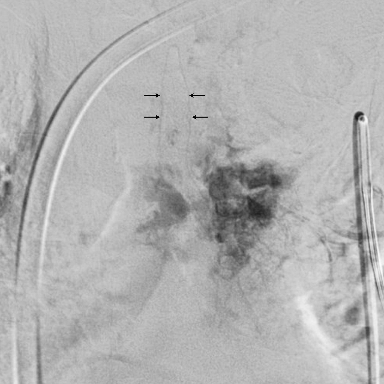
The patient continued to have progressive myelopathic symptoms, so surgical resection with preoperative embolization was planned. An angiogram was performed (Figure 2) at the level of the T5 intercostal artery, revealing a prominent radiculomedullary branch to the anterior spinal artery with its origin inseparable from the tumor nidus. Embolization was deemed to be unsafe because of the proximity of the origin of this artery in relation to the tumor. The patient elected to proceed with surgical resection despite the inability to embolize and underwent a T4-6 laminectomy for spinal decompression. Intraoperatively, this aggressive hemangioma had at this point infiltrated the ligamentum flavum and abutted the dura. Total blood loss was 600 mL. The patient did develop an epidural hematoma in the immediate postoperative period that was drained but did well postoperatively and was able to stand at postoperative day 2. She ultimately died 1 month postoperatively after requesting cessation of dialysis for her chronic renal failure.

Figure 2
  • Download figure
  • Open in new tab
  • Download powerpoint
Figure 2

Angiography via T5 intercostal artery. Tumor blush is visualized following contrast injection. A prominent hairpin radiculomedullary artery is noted emerging from the tumor nidus (arrows).

DISCUSSION

Vertebral hemangiomas are benign vascular hamartomas comprised of a dense network of thin-walled vessels lined by endothelial cells tending to orient along the longitudinal trabeculae of the vertebral body. They are the most common tumor of the spine, found in up to 10% of the population.7 Typically, hemangiomas are asymptomatic, incidental findings limited to the vertebral bodies.9 Neurological compromise due to vertebral hemangioma is rare.14 Hemangiomas can uncommonly involve the entire vertebral body with spread to the posterior elements and soft tissue, which is a risk factor for extraosseous extension and subsequent spinal cord compression.10,14 Only very rarely have hemangiomas limited to the posterior elements been reported, with 4 prior cases in the English literature.6,16,17

Vertebral hemangiomas have several typical radiologic findings. Plain radiographs reveal a “corduroy cloth” appearance due to the longitudinally oriented trabecular capillary network. Similarly, this trabecular sclerosis creates a polka-dot appearance on axial computed tomography. Findings from magnetic resonance imaging are typically intermediate T1 signal, hyperintense T2 signal, and avid enhancement with gadolinium.10 Interestingly, when compared to the observable vertebral body hemangiomas seen at multiple levels in Figure 1, the aggressive hemangiomas in the posterior elements have more pronounced hypointense T1 signal and a more solid appearance. A similar appearance is seen in other cases of posterior element hemangioma,6 and it is known that more aggressive hemangiomas often do not exhibit typical imaging characteristics.11 This could be due to the comparatively condensed space within the posterior elements, leading to early burst of the vertebral hemangioma. Notably, although the appearance was not typical for hemangioma, the presence of flow voids within the tumor suggest a highly vascular lesion.

In this case, the proximity of a prominent radiculomedullary artery precluded embolization as a safe option. We did not consider direct transpedicular percutaneous onyx embolization, but utilization of this technique may have been useful in this case due to the dangerous angioarchitecture of the tumor.15 Involvement of radiculomedullary feeders to the anterior spinal artery are a known risk of spinal vascular lesions and a contraindication to embolization due to the possibility of anterior spinal artery embolization. Interestingly, the appearance and size of this artery closely resemble the hairpin appearance of the artery of Adamkiewicz, which is the major contributor to the anterior spinal artery.1 Typically, the artery of Adamkiewicz is located on the left-hand side at the T9-12 intercostal artery (∼70% of cases), but origin from the T5-8 intercostal arteries has been noted in approximately 15% of cases. The lower intercostal arteries of our case were not visualized, so whether this was truly an aberrant artery of Adamkiewicz is uncertain.

Treatment of hemangioma is based on presentation.8 The vast majority of these tumors do not require treatment. Several methods of treatment have been investigated, including ethanol embolization,3 radiosurgical ablation,19 transarterial embolization,4 vertebral kyphoplasty,2 and surgical intervention, generally with posterior decompression or spinal stabilization. These tumors are highly vascular, and surgery can be associated with considerable blood loss. Several approaches to effective treatment of these tumors have been more recently developed.12,18 Although these tumors carry a high risk of bleeding, outcomes of treatment have generally been reported as favorable.8 In our case, the patient progressively regained neurological function, with the ability to stand on postoperative day 2.

CONCLUSIONS

We present a rare case of symptomatic vertebral hemangioma originating in the posterior elements causing myelopathy. This lesion was in close proximity to a radiculomedullary artery, precluding embolization. Still, safe resection was possible, and the immediate postoperative outcome was favorable.

  • ©International Society for the Advancement of Spine Surgery

REFERENCES

  1. 1 .↵
    1. Boll DT,
    2. Bulow H,
    3. Blackham KA, et al.
    MDCT angiography of the spinal vasculature and the artery of Adamkiewicz. Am J Roentgenol. 2006;187(4):1054–1060.
    OpenUrlCrossRefPubMed
  2. 2 .↵
    1. Brunot S,
    2. Berge J,
    3. Barreau X, et al.
    Long term clinical follow up of vertebral hemangiomas treated by percutaneous vertebroplasty. J Radiol. 2005;86:41–47.
    OpenUrlPubMed
  3. 3 .↵
    1. Heiss JD,
    2. Doppman JL,
    3. Oldfield EH.
    Brief report: relief of spinal cord compression from vertebral hemangioma by intralesional injection of absolute ethanol. N Engl J Med. 1994;331: 508–511.
    OpenUrlCrossRefPubMed
  4. 4 .↵
    1. Hekster RE,
    2. Endtz LJ.
    Spinal-cord compression caused by vertebral haemangioma relieved by percutaneous catheter embolisation: 15 years later. Neuroradiology. 1987;29(1):101.
    OpenUrlCrossRefPubMed
  5. 5 .↵
    1. Jayakumar PN,
    2. Vasudev MK,
    3. Srikanth SG.
    Symptomatic vertebral haemangioma: endovascular treatment of 12 patients. Spinal Cord. 1997;35(9):624–628.
    OpenUrlCrossRefPubMed
  6. 6 .↵
    1. Laredo JD,
    2. Reizine D,
    3. Bard M, et al.
    Vertebral hemangiomas: radiologic evaluation. Radiology. 1986;161(1):183–189.
    OpenUrlCrossRefPubMed
  7. 7 .↵
    1. McAllister VL,
    2. Kendall BE,
    3. Bull JW.
    Symptomatic vertebral haemangiomas. Brain. 1975;98(1):71–80.
    OpenUrlCrossRefPubMed
  8. 8 .↵
    1. Otten M,
    2. McCormick P.
    Natural history of spinal cavernous malformations. Handb Clin Neurol. 2017;143:233–239.
    OpenUrl
  9. 9 .↵
    1. Resnick D,
    1. Resnick D,
    2. Kyraikos M,
    3. Greenway GD.
    Tumors and tumor-like lesions of bone: imaging and pathology of specific lesions. In: Resnick D, ed. Diagnosis of Bone and Joint Disorders, 3rd ed. Philadelphia, PA: Saunders; 1995:3821–3840.
  10. 10 .↵
    1. Rudnick J,
    2. Stern M.
    Symptomatic thoracic vertebral hemangioma: a case report and literature review. Arch Phys Med Rehabil. 2004;85(9):1544–1547.
    OpenUrlPubMed
  11. 11 .↵
    1. Schnyder P,
    2. Fankhauser H,
    3. Mansouri B.
    Computed tomography in spinal hemangioma with cord compression. Skeletal Radiol. 1986;15:372–375.
    OpenUrlPubMed
  12. 12 .↵
    1. Singh PK,
    2. Chandra PS,
    3. Vaghani G, et al.
    Management of pediatric single-level vertebral hemangiomas presenting with myelopathy by three-pronged approach (ethanol embolization, laminectomy, and instrumentation): a single-institute experience. Childs Nerv Syst. 2016;32(2):307–314.
    OpenUrl
  13. 13 .↵
    1. Vinay S,
    2. Khan SK,
    3. Braybrooke JR.
    Lumbar vertebral haemangioma causing pathological fracture, epidural haemorrhage, and cord compression: a case report and review of literature. J Spinal Cord Med. 2011;34(3):335–339.
    OpenUrlPubMed
  14. 14 .↵
    1. Wang L,
    2. Song Y.
    A rare case of symptomatic hemangioma of the lumbar spine involving the spinous process. Spine J. 2016;16(3):e191–e192.
    OpenUrl
  15. 15 .↵
    1. Yao KC,
    2. Malek AM.
    Transpedicular N-butyl cyanoacrylate-mediated percutaneous embolization of symptomatic vertebral hemangiomas. J Neurosurg Spine. 2013;18(5):450–455.
    OpenUrl
  16. 16 .↵
    1. Yochum TR,
    2. Lile RL,
    3. Schultz GD, et al.
    Acquired spinal stenosis secondary to an expanding thoracic vertebral hemangioma. Spine. 1993;18:299–305.
    OpenUrlCrossRefPubMed
  17. 17 .↵
    1. Yung BC,
    2. Loke TK,
    3. Yuen NW, et al.
    Spinal cord compression caused by thoracic vertebral hemangioma involving only the posterior elements of two contiguous vertebrae. Skeletal Radiol. 1998;27(3):169–172.
    OpenUrlCrossRefPubMed
  18. 18 .↵
    1. Zhang HL,
    2. Hu YC,
    3. Aryal R, et al.
    Thoracic vertebral hemangioma with spinal cord compression: multidisciplinary surgical treatment and follow-up of six patients. Orthop Surg. 2016;8(4):462–467.
    OpenUrl
  19. 19 .↵
    1. Zhang M,
    2. Chen YR,
    3. Chang SD, et al.
    CyberKnife stereotactic radiosurgery for the treatment of symptomatic vertebral hemangiomas: a single-institution experience. Neurosurg Focus. 2017;42(1):E13.
    OpenUrl
PreviousNext
Back to top

In this issue

International Journal of Spine Surgery
Vol. 12, Issue 4
1 Aug 2018
  • Table of Contents
  • Index by author

Print
Download PDF
Article Alerts
Sign In to Email Alerts with your Email Address
Email Article

Thank you for your interest in spreading the word on International Journal of Spine Surgery.

NOTE: We only request your email address so that the person you are recommending the page to knows that you wanted them to see it, and that it is not junk mail. We do not capture any email address.

Enter multiple addresses on separate lines or separate them with commas.
Symptomatic Vertebral Hemangioma of the Posterior Elements Sharing Blood Supply with a Radiculomedullary Artery
(Your Name) has sent you a message from International Journal of Spine Surgery
(Your Name) thought you would like to see the International Journal of Spine Surgery web site.
CAPTCHA
This question is for testing whether or not you are a human visitor and to prevent automated spam submissions.
Citation Tools
Symptomatic Vertebral Hemangioma of the Posterior Elements Sharing Blood Supply with a Radiculomedullary Artery
AMIT R. PERSAD, RICHARD J. FOX, JEREMY L. REMPEL
International Journal of Spine Surgery Aug 2018, 12 (4) 415-418; DOI: 10.14444/5049

Citation Manager Formats

  • BibTeX
  • Bookends
  • EasyBib
  • EndNote (tagged)
  • EndNote 8 (xml)
  • Medlars
  • Mendeley
  • Papers
  • RefWorks Tagged
  • Ref Manager
  • RIS
  • Zotero
Share
Symptomatic Vertebral Hemangioma of the Posterior Elements Sharing Blood Supply with a Radiculomedullary Artery
AMIT R. PERSAD, RICHARD J. FOX, JEREMY L. REMPEL
International Journal of Spine Surgery Aug 2018, 12 (4) 415-418; DOI: 10.14444/5049
Twitter logo Facebook logo Mendeley logo
  • Tweet Widget
  • Facebook Like
  • Google Plus One

Jump to section

  • Article
    • ABSTRACT
    • INTRODUCTION
    • CASE
    • DISCUSSION
    • CONCLUSIONS
    • REFERENCES
  • Figures & Data
  • Info & Metrics
  • PDF

Related Articles

  • No related articles found.
  • PubMed
  • Google Scholar

Cited By...

  • No citing articles found.
  • Google Scholar

More in this TOC Section

  • Hemivertebra Resection and Spinal Arthrodesis by Single-Stage Posterior Approach in Congenital Scoliosis and Kyphoscoliosis: Results at 9.6 Years Mean Follow-up
  • Prevalence and Significance of Vitamin D Deficiency in Patients Undergoing Corrective Surgery for Adolescent Idiopathic Scoliosis
  • Adult Congenital Spine Deformity: Clinical Features and Motivations for Surgical Treatment
Show more Other & Special Categories

Similar Articles

Keywords

  • posterior elements
  • radiculomedullary artery
  • symptomatic vertebral hemangioma

Content

  • Current Issue
  • Latest Content
  • Archive

More Information

  • About IJSS
  • About ISASS
  • Privacy Policy

More

  • Subscribe
  • Alerts
  • Feedback

Other Services

  • Author Instructions
  • Join ISASS
  • Reprints & Permissions

© 2025 International Journal of Spine Surgery

International Journal of Spine Surgery Online ISSN: 2211-4599

Powered by HighWire