


Abstract
Objective This study aims to investigate the effect of methylprednisolone (MP) intrathecal injection on a rat model of acute spinal cord injury (ASCI).
Methods Allen’s frame was used to establish a rat model of ASCI. MP and normal saline were intrathecally injected to Sprague–Dawley rats at 0, 3, 6, 8, 12, and 24 hours after ASCI, and injured spinal cord tissues were sterilely extracted after 24 hours of treatment. Isobaric tags for relative and absolute quantitation (iTRAQ) were coupled with 2-dimensional liquid chromatography tandem mass spectrometry to separate and identify differentially expressed proteins.
Results The expression of growth factor receptor-bound protein 2 (Grb2) was downregulated in the MP groups at 0 hours (iTRAQ ratio = 0.996), 3 hours (iTRAQ ratio = 0.737), 8 hours (iTRAQ ratio = 0.763), and 24 hours (iTRAQ ratio = 0.908) after injury compared with that in the control groups. No significant difference in Grb2 expression was observed between the control groups at 6 and 12 hours after ASCI.
Conclusions Standardized MP intrathecal injection after ASCI treatment reduces Grb2 activation in a rat ASCI model. Further studies should determine whether or not the same effect can be observed in human ASCIs.
- methylprednisolone
- growth factor receptor-bound protein 2
- acute spinal cord injury
- isobaric tags for relative and absolute quantitation
INTRODUCTION
With the rapid development of traffic and construction businesses, acute spinal cord injury (ASCI) resulting from spinal fracture has become common in the orthopedic field. The incidence rate of ASCI has annually increased to 28.3 to 45 per 1 million people in developed countries and approximately 6.7 per 1 million people (ie, >10,000 people) in China. Approximately 10,000 new cases of ASCI, mostly young individuals, are reported each year in the United States; patients need more than $2 million for the treatment.1 Clinical workers have exerted considerable efforts to reduce the mortality from 50% in the early 20th century to 6% at present.2 However, the functional recovery after ASCI remains unsatisfactory.
Despite the volume of studies that concentrated on ASCI, substantial progress on ASCI treatment has yet to be achieved.3–5 ASCI treatment remains a worldwide medical problem because of the lack of a satisfactory method for neural functional recovery.6 Although animal studies cannot absolutely explain the beneficial effects of steroid use, the results of many animal studies support the positive effects of steroid use in ASCI. Corticosteroids are known to have neuroprotective effects, including improvement of vascular perfusion, prevention of calcium influx and accumulation, modulation of the inflammatory cells, prevention of the loss of spinal cord neurofilament proteins that facilitate neuronal excitability and impulse conduction, and inhibition of lipid peroxidation and inflammatory cytokines.7
In 2017, AOSpine practice guideline suggested that 24-hour infusion of high-dose methylprednisolone sodium succinate should be provided in patients with ASCI within 8 hours as a treatment option. However, subsequent meta-analyses have consistently shown that high-dose methylprednisolone sodium succinate used in ASCI patients has no effect on neurological improvement. Therefore, it is necessary to fully understand the effects and side effects of steroid use and to apply them appropriately in clinical practice.7 Allen’s frame was used to establish a rat model of ASCI. Methylprednisolone (MP) and normal saline (NS) were intrathecally injected into Sprague–Dawley (SD) rats at 0, 3, 6, 8, 12, and 24 hours after ASCI. Isobaric tags for relative and absolute quantitation (iTRAQ) were coupled with 2-dimensional (2D) liquid chromatography tandem mass spectrometry (2D-LC-MS/MS) to separate and identify differentially expressed proteins.
MATERIALS AND METHODS
Ethics Statement
All animal procedures were performed in accordance with the guidelines of the China Council for Animal Care and approved by the Central South University Animal Care Committee (Protocol No: 20200220005). The animals were housed in specific pathogen-free facilities at the Central South University. All surgeries and imaging were performed under anesthesia using ketamine, and all efforts were exerted to minimize suffering. Mice were humanely euthanized using CO2 at a flow rate of 20% of the chamber volume/min following the guidelines of the China Council for Animal Care.
Animals
In total, 120 8- to 12-week-old male SD rats (Central South University Laboratory) weighing 300 to 400 g were used for this study. All animals were maintained in a standard housing environment with ad libitum access to food and water. Samples of spinal cord tissues were collected to search for biomarkers using iTRAQ coupled with 2D-LC-MS/MS. In all experiments, the animals were randomly and equally divided into the following 12 experimental groups (n = 10): MP-0, MP-3, MP-6, MP-8, MP-12, and MP-24 (MP intrathecal injection after ASCI at 0, 3, 6, 8, 12, and 24 hours); C-0, C-3, C-6, C-8, C-12, and C-24 (NS intrathecal injection after ASCI at 0, 3, 6, 8, 12, and 24 hours).
Surgery
The animals were anesthetized using an intraperitoneal injection of ketamine (100 mg/kg), atropine (10 mg/kg), and diazepam (5 mg/kg). After inducing a surgical plane of anesthesia (anesthesia endpoint was assessed by toe pinch and eye blink reflex), the surgical areas were shaved and disinfected. The animals were placed in a stereotaxic apparatus, and dorsal midline incisions were made from T6 to T12. After the thoracic vertebrae were carefully exposed, laminectomy was performed using a fine rongeur at the T8/10 level to expose the spinal cord. Following laminectomy, the spine was stabilized by clamping the transverse processes of one segment above and below the lesion site.3 A contusion was generated through the displacement-controlled Allen’s frame.4 The impactor tip was placed at the T9 center. A contusion was induced with displacement in the 2 groups. Afterward, MP and NS were intrathecally injected at 0, 3, 6, 8, 12, and 24 hours after ASCI. After surgery, the animals were kept in a temperature-controlled incubator until completely awake. Morphine (0.03 mg/kg) was subcutaneously administered to prevent postoperative pain.
Spinal Cord Tissue Collection
Spinal cord tissues were collected at 24 hours after MP and NS intrathecal injection. The T8/10 spinal cords (approximately 1 cm long and 500 mg) were dissected, the meninges were carefully removed, and the tissues were thoroughly rinsed with NS to remove blood. The spinal cord tissues were immediately frozen in liquid nitrogen and preserved at −80°C until analysis.
Separation and Identification of Differentially Expressed Proteins
Only 8 samples can be studied at a time using iTRAQ, and the samples should be simultaneously evaluated. Thus, the 6 control groups (C-0, C-3, C-6, C-8, C-12, and C-24) were analyzed using iTRAQ and 2D-LC-MS/MS, and the 4 groups with significant expression levels were selected. The 4 control groups and 4 MP groups were simultaneously analyzed.
iTRAQ Labeling and Strong Cation-Exchange Chromatography Fractionation
Spinal cord tissues were seeded in 6- or 8-well plates and transfected with 5 pmol miR-451 mimics using Lipofectamine 2000 (Invitrogen). After 48 hours of transfection, cells were harvested, and proteins were prepared and digested as previously described. The digested peptides were labeled with different iTRAQ reagents (Applied Biosystems), including 114, 115, 116, 117, 118, and 119. Subsequently, the labeled peptides were equally mixed and were fractionated using Poly SULFOETHYL ATM Strong Cation-Exchange column (200 × 4.6 mm, 5 μm particle size, and 200 Å pore size) using a high-performance liquid chromatography (HPLC) system (Shimadzu, Japan). The HPLC gradient consisted of 100% buffer A (10 mM KH2PO4, 25% acetonitrile, and pH 2.85) for 5 minutes; 0% to 20% buffer B (10 mM KH2PO4, 25% ACN, 500 mM KCL, and pH 3.0) for 15 minutes; 20% to 40% buffer B for 10 minutes; 40% to 100% buffer B for 5 minutes; and 100% buffer A for 10 minutes. The chromatograms were obtained at 218 nm. A total of 12 fractions were collected, desalted using Sep-Pak Vac C18 cartridges (Waters, Milford, MA), concentrated to dryness using vacuum centrifuge, and then reconstituted in 0.1% formic acid for 2D-LC-MS/MS analysis.
2D Liquid Chromatography Tandem Mass Spectrometry
LC-MS/MS analysis of the samples was performed on an AB SCIEX TripleTOF 5600 mass spectrometer (AB SCIEX, Framingham, MA, USA) coupled with an online micro flow HPLC system (Shimadzu, JAPAN). The HPLC system consisted of a trap column (200 µm × 2 cm, C18 material 5–10 µm, and 120 A°) and an analytical column (75 µm × 10 cm, C18 material 5 µm, and 120 A°). The peptides were separated with a nanobored C18 column with a picofrit nanospray tip (75 μm ID × 15 cm, and 5 μm particles; New Objectives, Wubrun, MA) at a constant flow rate of 20 μL/min. A splitter was used to obtain an effective flow rate of 0.2 μL/min. The MS spectra were acquired in the positive ion mode, and the peptides with +2 to +4 ions in the survey scan were targeted with a selected mass range of 300 to 2000 m/z. The 3 most abundantly charged peptides above a count threshold were selected for MS/MS and dynamically excluded for 30 seconds with ±30 mDa mass tolerance. The fragment intensity multiplier was set to 20, and the maximum accumulation time was 2 seconds. The peak areas of the iTRAQ reporter ions reflected the relative abundance of the proteins in the samples.
Bioinformatics Analysis
Mascot software (Matrix Science) version 2.3.02 was used to simultaneously identify and quantify proteins. In this version, only unique peptides used for protein quantification were selected. The spectra were combined into a single MGF (Mascot generic format) file after loading the raw data, and the MGF file was searched. The search parameters were as follows: trypsin was selected as the enzyme with one missed cleavage allowed; fixed modifications of carbamidomethylation at Cys, variable modifications of oxidation at Met; peptide tolerance was set to 10 ppm; and MS/MS tolerance was set to 0.05 Da. The peptide charge was set as Mr, and monoisotopic mass was selected. iTRAQ 6 or 8 plex was simultaneously selected for quantification during the search.
The search results were evaluated through additional filters before exporting the data. The filters for protein identification were set as follows: significance threshold P < 0.05 (with 95% confidence) and ion score or expected cut off less than 0.05 (with 95% confidence). For protein quantitation, the filters were set as follows: “median” was selected for the protein ratio type (http://www.matrixscience.com/help/quant_config_help.html); minimum precursor charge was set to +2 and minimum peptide was set to 2; and only unique peptides were used to quantify proteins. Median intensities were set to normal, and outliers were automatically removed. The peptide threshold was set as described above for identity.
Proteins with at least 1.2-fold changes between the treatment and control samples at P < 0.05 were determined as differentially expressed proteins. Quantitation was performed at the peptide level by following the procedures described in http://www.matrixscience.com/help/quant_statistics_help.html. Student’s t test was performed using Mascot 2.3.02 software.
RESULTS
In this study, no severe vomiting, incision infection, or death was recorded among the 120 male SD rats, of which 60 male were intrathecally injected with high-dose MP after injury.
iTRAQ analysis of proteins in the spinal cord tissues was performed. A total of 220,568 MS/MS spectra were acquired from all iTRAQ runs, of which 13,742 spectra were utilized to assign 11,972 unique peptides that represent 2203 proteins. All spectra were searched against a decoy database with all Swiss-Prot sequences reversed. Only proteins that were present at a confidence interval of 95% or higher and were identified through at least 1 peptide were considered. No significant difference in the expression of growth factor receptor-bound protein 2 (Grb2) was detected between the control groups at 6 hours (C-6 vs C-8 iTRAQ ratio = 0.993) and 12 hours (C-12 vs C-24 iTRAQ ratio = 0.982) after ASCI. Therefore, the control groups (C-0, C-3, C-8, and C-24) and 4 MP groups (MP-0, MP-3, MP-8, and MP-24) were simultaneously identified (Table 1 and Figure).
Mean expression of Grb2 by group at each time point.
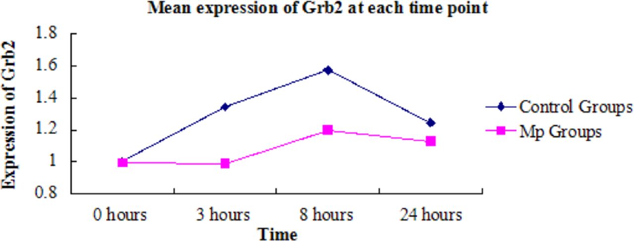
Mean expression of Grb2 at each time point. Abbrevations: Grb2, growth factor receptor-bound protein 2; Mp, methylprednisolone.
The mean relative Grb2 expression in the control groups gradually increased, peaked at 8 hours (iTRAQ ratio = 1.573, indicating that Grb2 in C-8 increased more than 1.573-fold from 0–8 hours after injury) and then subsequently decreased at 24 hours (iTRAQ ratio = 1.245; Table 2 and Figure). In the MP group, the mean relative expression of Grb2 diminished at 0 hour (C-0 vs MP-0 iTRAQ ratio = 0.937), 3 hours (C-3 vs MP-3 iTRAQ ratio = 0.737), 8 hours (C-8 vs MP-8 iTRAQ ratio = 0.763), and 24 hours (C-24 vs MP-24 iTRAQ ratio = 0.908) after injury compared with the control groups. The expression level of Grb2 significantly decreased at 3 hours (P < 0.005) and 8 hours (P < 0.005) after injury in the animals that received MP intrathecal injection compared with those that received NS.
iTRAQ ratios by group comparisons.
DISCUSSION
Recent studies have shown that the intrathecal injection of drugs is an effective ASCI treatment; in particular, the intrathecal injection of MP significantly reduces the systemic side effects of intravenous MP.8–12 Kohmura11 reported that transforming growth factor beta-1 can promote motor function recovery by intrathecal injection treatment of ASCI in the thoracic segment in rats. Brynes2 treated moderate ASCI in the ninth thoracic vertebra of rats with the selective intrathecal infusion of metabotropic glutamate receptor 5-antagonized (RS)-2-chloro-5-hydroxyphenylglycine for 7 days. After the 28th day, the movement functions of the rats were significantly restored. The present study is the first to treat ASCI through MP intrathecal injections at different timepoints (0, 3, 6, 8, 12, and 24 hours). Similar to a previous study, the present study showed that ASCI treatment via MP intrathecal injection can reduce systemic side effects. However, MP cannot promote the recovery of injured nerve cells in a short time because Grb2 expression significantly decreased after injury in the rats that received MP intrathecal injection compared with those that received NS intrathecal injection.
The Grb2-associated binder (Gab) adapter/scaffolding protein family consists of conserved proteins, namely, mammalian Gab1, Gab2, and Gab3. Grb2 binds with guanine nucleotide exchange factors via its SH3 domains and activates the Ras-dependent signaling pathway. The direct binding of Grb2 with TrkA (the nerve growth factor [NGF] receptor tyrosine kinase) via its SH2 domain can facilitate the subsequent Grb2/SH3 domain-mediated recruitment and activation of other signaling molecules to the activated TrkA receptor complex.13 NGF/TrkA influences NGF-stimulated differentiation and mitogenesis in neuronal cells. Giuliana et al14 reported that the recruitment of the Grb2/SOS complex by p52/46Shc results in SOS membrane relocalization, an event sufficient to induce Ras activation. Consistent with this model, p52/p46Shc overexpression enhances granulocyte–macrophage colony-stimulating factor-induced Ras downstream signaling events,2 such as mitogen-activated protein kinase (MAPK) activation.15 However, Rai potentiates the MAPK and PI3K signaling pathways and regulates Ret-dependent and -independent survival signals. Rai overexpression in neuronal cell lines promotes survival by reducing apoptosis. Grb2 serves as a multipurpose adapter molecule that recruits the guanine nucleotide-releasing factor (SOS) to the membrane and thus activates the Ras/Raf/MEK/ERK cascade. This cascade can stimulate neurite outgrowth.16,17
National ASCI Studies II and III were double-blind randomized controlled trials that investigated the clinical effects of a high-dose intravenous MP regime on spinal cord recovery against placebo after ASCI. Although the preliminary data demonstrated no difference between the intravenous MP and placebo, a post hoc analysis demonstrated a statistically significant improvement in motor scores at 1 year after injury when the therapy was initiated within 8 hours of injury.18–20 After the National Acute Spinal Cord Injury Study (NASCIS), the high-dose intravenous MP became the “standard of care” for patients within 8 hours of ASCI.19 Subsequent analysis of the NASCIS methodology, including the post hoc nature of its clinical significance, led some investigators to question the conclusions of NASCIS.6,19,21–24 Additional randomized placebo-controlled studies have failed to replicate the results of NASCIS.25 In addition, patients who received the NASCIS MP protocol suffered from increased rates of complications, such as pulmonary emboli, wound infections, severe pneumonia, and sepsis.25–27 In our studies, the MP dose equivalent to the NASCIS protocol inhibited Grb2 expression in the rat model, suggesting the possible inhibition of the neuroprotective cascade after ASCI in the short period.
In the present study, the mean relative expression of Grb2 in the control groups gradually increased and then peaked at 8 hours. This phenomenon may account for the therapeutic window restriction of 8 hours after injury. Although the results reached statistical significance, the present study was limited by the small number of rats in each group. Grb2 expression might be an important potential neuroprotective factor, but it may not be in parallel with the entire neuroprotective cascade in humans. Therefore, transposing the data onto human ASCI is difficult.
CONCLUSIONS
A standardized dose of intravenous MP based on the NASCIS protocol reduces Grb2 expression and is a potential neuroprotective cytokine in a rat ASCI model. Additional studies must be conducted to determine whether the same effects can be observed in human ASCIs and whether the suppressive effects of MP can be observed in other portions of the neuroprotective cascade after ASCI.
Footnotes
Funding This work was funded by Science and Technology Bureau Technology Research and Development Foundation of Changde (2019S176 to XB), Science and Technology Inovation Development Guiding Plan Project of Changde (2020ZDSK86 to YZ).
Declaration of Conflicting Interests The authors report no conflicts of interest in this work.
- This manuscript is generously published free of charge by ISASS, the International Society for the Advancement of Spine Surgery. Copyright © 2023 ISASS. To see more or order reprints or permissions, see http://ijssurgery.com.
References
In this issue


Jump to section
Related Articles
Cited By...
- No citing articles found.