


Article Figures & Data
Figures
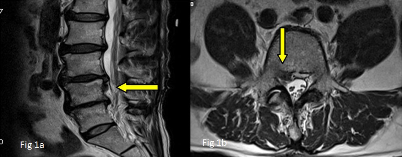
- Fig. 1
Preoperative MRI. 1a: saggital T2W down migrated disc, 1b: axial T2W with down migrated disc herniation upto the inferior pedicle level.
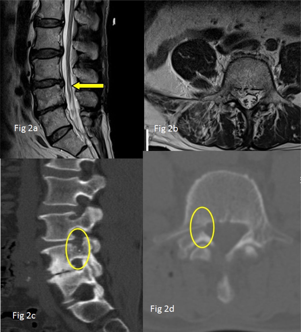
- Fig. 2
Postoperative MRI and CT. 2a: saggital T2W removal of herniated disc. 2b: axial T2W showing complete decompression upto the inferior level of the pedicle. 2c &2d: saggital and axial CT shows the entry of transpedicular approach through the pedicle (yellow circle).
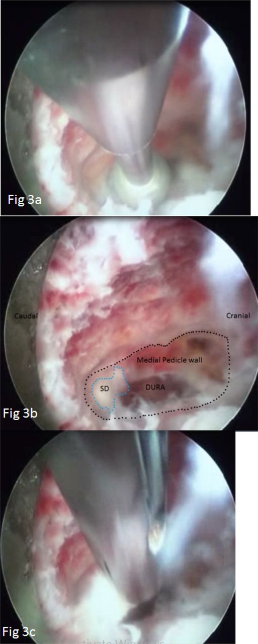
- Fig. 3
Endoscopic view. 3a: drilling of medial pedicular wall 3b: tail of sequestered disc (SD); dotted line demarcates the medial pedicle wall. 3c: removal of herniated fragment with an endoscopic forceps.
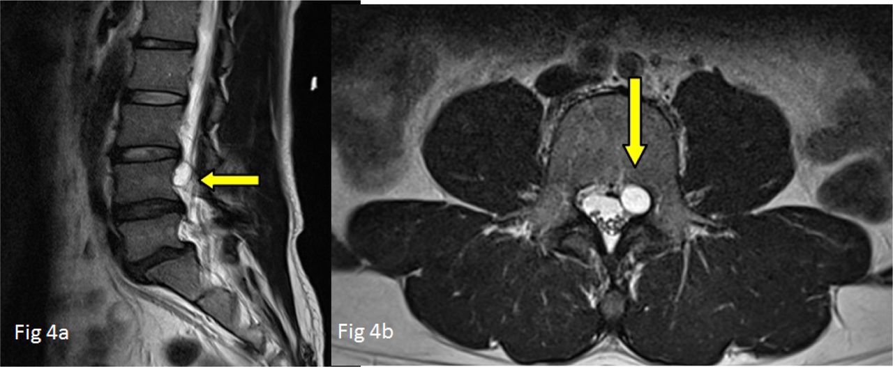
- Fig. 4
Preoperative MRI. 4a: saggital T2W discal cyst, 4b: T2W axial showing discal cyst at the medial aspect of L4 left pedicle.
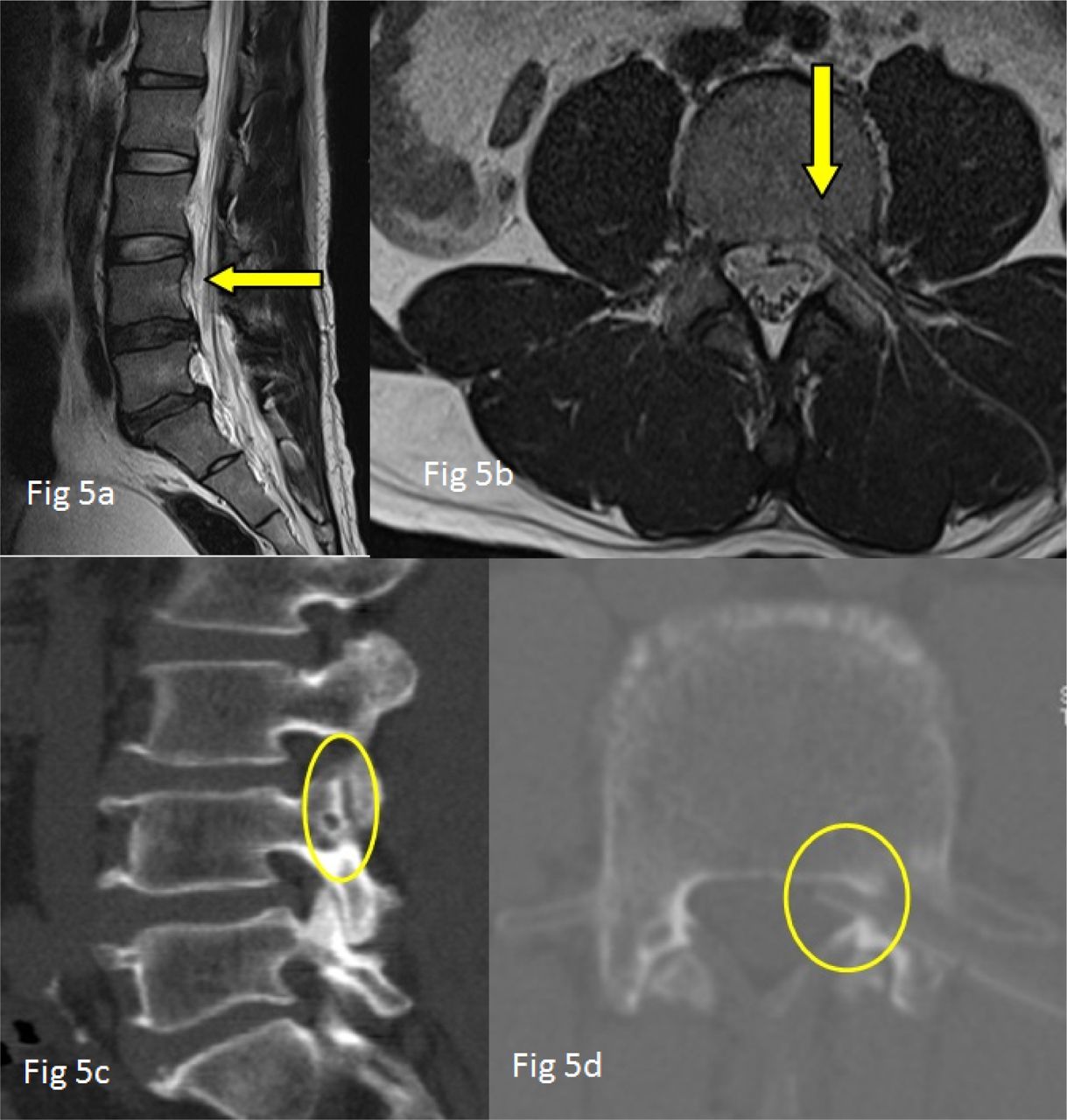
- Fig. 5
Postoperative MRI & CT. 5a: Complete removal of discal cyst on sagg T2W; 5b: T2W axial complete decompression and drain placed in situ; 5c & 5d: saggital and axial CT shows the transpedicular entry point and trajectory through the pedicle (yellow circle).
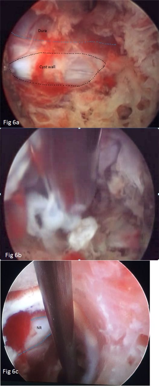
- Fig. 6
Endoscopic view. 6a: the black dotted line shows the ruptured and collapsed discal cyst wall, the blue dotted line demarcates dura. 6b: tail of herniated disc fragment being grabbed by an endoscopic forceps. 6c: canal space after removal of discal cyst (NR – nerve root).
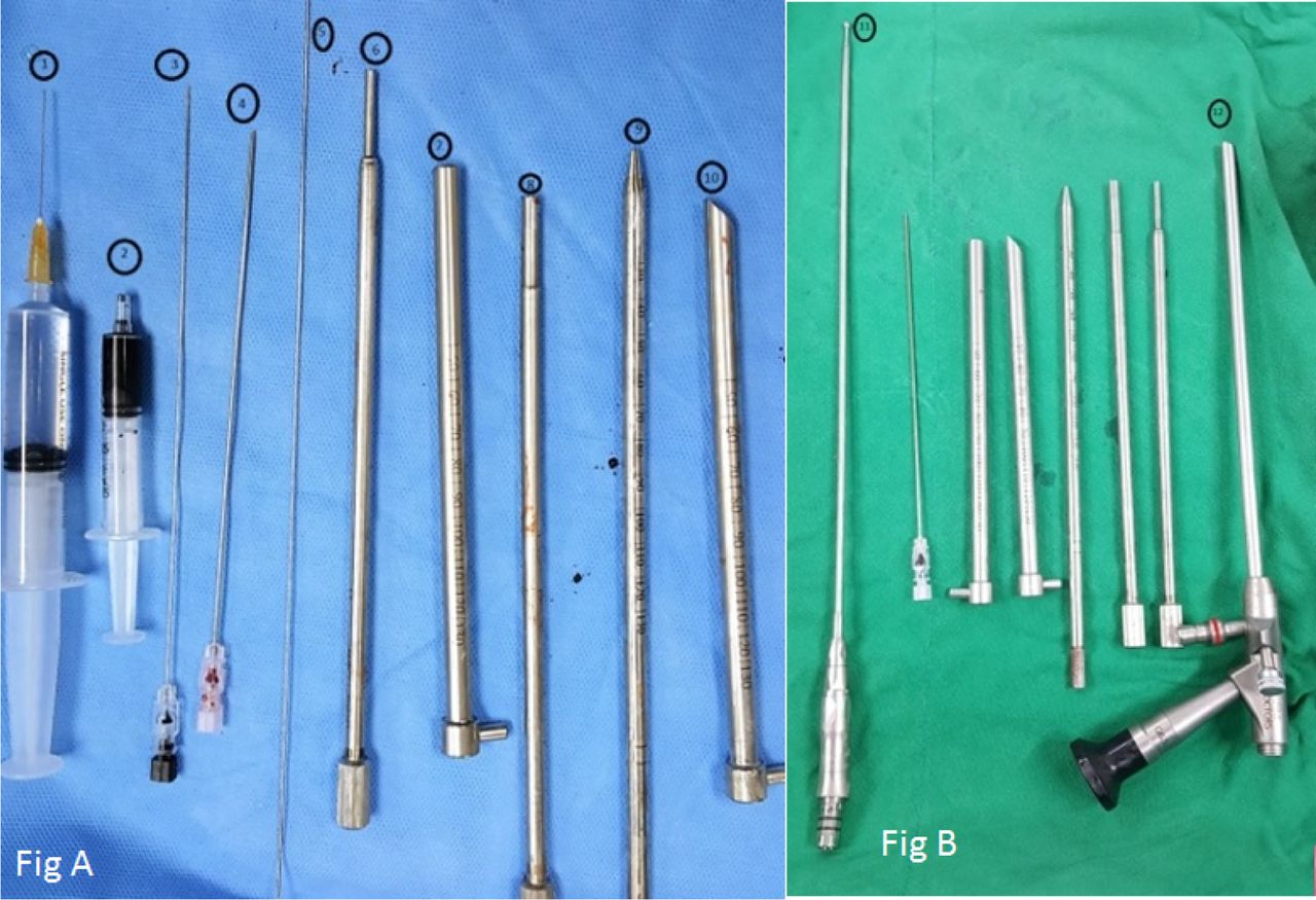
- Fig. 7
1) Local anesthetic 2) Dye for discography 3) 23 G discography needle 4) 18 G endoscopy needle 5) guide wire, 6 ) & 8) Triphines, 7) & 10) cannula 9) obturator 11) Endoscopic Drill 12) Endoscope.
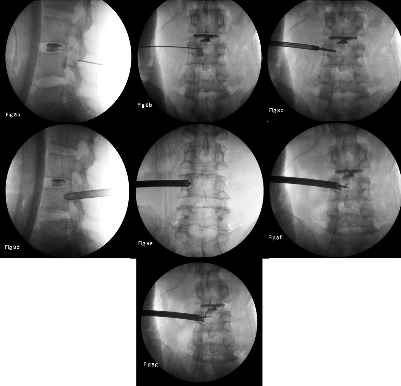
- Fig. 8
Intraoperative fluoroscopy. 8a: discography with extravasation of the dye; 8b: needle placement transpedicularly; 8c: reaming by triphine; 8d: final cannula placement; 8e: endoscopic drilling of medial pedicle wall, 8f & 8g: endoscopic forceps placement during removal of herniated disc fragment.
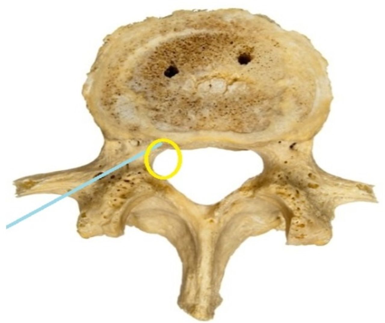
- Fig. 9
The blue line indicates the trajectory of transpedicular endoscopy and yellow circle denotes the target point which has to be drilled endoscopically.


Tables
Level Mean transverse diameter (millimetres) Mean medial an-gulation (degrees) Mean sagittal (caudal) angulation (degrees) L1 8 14.5 2.6 L2 7.8 14.2 2.7 L3 10.2 18.5 2.7 L4 13.4 16.6 3.9 L5 18.0 24.6 5.5
In this issue


Jump to section
Related Articles
Cited By...
- No citing articles found.