


Article Figures & Data
Figures
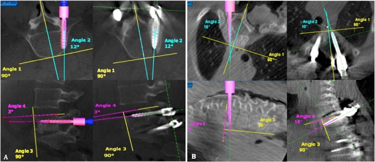
- Fig. 1
Axial and sagittal images of the virtual screw and actual screw angles relative to the mid sagittal line and superior endplate, respectively. (A) Example of most accurate screw placement (B) Example of least accurate screw placement.
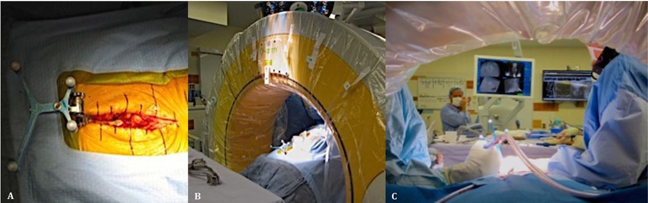
- Fig. 2
(A) The patient is in a prone position, and a reference frame is attached to a spinous process. (B) The O-arm is brought into the field and a CT scan obtained. (C) The position of the navigated instruments is projected onto the CT images on a monitor visible to the surgeon.
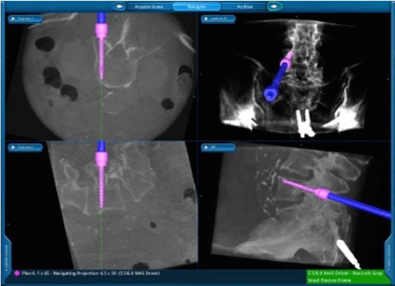
- Fig. 3
Image of the Stealth Station navigation screen showing the axial, sagittal, and coronal virtual screw projection.
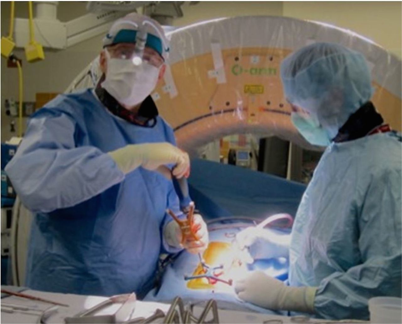
- Fig. 4
Using a navigated screwdriver, the surgeon is able visualize the real-time trajectory of the screw on the Stealth Station screen during placement.
- Fig. 5
AP and lateral spinal preoperative (A, B) and postoperative (C, D) x-rays after a Stealth guided posterior spinal instrumentation.
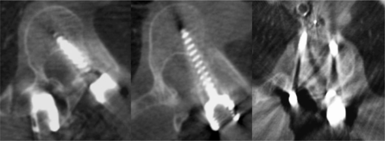
- Fig. 6
Axial intraoperative CT scan of the three pedicle screws which were removed or revised. Two screws breached medially and one screw was too long.
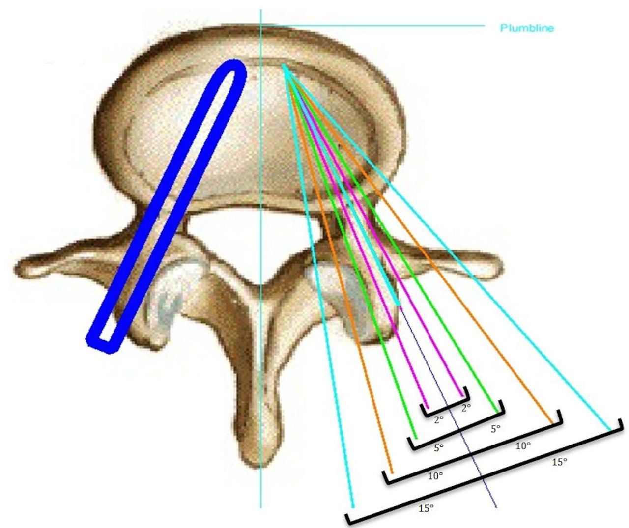
- Fig. 7
Varying degrees of accuracy from an ideal screw trajectory in the lumbar spine.


Tables
Patient Demographics Mean age (range), yr 44 (6-85) Sex, no. (%) Female 17 (55) Male 14 (45) Primary Diagnosis, no. (%) Degenerative disk disease 8 (26) Scoliosis 7 (23) Spondylolithesis 6 (19) Pseudarthrosis 3 (10) Trauma 3 (10) Kyphosis 2 (6) Tumor/metastatic disease 2 (6) Fusion procedure, no (%) PSF 16 (52) TLIF 7 (22) Combined AP 4 (13) Combined TLIF/PSF 4 (13) Level No. of screws Axial Sagittal Mean virtual angle (°) Mean actual angle (°) Mean angle difference (°) Axial Percent error Mean virtual angle (°) Mean actual angle (°) Mean angle difference (°) Sagittal Percent error T1 9 22.4 21.2 2.56 1.25% 4.56 1.78 3.00 1.59% T2 11 17.8 17.3 2.73 1.38% 4.00 4.45 2.82 1.52% T3 11 15.5 14.8 3.27 1.66% 2.45 2.18 2.09 1.14% T4 8 18.1 15.8 3.13 1.57% 4.38 2.63 2.00 1.08% T5 15 16.1 15.1 2.53 1.30% 3.47 2.47 1.93 1.04% T6 12 13.8 12.1 2.00 1.03% 3.25 3.25 1.00 0.55% T7 12 12.8 12.3 2.00 1.04% 4.17 3.33 1.83 0.99% T8 14 13.9 13.5 1.71 0.88% 4.00 4.79 1.79 0.97% T9 15 13.1 13.3 1.40 0.73% 3.20 4.20 1.67 0.91% T10 13 11.1 10.1 1.77 0.93% 1.69 0.77 1.69 0.92% T11 16 9.9 9.4 1.94 1.02% 5.69 5.38 1.69 0.91% T12 10 8.7 9.0 1.30 0.69% 2.44 2.33 1.44 0.75% L1 11 15.1 14.4 1.64 0.84% 3.91 4.18 1.91 1.03% L2 14 20.7 19.1 2.57 1.25% 3.71 3.64 2.50 1.35% L3 12 18.8 18.7 2.00 1.00% 4.58 3.67 1.75 0.95% L4 18 16.9 16.8 2.67 1.33% 3.86 4.14 2.19 1.22% L5 22 14.6 14.8 2.55 1.29% 3.05 4.32 2.45 1.34% S1 17 11.2 10.7 1.59 0.83% 3.94 3.24 4.35 2.32% Total 240 14.8 14.2 2.17 1.11 3.67 3.49 2.16 1.17% P value 0.0016 P value = 0.19 Level No. of screws Axial Sagittal Mean virtual angle (°) Mean actual angle (°) Mean angle difference (°) Percent error Mean virtual angle (°) Mean actual angle (°) Mean angle difference (°) Percent error Thoracic 146 14.1 13.4 2.14 1.10% 3.63 3.26 1.88 1.02% Lumbosacral 94 15.9 15.5- 2.22 1.12% 3.73 3.86 2.61 1.41% P value = 0.882 P value = 0.024 Level from reference frame No. of screws Axial Sagittal Mean virtual angle (°) Mean actual angle (°) Mean angle difference (°) Percent error Mean virtual angle (°) Mean actual angle (°) Mean angle difference (°) Percent error 0 45 14.1 14.3 1.83 0.9% 3.0 3.6 2.02 1.1% 1 64 16.5 16.6 1.50 0.8% 3.8 3.2 1.50 1.4% 2 37 15.5 14.6 2.22 1.1% 3.4 3.6 2.22 0.9% 3 34 19.2 18.1 2.44 1.2% 4.5 4.0 2.44 0.9% 4 23 14.1 13.3 2.13 1.1% 3.7 3.1 2.13 1.3% 5 19 18.4 17.3 2.68 1.3% 2.2 1.4 2.68 1.3% 6 9 36.2 35.4 2.56 1.3% 5.4 5.7 2.56 1.2% 7-10 9 19.0 15.0 6.00 3.0% 5.4 5.6 6.00 1.1%
In this issue


Jump to section
Related Articles
Cited By...
- No citing articles found.