


Article Figures & Data
Figures
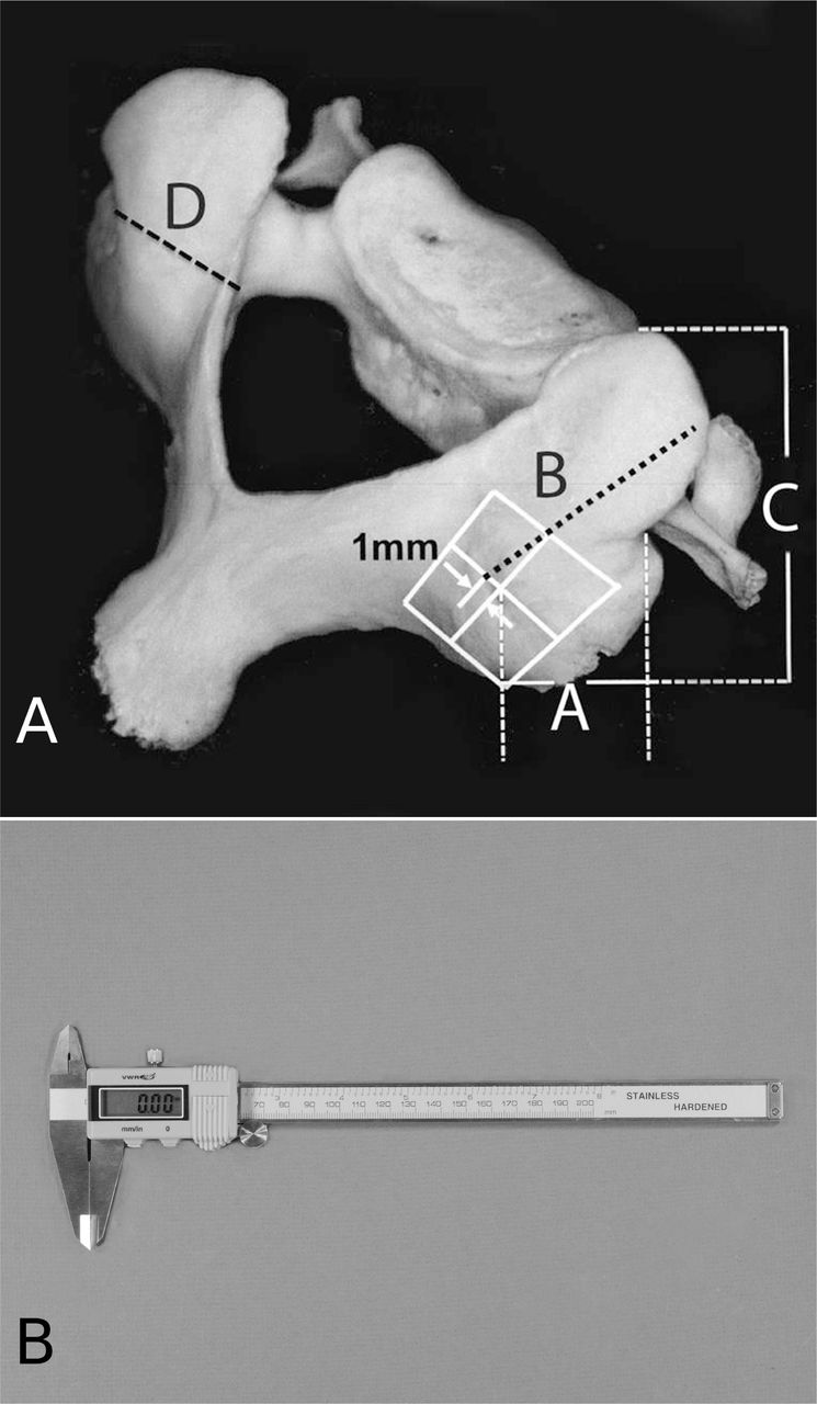
- Fig. 1A
Typical cervical vertebra showing various measurements of the Articular pillar by Vernier calipers: A = Antero-posterior diameter (Roy-Camille method), B = Oblique antero-posterior diameter (Magerl method), C = Height of the articular pillar, D = Transverse diameter. Fig. 1B. VWR (Van Waters and Rogers) stainless steel digital Vernier calipers used to measure various dimensions on dry cervical vertebrae.
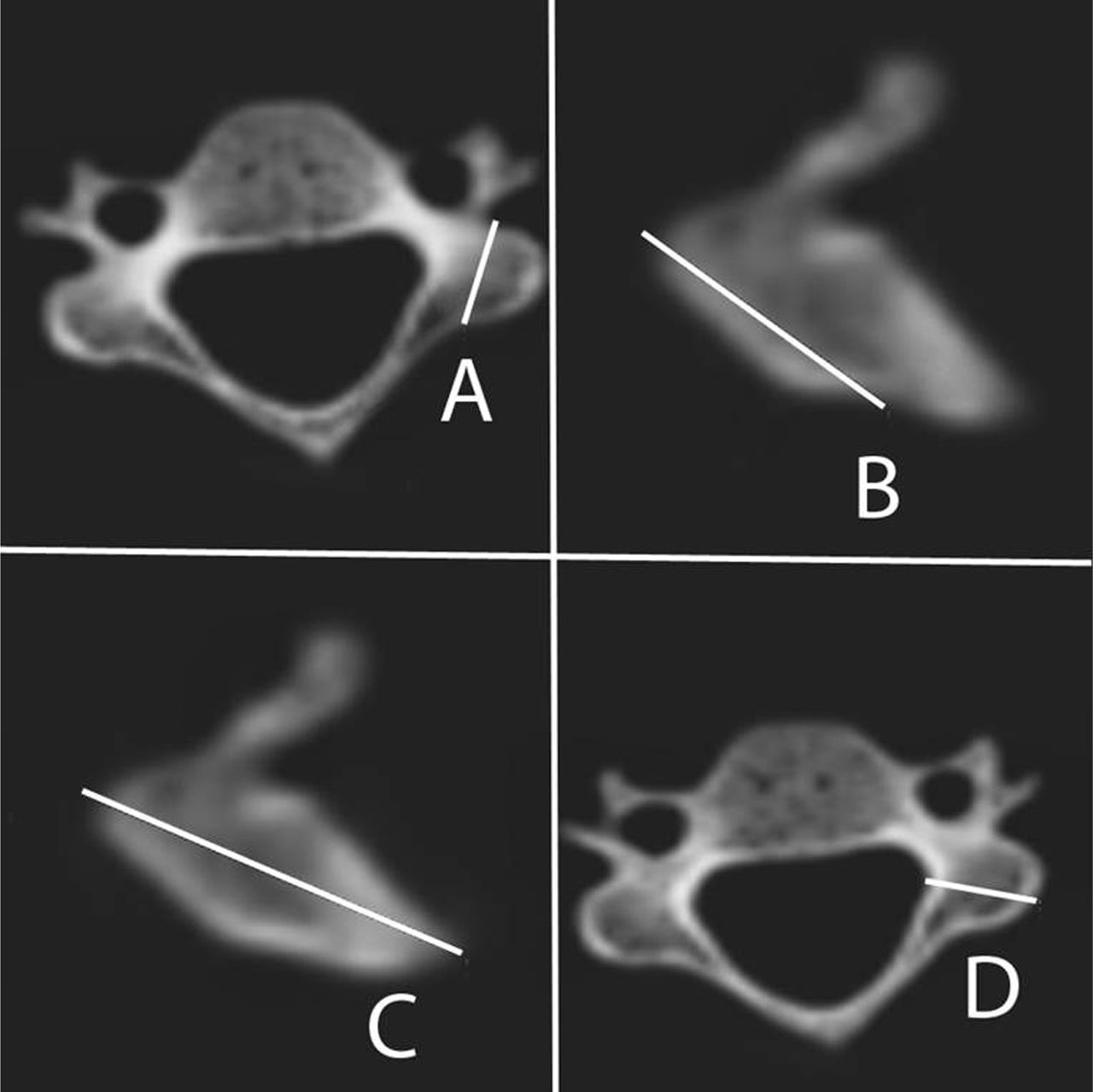
- Fig. 2
Typical cervical vertebra showing various measurements of the Articular pillar by CT scan software: A = Antero-posterior diameter (Roy-Camille method), B = Oblique antero-posterior diameter (Magerl method), C = Height of the articular pillar, D = Transverse diameter.
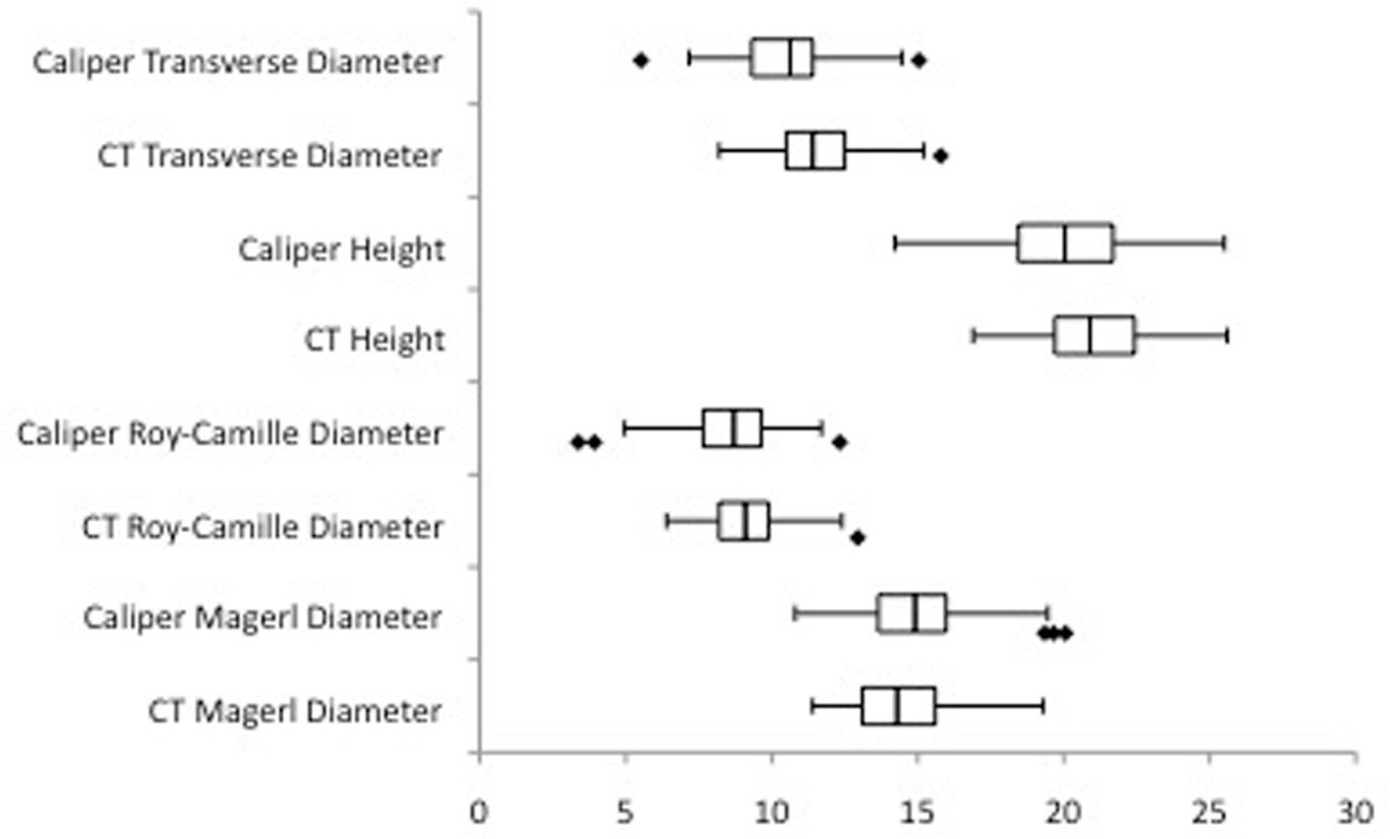
- Fig. 3
Box-and-whisker plot showing various dimensions of the articular pillars in millimeters (mm). Each plot shows the median, quantification of data and outliers.
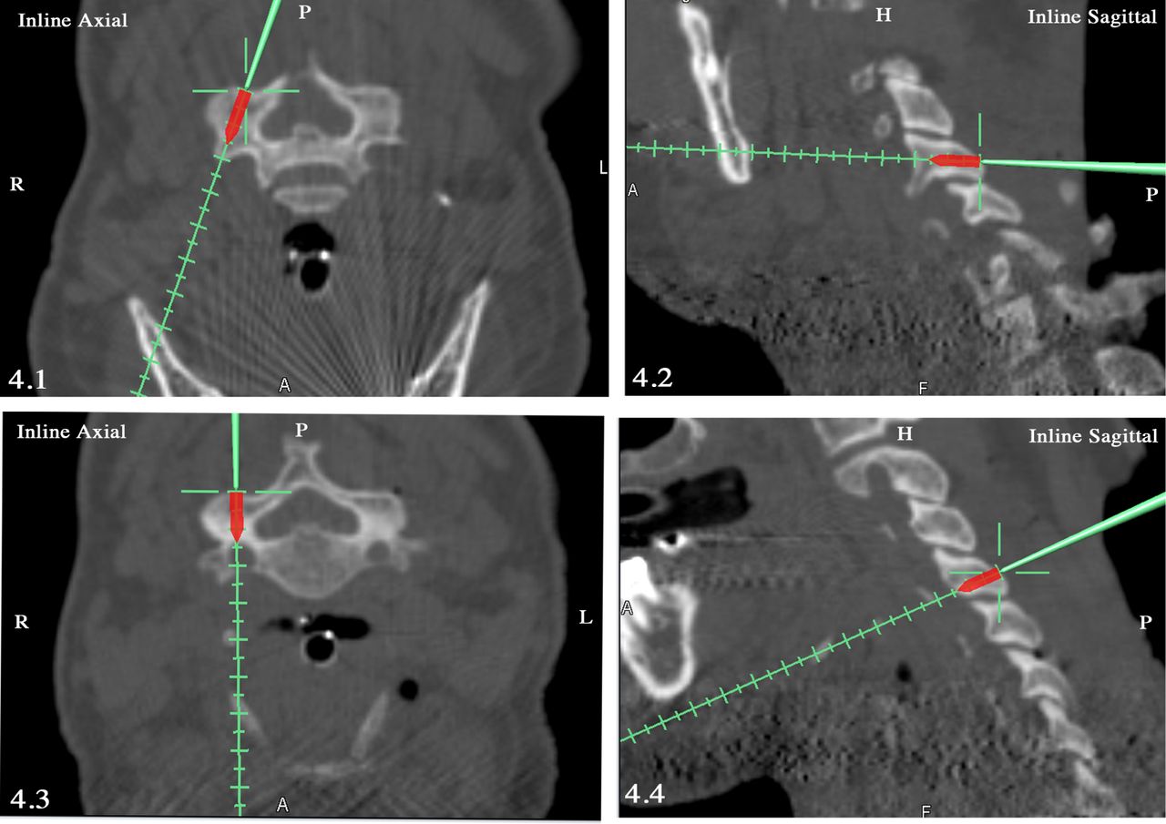
- Fig. 4
Most commonly used articular pillar screw trajectories: Magerl technique (4.1, 4.2) and Roy-Camille technique (4.3, 4.4).


Tables
- Table 1
Diameters of Articular pillars of typical cervical vertebrae as measured with Vernier calipers and CT measurement software (mm).
Transverse diameter (mm) Height (mm) Antero-posterior diameter † (mm) Oblique Antero-posterior diameter ‡ (mm) Mean (SD) Range Mean (SD) Range Mean (SD) Range Mean (SD) Range Right Articular pillars Vernier caliper 10.6 (1.5) 7.5 – 15.4 20.0 (2.3) 14.2 – 25.5 8.7 (1.5) 4.4 – 12.7 15.0 (1.8) 11.9 – 20.3 CT software 11.5 (1.4) 8.2 – 16.1 21.1 (1.9) 17.1 – 25.4 9.1 (1.2) 6.4 – 11.5 14.3 (1.8) 11.5 – 19.3 Left Articular pillars Vernier caliper 10.3 (1.3) 6.0 – 13.2 20.1 (2.0) 14.6 – 24.5 8.5 (1.6) 3.9 – 11.7 14.8 (1.8) 10.8 – 19.6 CT software 11.7 (1.4) 9.4 – 14.9 20.9 (1.9) 16.9 – 25.6 9.2 (1.2) 7.0 – 13.3 14.7 (1.7) 11.4 – 18.7 All Articular pillars Vernier caliper 10.5 (1.5) 6.0 – 15.4 20.1 (2.1) 14.2 – 25.5 8.6 (1.6) 3.9 – 12.7 14.9 (1.8) 10.8 – 20.3 CT software 11.6 (1.4) 8.2 – 16.1 21.0 (1.9) 16.9 – 25.6 9.1 (1.2) 6.4 – 13.3 14.5 (1.7) 11.4 – 19.3 - Table 2
Unpaired t-test comparing measurements of Articular pillar diameters made using Vernier calipers and CT measurement software.
Transverse diameter (p value) Height (p value) Antero-posterior diameter † (p value) Oblique anteroposterior diameter ‡ (p value) Right Articular pillars <0.001 <0.001 0.04 0.02 Left Articular pillars <0.001 0.01 0.004 0.76 All Articular pillars <0.001 <0.001 <0.001 0.06
In this issue


