
Main menu

  • Home
  • Content
    • Current Issue
    • Advance Online Publication
    • Archive
  • About Us
    • About ISASS
    • About the Journal
    • Author Instructions
    • Editorial Board
    • Reviewer Guidelines & Publication Criteria
  • More
    • Advertise
    • Subscribe
    • Alerts
    • Feedback
  • Join Us
  • Reprints & Permissions
  • Sponsored Content
  • Other Publications
    • ijss

User menu

  • My alerts

Search

  • Advanced search
International Journal of Spine Surgery
  • My alerts
International Journal of Spine Surgery

Advanced Search

  • Home
  • Content
    • Current Issue
    • Advance Online Publication
    • Archive
  • About Us
    • About ISASS
    • About the Journal
    • Author Instructions
    • Editorial Board
    • Reviewer Guidelines & Publication Criteria
  • More
    • Advertise
    • Subscribe
    • Alerts
    • Feedback
  • Join Us
  • Reprints & Permissions
  • Sponsored Content
  • Follow ijss on Twitter
  • Visit ijss on Facebook
Research ArticleBiomechanics

Comparing the Biomechanical Stability of Cortical Screw Trajectory Versus Standard Pedicle Screw Trajectory for Short- and Long-Segment Posterior Fixation in 3-Column Thoracic Spinal Injury

AMEY R. SAVARDEKAR, NESTOR G. RODRIGUEZ-MARTINEZ, ANNA G.U.S. NEWCOMB, PHILLIP M. REYES, HECTOR SORIANO-BARON, STEVE W. CHANG, BRIAN P. KELLY and NEIL R. CRAWFORD
International Journal of Spine Surgery June 2019, 13 (3) 245-251; DOI: https://doi.org/10.14444/6033
AMEY R. SAVARDEKAR
Department of Neurosurgery, Barrow Neurological Institute, St. Joseph's Hospital and Medical Center, Phoenix, Arizona
MD, MCH
  • Find this author on Google Scholar
  • Find this author on PubMed
  • Search for this author on this site
NESTOR G. RODRIGUEZ-MARTINEZ
Department of Neurosurgery, Barrow Neurological Institute, St. Joseph's Hospital and Medical Center, Phoenix, Arizona
MD
  • Find this author on Google Scholar
  • Find this author on PubMed
  • Search for this author on this site
ANNA G.U.S. NEWCOMB
Department of Neurosurgery, Barrow Neurological Institute, St. Joseph's Hospital and Medical Center, Phoenix, Arizona
MS
  • Find this author on Google Scholar
  • Find this author on PubMed
  • Search for this author on this site
PHILLIP M. REYES
Department of Neurosurgery, Barrow Neurological Institute, St. Joseph's Hospital and Medical Center, Phoenix, Arizona
BSE
  • Find this author on Google Scholar
  • Find this author on PubMed
  • Search for this author on this site
HECTOR SORIANO-BARON
Department of Neurosurgery, Barrow Neurological Institute, St. Joseph's Hospital and Medical Center, Phoenix, Arizona
MD
  • Find this author on Google Scholar
  • Find this author on PubMed
  • Search for this author on this site
STEVE W. CHANG
Department of Neurosurgery, Barrow Neurological Institute, St. Joseph's Hospital and Medical Center, Phoenix, Arizona
MD
  • Find this author on Google Scholar
  • Find this author on PubMed
  • Search for this author on this site
BRIAN P. KELLY
Department of Neurosurgery, Barrow Neurological Institute, St. Joseph's Hospital and Medical Center, Phoenix, Arizona
PHD
  • Find this author on Google Scholar
  • Find this author on PubMed
  • Search for this author on this site
NEIL R. CRAWFORD
Department of Neurosurgery, Barrow Neurological Institute, St. Joseph's Hospital and Medical Center, Phoenix, Arizona
PHD
  • Find this author on Google Scholar
  • Find this author on PubMed
  • Search for this author on this site
  • Article
  • Figures & Data
  • Info & Metrics
  • PDF
Loading

Article Figures & Data

Figures

  • Tables
  • Figure 1
    • Download figure
    • Open in new tab
    • Download powerpoint
    Figure 1

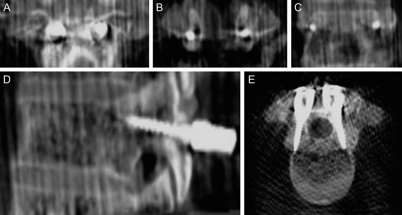
    Coronal (A, plane of entry; B, inside pedicles; and C, plane at screw tip), (D) lateral, and (E) axial views of the cortical screw trajectory at T1. Used with permission from Barrow Neurological Institute, Phoenix, Arizona.

  • Figure 2
    • Download figure
    • Open in new tab
    • Download powerpoint
    Figure 2

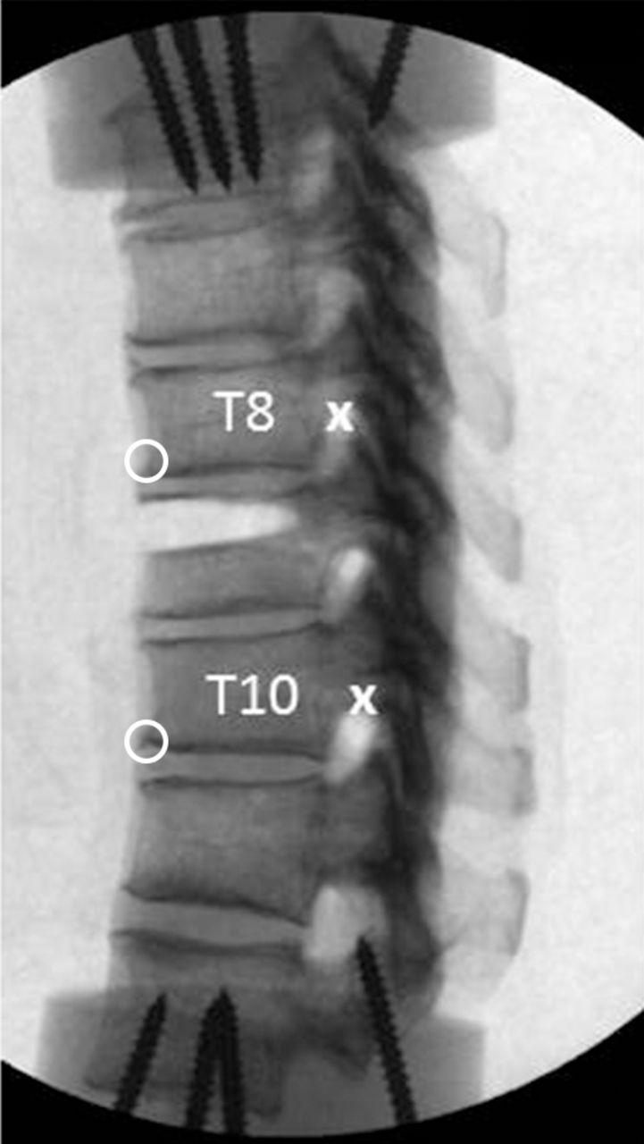
    Lateral view radiograph shows the modeled burst fracture at T9. Anterior (O) and posterior (X) anatomic landmarks were used for tracking during motion analysis and for calculations of vertical displacement during compressive loading. Bone at the superior one-third of the vertebral body, including the posterior cortex and proceeding laterally to the pedicles, was removed using a high-speed drill. The posterior longitudinal ligament was kept intact. Used with permission from Barrow Neurological Institute, Phoenix, Arizona.

  • Figure 3
    • Download figure
    • Open in new tab
    • Download powerpoint
    Figure 3

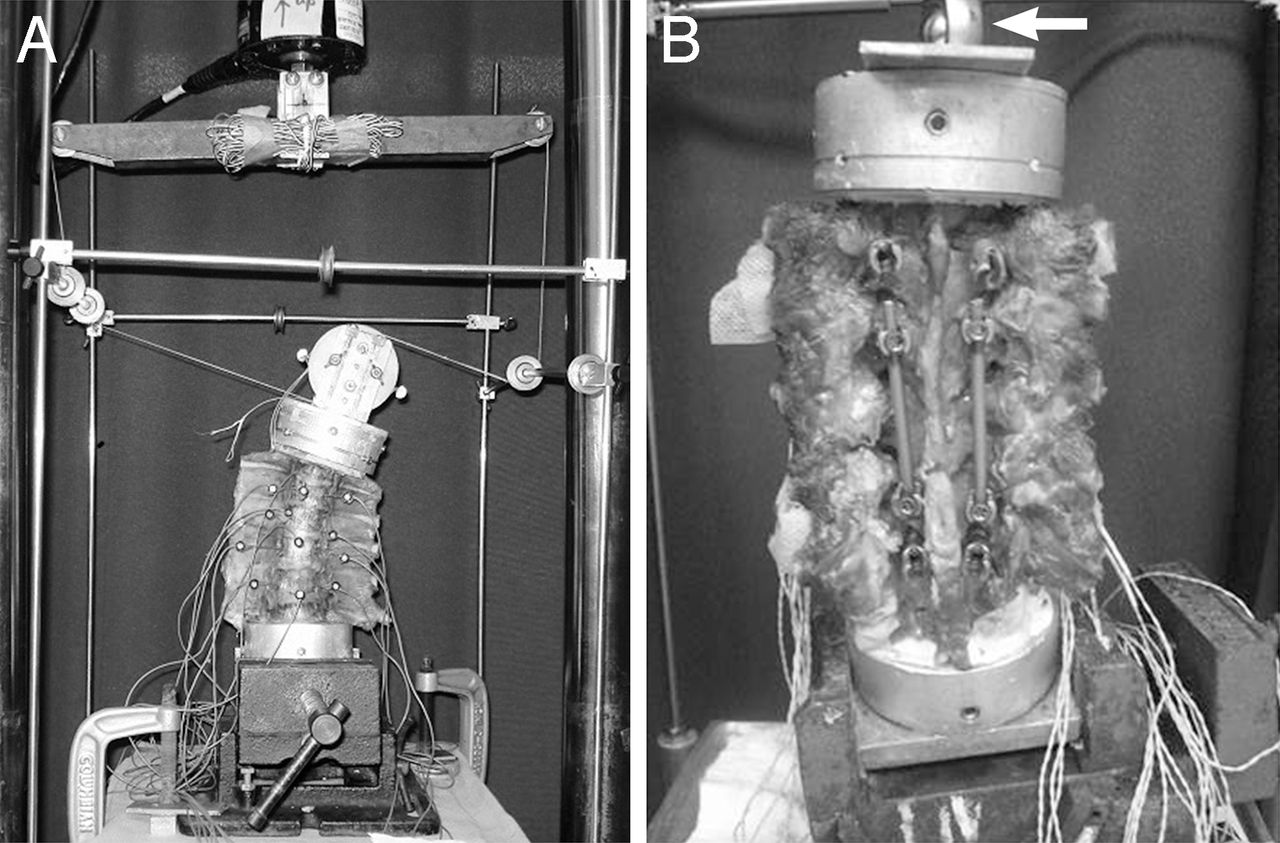
    Test configurations for pure moment (A) angular flexibility loading and (B) compression loading performed in a standard servo-hydraulic test frame (MTS Systems Corp., Eden Prairie, Minnesota). During flexibility tests, adjustable pulleys were used, enabling 2 equal and opposite forces separated by a small distance (pure moment) to be applied via a loop of string when the piston advanced upward. Optical markers for tracking specimen motion are also visible. Shown for (A) left lateral bending; reconfiguring the string and pulleys allows for flexion, extension, or axial rotation. The angle vise securing (B) the construct was repositioned on the base of the test frame in the transverse plane until minimal intervertebral rotations were observed during application of the compressive load via the piston (arrow). Shown for short-segment fixation with cortical screws and rods (distal and proximal screws in place but not attached to rods, posterior view). Used with permission from Barrow Neurological Institute, Phoenix, Arizona.

  • Figure 4
    • Download figure
    • Open in new tab
    • Download powerpoint
    Figure 4

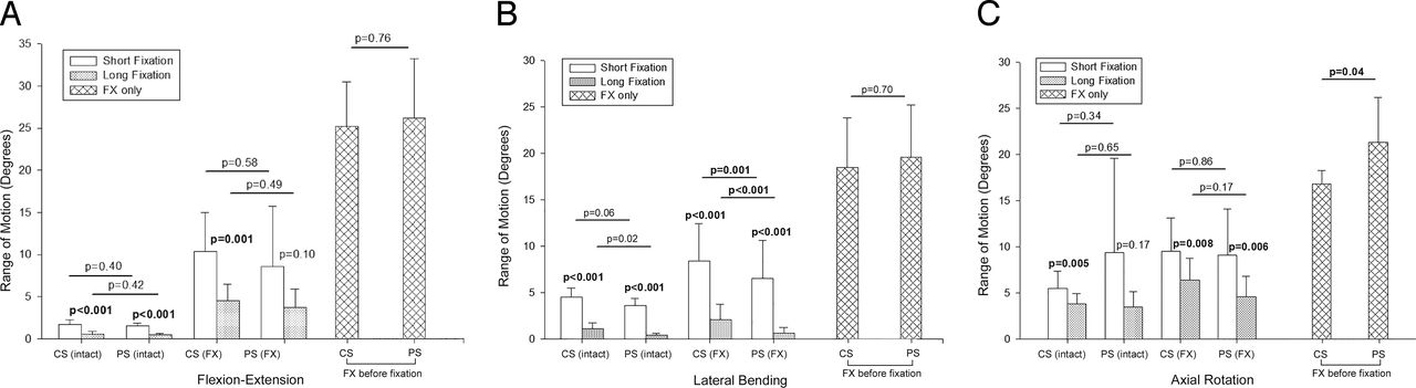
    Mean range of motion across T8-T10 (or T7-T9) for all conditions (intact vertebrae, FX with short-segment and long-segment fixation, and FX before fixation). (A) Flexion-extension, (B) lateral bending, and (C) axial rotation. P values are based on statistical analysis of raw values of range of motion. Error bars indicate SD. Abbreviations: CS, cortical screw; FX, burst fracture; PS, pedicle screw. Used with permission from Barrow Neurological Institute, Phoenix, Arizona.

  • Figure 5
    • Download figure
    • Open in new tab
    • Download powerpoint
    Figure 5

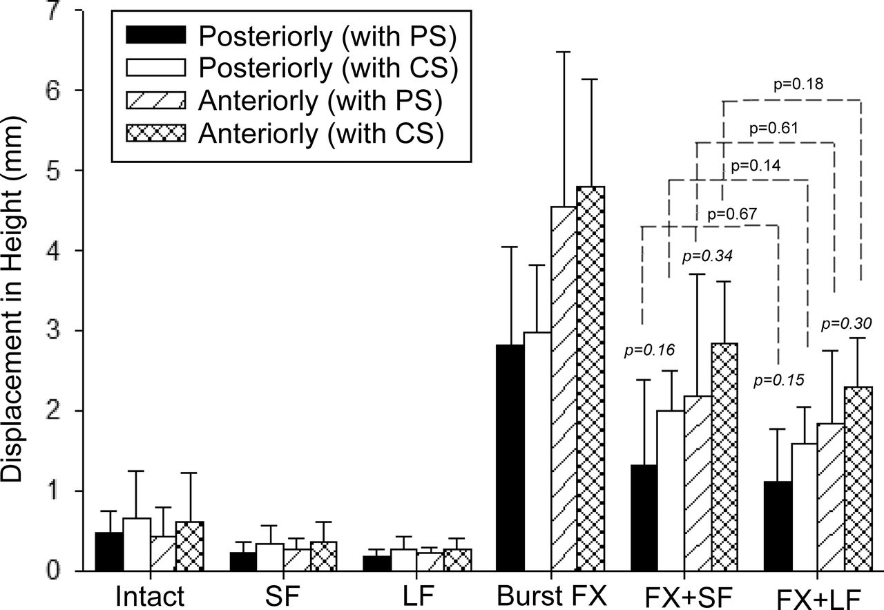
    Mean changes in rostrocaudal height across T8-T10 (or T7-T9), anteriorly and posteriorly, during 300-N axial compression for all conditions. P values shown in italics are for differences between PS and CS with fracture plus fixation (P ≥ .15). P values shown atop dashed lines are for differences between short and long fixations within CS and PS groups (P > .14). Error bars indicate SD; CS, cortical screw; FX, burst fracture; LF, long fixation; PS, pedicle screw; SF, short fixation. Used with permission from Barrow Neurological Institute, Phoenix, Arizona.

Tables

  • Figures
  • Table 1
PreviousNext
Back to top

In this issue

International Journal of Spine Surgery
Vol. 13, Issue 3
1 Jun 2019
  • Table of Contents
  • Index by author

Print
Download PDF
Article Alerts
Sign In to Email Alerts with your Email Address
Email Article

Thank you for your interest in spreading the word on International Journal of Spine Surgery.

NOTE: We only request your email address so that the person you are recommending the page to knows that you wanted them to see it, and that it is not junk mail. We do not capture any email address.

Enter multiple addresses on separate lines or separate them with commas.
Comparing the Biomechanical Stability of Cortical Screw Trajectory Versus Standard Pedicle Screw Trajectory for Short- and Long-Segment Posterior Fixation in 3-Column Thoracic Spinal Injury
(Your Name) has sent you a message from International Journal of Spine Surgery
(Your Name) thought you would like to see the International Journal of Spine Surgery web site.
CAPTCHA
This question is for testing whether or not you are a human visitor and to prevent automated spam submissions.
Citation Tools
Comparing the Biomechanical Stability of Cortical Screw Trajectory Versus Standard Pedicle Screw Trajectory for Short- and Long-Segment Posterior Fixation in 3-Column Thoracic Spinal Injury
AMEY R. SAVARDEKAR, NESTOR G. RODRIGUEZ-MARTINEZ, ANNA G.U.S. NEWCOMB, PHILLIP M. REYES, HECTOR SORIANO-BARON, STEVE W. CHANG, BRIAN P. KELLY, NEIL R. CRAWFORD
International Journal of Spine Surgery Jun 2019, 13 (3) 245-251; DOI: 10.14444/6033

Citation Manager Formats

  • BibTeX
  • Bookends
  • EasyBib
  • EndNote (tagged)
  • EndNote 8 (xml)
  • Medlars
  • Mendeley
  • Papers
  • RefWorks Tagged
  • Ref Manager
  • RIS
  • Zotero
Share
Comparing the Biomechanical Stability of Cortical Screw Trajectory Versus Standard Pedicle Screw Trajectory for Short- and Long-Segment Posterior Fixation in 3-Column Thoracic Spinal Injury
AMEY R. SAVARDEKAR, NESTOR G. RODRIGUEZ-MARTINEZ, ANNA G.U.S. NEWCOMB, PHILLIP M. REYES, HECTOR SORIANO-BARON, STEVE W. CHANG, BRIAN P. KELLY, NEIL R. CRAWFORD
International Journal of Spine Surgery Jun 2019, 13 (3) 245-251; DOI: 10.14444/6033
Twitter logo Facebook logo Mendeley logo
  • Tweet Widget
  • Facebook Like
  • Google Plus One

Jump to section

  • Article
    • ABSTRACT
    • INTRODUCTION
    • MATERIALS AND METHODS
    • RESULTS
    • DISCUSSION
    • ACKNOWLEDGMENTS
    • Footnotes
    • REFERENCES
  • Figures & Data
  • Info & Metrics
  • PDF

Related Articles

  • No related articles found.
  • PubMed
  • Google Scholar

Cited By...

  • No citing articles found.
  • Google Scholar

More in this TOC Section

  • Sequential Anterior Longitudinal Ligament Release With Expandable Spacers for Lordosis Correction in Anterior-to-Psoas Lumbar Interbody Fusion: A Radiographic and Biomechanical Study
  • Comparative Biomechanical Analysis of Anterior Lumbar Interbody Fusion and Bilateral Expandable Transforaminal Lumbar Interbody Fusion Cages: A Finite Element Analysis Study
  • Impact of Different Operative Techniques for Patients With Adolescent Idiopathic Scoliosis on Frontal Curve Correction and Sagittal Balance
Show more Biomechanics

Similar Articles

Keywords

  • burst fracture
  • construct length
  • cortical screw
  • flexibility
  • pedicle screw
  • thoracolumbar biomechanics

Content

  • Current Issue
  • Latest Content
  • Archive

More Information

  • About IJSS
  • About ISASS
  • Privacy Policy

More

  • Subscribe
  • Alerts
  • Feedback

Other Services

  • Author Instructions
  • Join ISASS
  • Reprints & Permissions

© 2025 International Journal of Spine Surgery

International Journal of Spine Surgery Online ISSN: 2211-4599

Powered by HighWire