


Article Figures & Data
Figures
- Figure 1
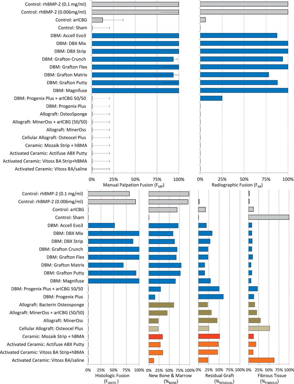
Upper graphs show percent fusion rate by manual palpation (FMP) and radiography (FXR); lower graphs show percent fusion rate by histology (FHISTO), as well as histologic analysis of average percentage of new bone and marrow formation (%BONE), residual graft (%RESIDUAL), and fibrous tissues (%FIBROUS) in the implant area, ex vivo at 8 weeks after PLF. Except for Progenix Plus (0%), FMP for DBMs (100% [CI: 80%–100%]; 94% [CI: 71%–98.5%]) was greater than that for allografts and cellular allografts, ceramics, and activated ceramics (0%). Excluding Progenix Plus, FMP was not significantly different among DBMs and rhBMP-2 (at 0.006 and 0.1 mg/mL concentrations). Histologic analysis is correlated with fusion rates as discussed in the Results section.
- Figure 2
Selected in vivo radiographs of rats at 0-, 4-, and 8-week time points with radiodense materials having FMP of 0%. Despite the lack of fusion as determined by manual palpation (FMP) and histology (FHISTO), these ceramic and activated ceramic materials have high radiodensity that mimics a fusion mass on radiography. Multifocal changes to the radiodensity pattern from 0 to 8 weeks suggest remodeling activity. Careful radiographic review is necessary to note the transverse radiolucent fissures and lack of continuous bone bridges.
- Figure 3
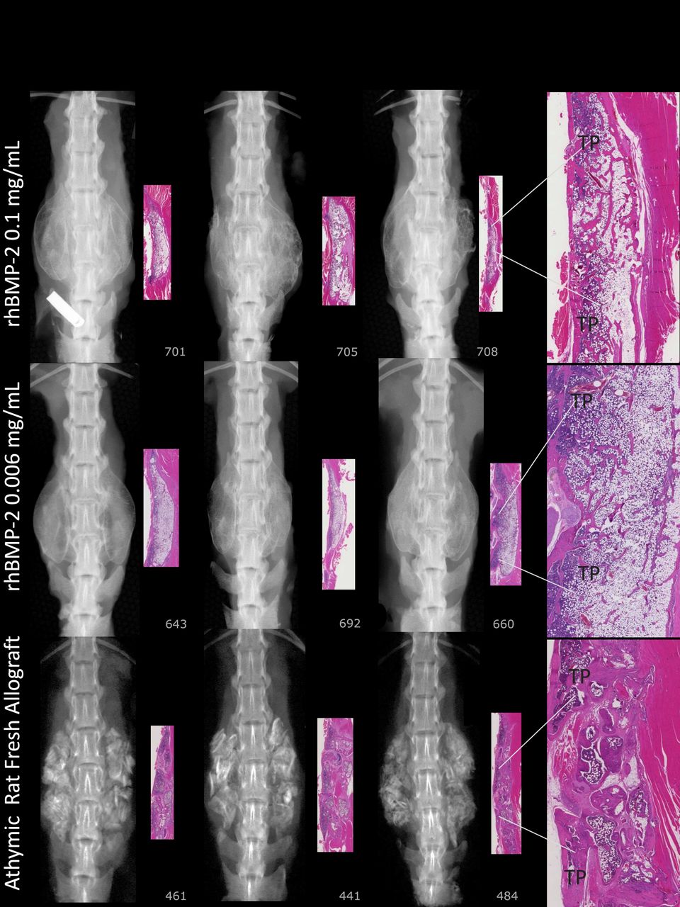
Bilateral robust mineralized fusion masses span L3 to L5 (0.1 mg/mL rhBMP-2/ACS). Radiography shows uniform density with defined margins demonstrating formed bone with cortex (0.006 mg/mL rhBMP-2/ACS). Number 692: histology shows a thin tract of wispy basophilic material (scored as residual material with mineralization) infiltrated with fibrous connective tissue and a few multinucleated giant cells. Radiography shows a fusion region with a thin rim of lamellar bone that forms a complete border continuous with TPs L3 to L4 to L5. Number 660: on histology the fusion is robust with lamellar bone continuous with L3 to L4 to L5 TPs. Trabeculae near TPs are mildly robust, and some portions of the mass are devoid of trabeculae. Particulate dense mineral exists in the L3 to L4 to L5 TP interspaces on radiography (arICBG). Histology shows 69% new bone and marrow formation, suggesting bioactivity and early remodeling.
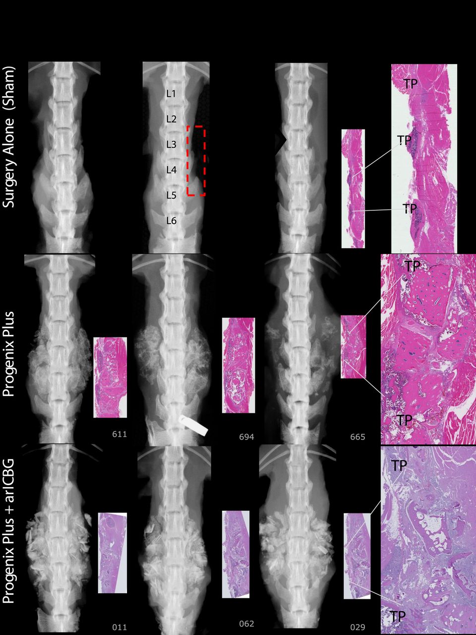
- Figure 4
Surgery Alone (Sham): No dense mineral exists in the TPs interspace of L3 to L4 to L5 regions of interest (ROI), outlined in red rectangle on radiographic image. Paired histology indicates TP interspaces filled with fibrous tissues. Progenix Plus: No evidence of fusion on histology; exhibits variably sized bony residual implant pieces, encapsulated with fibrous connective tissue. There are scattered regions of mineralization in the fibrotic areas. Radiography and histology show limited foci of lamellar bone extending from TP. Progenix Plus + arICBG, numbers 029 and 062: No evidence of fusion on histology, and demonstrates large pieces of residual implant material surrounded by fibrous tissue. Trabeculae are sparse, with little evidence of connections. Areas of fatty marrow show basophilic staining consistent with saponified fat.
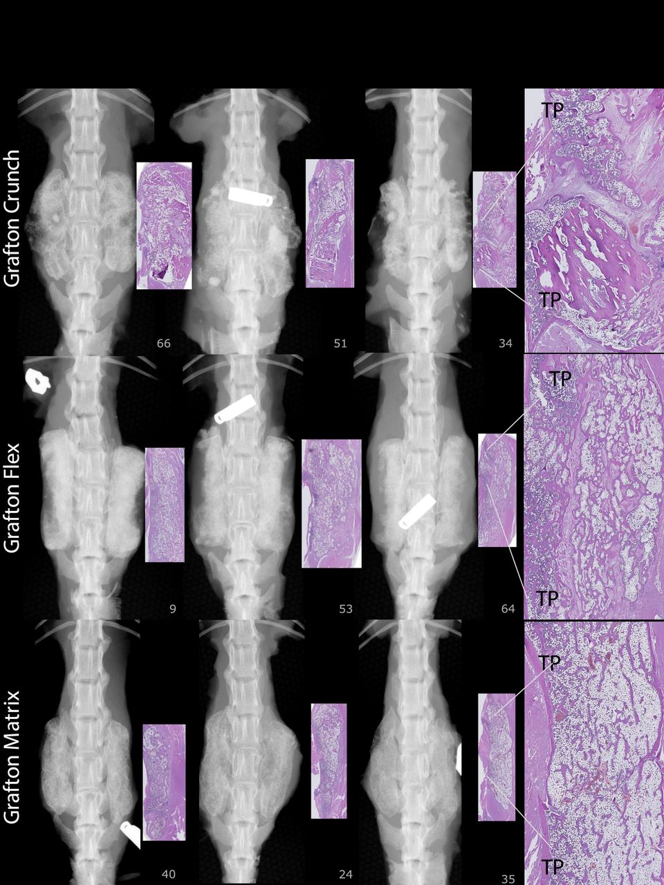
- Figure 5
Grafton-Crunch: Histology shows 65% new bone, with uniform spacing and orientation of lamellar trabeculae, and bony union across TP interspaces with connectivity to TPs L3 to L4 to L5. Number 034: Residual graft (RG) from implanted material. Grafton-Flex, number 009: Robust trabeculae and marrow areas connecting to 3 TPs. Number 064: Evidence of bony union across TP interspaces; however, RG is present. Grafton-Matrix: Histology shows a large amount of lamellar bone with regular sized trabeculae, oriented relatively uniformly throughout the implant area and connected to TPs, 79% new bone.
- Figure 6
Accell Evo3: Histology demonstrates trabecular and lamellar bone with low residual implant material and 72% new bone. DBX Mix: Histology shows irregularly shaped but robust trabeculae are laced between implant pieces throughout the implant area with continuous cortex and connectivity with TPs. Some regions are devoid of trabecular bone. Most demonstrate an outer rim of lamellar bone. DBX Strip: Trabeculae are generally robust; however, many regions demonstrate partial residual graft as short trabeculae without viable osteocytes. Number 027: Tenuous fusion due to clumped pieces of implant material surrounded by fibrosis. Some sections have an outer rim of lamellar bone.
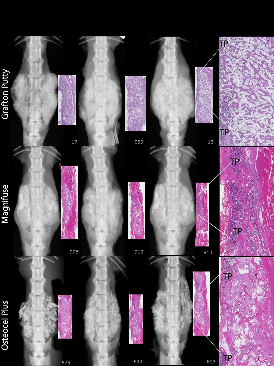
- Figure 7
Grafton Putty: Histology shows 77% new bone, typically with an outer rim of lamellar bone with regular trabeculae, oriented relatively uniformly throughout. Fibrosis is present near clumps of RG. Number 059R: Mildly robust trabeculae are interrupted by a central fibrous tract bisecting the right implant site; histologically not fused on the right but fused on the left. This “nonunion” restricted motion sufficiently that, coupled with the left sided fusion, this rat was evaluated as fused by manual palpation. Magnifuse: There was a rim of lamellar bone surrounding margins of the areas of interest, 65% new bone with broad, variably shaped trabeculae. Numbers 908 and 910: A thin track of braided material defined the border sites. Number 913: Residual implant material midsection. Osteocel Plus, Number #613: Shows spaced spicules of residual implant material surrounded by moderate amounts of fibrosis. Scattered multinucleated, giant cells and lymphocytes evenly spaced between TPs. There are no trabeculae of viable lamellar bone bridging TPs. Numerous trabeculae of acellular bone present. Number 675: Small regions of viable trabeculae sprouting out of the original TPs partially encasing spicules of residual implant material, most are nonviable bone surrounded by fibrosis.
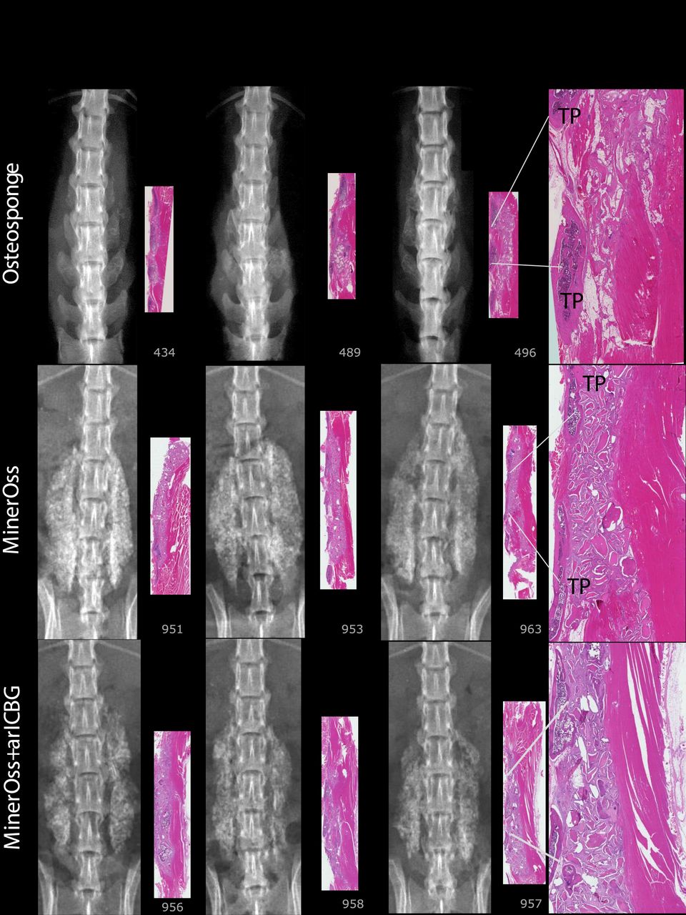
- Figure 8
OsteoSponge: There is a subjective compression of the graft material into a small area. Lamellar compact bone matrix was present, yet no woven bone. There is little to no union with TPs. MinerOss, Number 951: Histologically, there is some ongoing endochondral bone formation; however, the majority of the implant site is composed of residual implant and surrounding soft tissue associated with macrophages and giant cells. Number 963 demonstrates significant residual implant with fibrous tissue, macrophages, and giant cells. MinerOss + arICBG: The addition of arICBG increased histologic new bone formation from 23% to 45% overall; however, this did not result in fusion. Number 956: Islands of bone formation are seen within residual implanted material. Number 958: Lamellar bone formation and endochondral bone formation are ongoing.
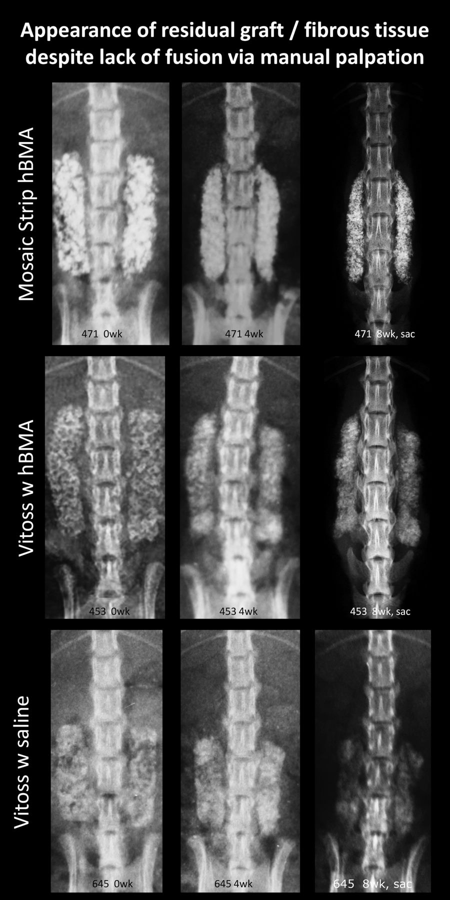
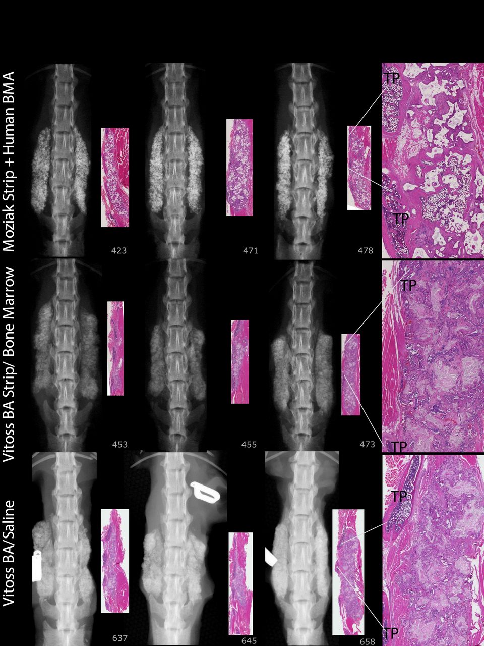
- Figure 9
Mozaik Strip + hBMA: The predominant feature is residual implant material with a cellular response composed of macrophages and giant cells. Vitoss BA Strip + hBMA: The predominant cellular response was composed of macrophages and giant cells for all specimens. The addition of hBMA increased histologic new bone formation from 12% to 34%, and reduced fibrous tissue from 63% to 11%; however, there was no fusion. Vitoss BA Strip + Saline: Sections reveal finely stippled amphiphilic residual material surrounded by mineralization, scattered multinucleated giant cells, and macrophages. Lamellar bone present was limited to the region close to the decorticated TPs. Number 658 demonstrates 75% fibrous tissue.
- Figure 10
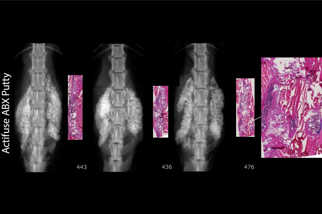
Actifuse ABX Putty number 476 is typical, with residual implant material and minimal lamellar bone/cartilage matrix. No bony fusion connecting TPs.
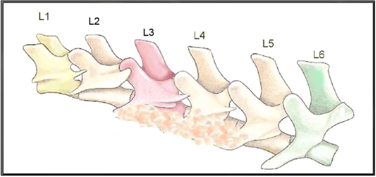
- Appendix 1
This represents an athymic rat spine with the typical 6 lumbar vertebral levels (L1–L6), depicting graft material on the TPs of L3 to L4 and L4 to L5.


Tables
In this issue


Jump to section
Related Articles
Cited By...
More in this TOC Section
Similar Articles
Keywords
- bone
- bone grafts
- substitute
- expander
- demineralized bone matrix (DBM)
- demineralized bone matrix-based products (DBM-based products)
- allografts
- cellular allografts
- autograft
- ceramic
- activated ceramic
- bone morphogenetic protein
- recombinant human bone morphogenetic protein
- rhBMP
- rhBMP-2
- peptide
- differentiation factor
- posterolateral fusion
- muscle pouch
- rat model
- rats
- athymic rats