
Main menu

  • Home
  • Content
    • Current Issue
    • Advance Online Publication
    • Archive
  • About Us
    • About ISASS
    • About the Journal
    • Author Instructions
    • Editorial Board
    • Reviewer Guidelines & Publication Criteria
  • More
    • Advertise
    • Subscribe
    • Alerts
    • Feedback
  • Join Us
  • Reprints & Permissions
  • Sponsored Content
  • Other Publications
    • ijss

User menu

  • My alerts

Search

  • Advanced search
International Journal of Spine Surgery
  • My alerts
International Journal of Spine Surgery

Advanced Search

  • Home
  • Content
    • Current Issue
    • Advance Online Publication
    • Archive
  • About Us
    • About ISASS
    • About the Journal
    • Author Instructions
    • Editorial Board
    • Reviewer Guidelines & Publication Criteria
  • More
    • Advertise
    • Subscribe
    • Alerts
    • Feedback
  • Join Us
  • Reprints & Permissions
  • Sponsored Content
  • Follow ijss on Twitter
  • Visit ijss on Facebook
Research ArticleCervical Spine

Morphometric Study of the Cervical Spinal Canal Content and the Vertebral Artery

EVREN AYDOĞMUŞ and SAFIYE ÇAVDAR
International Journal of Spine Surgery August 2020, 14 (4) 455-461; DOI: https://doi.org/10.14444/7060
EVREN AYDOĞMUŞ
1Department of Neurosurgery, Dr. Lütfi Kirdar Kartal Education and Research Hospital, Istanbul, Turkey
  • Find this author on Google Scholar
  • Find this author on PubMed
  • Search for this author on this site
SAFIYE ÇAVDAR
2Department of Anatomy, Koç University, School of Medicine Istanbul, Turkey
  • Find this author on Google Scholar
  • Find this author on PubMed
  • Search for this author on this site
  • Article
  • Figures & Data
  • Info & Metrics
  • PDF
Loading

Article Figures & Data

Figures

  • Tables
  • Figure 1
    • Download figure
    • Open in new tab
    • Download powerpoint
    Figure 1

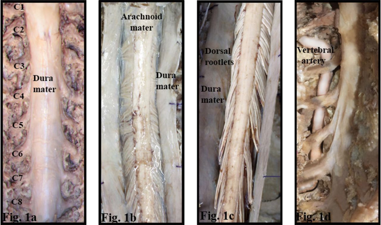
    The skin, subcutaneous tissue, muscles of the region, and the spinous processes and laminae of C1 to C7 vertebrae were removed. (a) The dura mater and the cervical spinal nerves within the dura were exposed. (b) The dura mater was longitudinally incised and the arachnoid matter was exposed. (c) The arachnoid matter was reflected and the dorsal rootlets were exposed. (d) Anterior to the spinal nerves the V2 segment of the vertebral artery was exposed.

  • Figure 2
    • Download figure
    • Open in new tab
    • Download powerpoint
    Figure 2

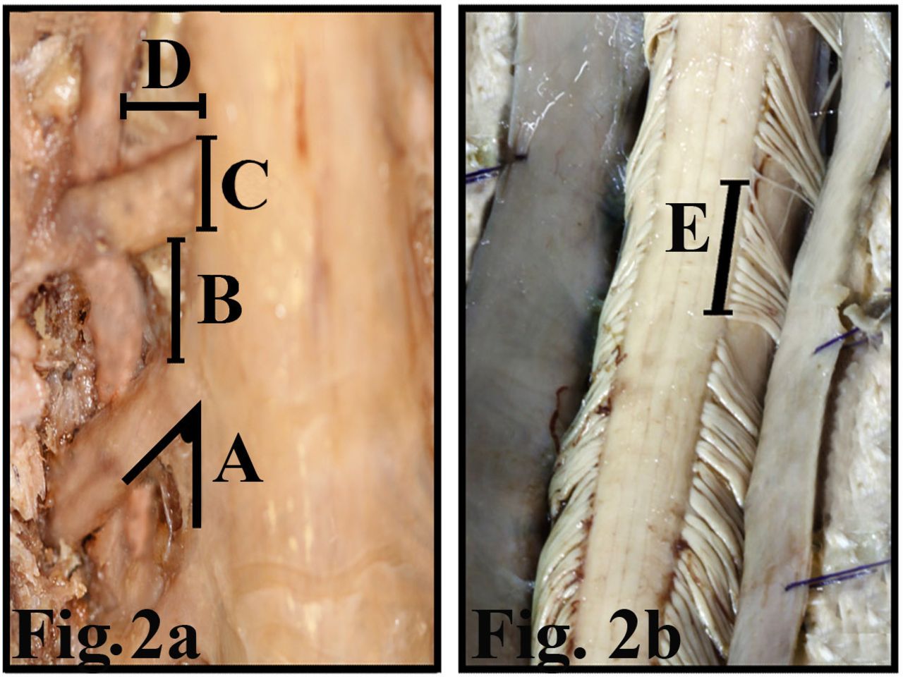
    (a,b) Measurements made on the cervical spinal nerve and the vertebral artery. A indicates angles between the spinal nerve and the spinal cord; B, distance between respective spinal nerves; C, width of spinal nerves; D, distance between of the spinal cord and the vertebral artery; E, the length of dorsal root entry zone.
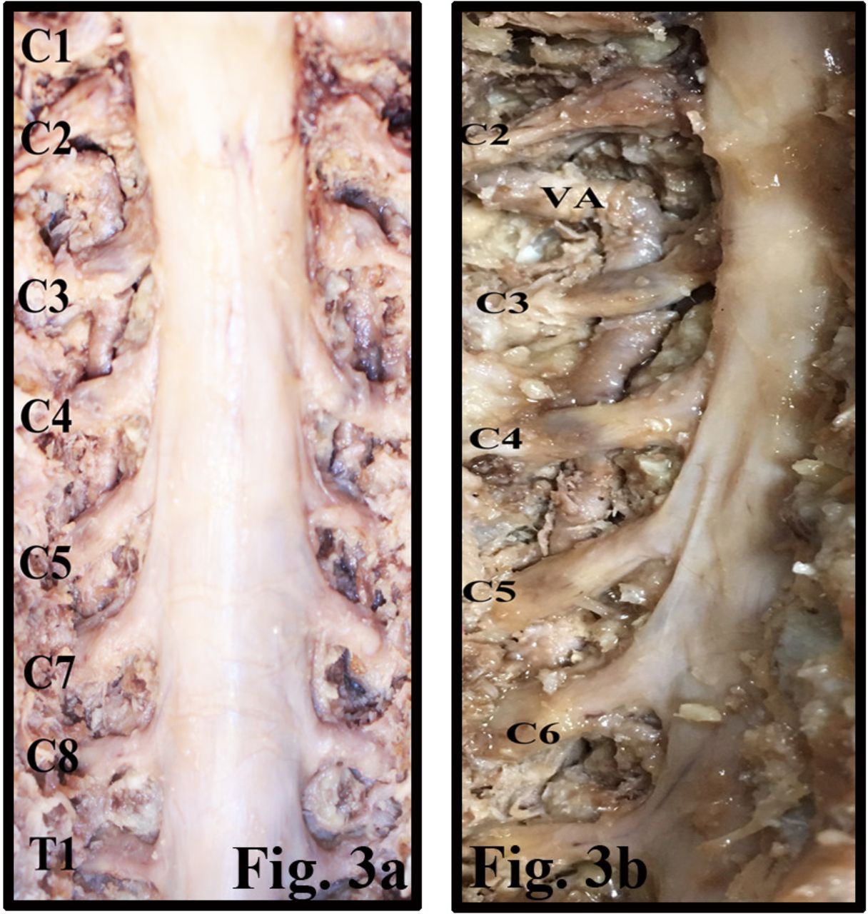
  • Figure 3
    • Download figure
    • Open in new tab
    • Download powerpoint
    Figure 3

    (a) The spinal cord and the cervical spinal nerves have been exposed within the vertebral canal. (b) The relation of the spinal nerve and the vertebral artery (VA) has been exposed.

Tables

  • Figures
  • Table 1
  • Table 2
PreviousNext
Back to top

In this issue

International Journal of Spine Surgery
Vol. 14, Issue 4
1 Aug 2020
  • Table of Contents
  • Index by author

Print
Download PDF
Article Alerts
Sign In to Email Alerts with your Email Address
Email Article

Thank you for your interest in spreading the word on International Journal of Spine Surgery.

NOTE: We only request your email address so that the person you are recommending the page to knows that you wanted them to see it, and that it is not junk mail. We do not capture any email address.

Enter multiple addresses on separate lines or separate them with commas.
Morphometric Study of the Cervical Spinal Canal Content and the Vertebral Artery
(Your Name) has sent you a message from International Journal of Spine Surgery
(Your Name) thought you would like to see the International Journal of Spine Surgery web site.
CAPTCHA
This question is for testing whether or not you are a human visitor and to prevent automated spam submissions.
Citation Tools
Morphometric Study of the Cervical Spinal Canal Content and the Vertebral Artery
EVREN AYDOĞMUŞ, SAFIYE ÇAVDAR
International Journal of Spine Surgery Aug 2020, 14 (4) 455-461; DOI: 10.14444/7060

Citation Manager Formats

  • BibTeX
  • Bookends
  • EasyBib
  • EndNote (tagged)
  • EndNote 8 (xml)
  • Medlars
  • Mendeley
  • Papers
  • RefWorks Tagged
  • Ref Manager
  • RIS
  • Zotero
Share
Morphometric Study of the Cervical Spinal Canal Content and the Vertebral Artery
EVREN AYDOĞMUŞ, SAFIYE ÇAVDAR
International Journal of Spine Surgery Aug 2020, 14 (4) 455-461; DOI: 10.14444/7060
Twitter logo Facebook logo Mendeley logo
  • Tweet Widget
  • Facebook Like
  • Google Plus One

Jump to section

  • Article
    • ABSTRACT
    • INTRODUCTION
    • METHODS
    • RESULTS
    • DISCUSSION
    • Footnotes
    • REFERENCES
  • Figures & Data
  • Info & Metrics
  • PDF

Related Articles

  • No related articles found.
  • PubMed
  • Google Scholar

Cited By...

  • No citing articles found.
  • Google Scholar

More in this TOC Section

  • Innovation and Adversity in Spine Surgery: A Retrospective
  • Cervical Myelopathy Secondary to Bilateral Atlantoaxial Pseudoarticulations in Rheumatoid Arthritis: A Case Report
  • Incidence of Traumatic Spinal Injury Following Public Policy Update on Moped Usage in South Carolina
Show more Cervical Spine

Similar Articles

Keywords

  • dorsal cervical nerve roots
  • spinal cord
  • vertebral artery

Content

  • Current Issue
  • Latest Content
  • Archive

More Information

  • About IJSS
  • About ISASS
  • Privacy Policy

More

  • Subscribe
  • Alerts
  • Feedback

Other Services

  • Author Instructions
  • Join ISASS
  • Reprints & Permissions

© 2025 International Journal of Spine Surgery

International Journal of Spine Surgery Online ISSN: 2211-4599

Powered by HighWire