
Main menu

  • Home
  • Content
    • Current Issue
    • Advance Online Publication
    • Archive
  • About Us
    • About ISASS
    • About the Journal
    • Author Instructions
    • Editorial Board
    • Reviewer Guidelines & Publication Criteria
  • More
    • Advertise
    • Subscribe
    • Alerts
    • Feedback
  • Join Us
  • Reprints & Permissions
  • Sponsored Content
  • Other Publications
    • ijss

User menu

  • My alerts

Search

  • Advanced search
International Journal of Spine Surgery
  • My alerts
International Journal of Spine Surgery

Advanced Search

  • Home
  • Content
    • Current Issue
    • Advance Online Publication
    • Archive
  • About Us
    • About ISASS
    • About the Journal
    • Author Instructions
    • Editorial Board
    • Reviewer Guidelines & Publication Criteria
  • More
    • Advertise
    • Subscribe
    • Alerts
    • Feedback
  • Join Us
  • Reprints & Permissions
  • Sponsored Content
  • Follow ijss on Twitter
  • Visit ijss on Facebook
Research ArticleSpecial Issue-Biomechanics

Biomechanics of Cervical Disc Arthroplasty—A Review of Concepts and Current Technology

Avinash G. Patwardhan and Robert M. Havey
International Journal of Spine Surgery August 2020, 14 (s2) S14-S28; DOI: https://doi.org/10.14444/7087
Avinash G. Patwardhan
1Musculoskeletal Biomechanics Laboratory, Edward Hines, Jr. VA Hospital, Hines, Illinois
2Department of Orthopaedic Surgery and Rehabilitation, Loyola University Medical Center, Maywood, Illinois
PHD
  • Find this author on Google Scholar
  • Find this author on PubMed
  • Search for this author on this site
Robert M. Havey
1Musculoskeletal Biomechanics Laboratory, Edward Hines, Jr. VA Hospital, Hines, Illinois
MS
  • Find this author on Google Scholar
  • Find this author on PubMed
  • Search for this author on this site
  • Article
  • Figures & Data
  • Info & Metrics
  • PDF
Loading

Article Figures & Data

Figures

  • Figure 1
    • Download figure
    • Open in new tab
    • Download powerpoint
    Figure 1

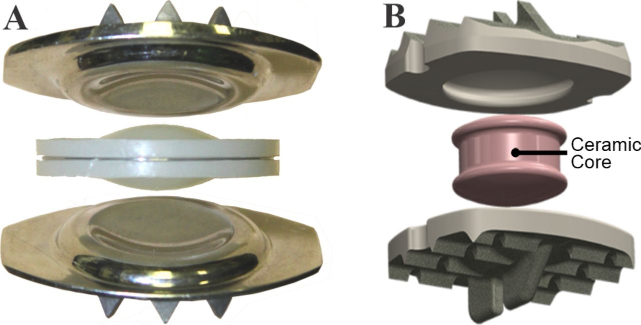
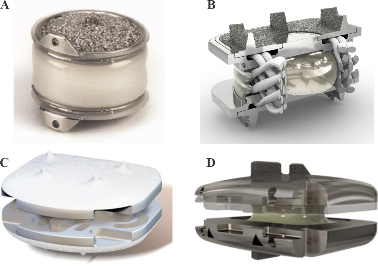
    A disc prosthesis with 3 components including a biconvex mobile core that articulate in 2 spherical bearings (ball-and-socket joints). (A) The Charité lumbar disc. (B) The Simplify cervical disc prosthesis (Source: Simplify Medical, Inc).

  • Figure 2
    • Download figure
    • Open in new tab
    • Download powerpoint
    Figure 2

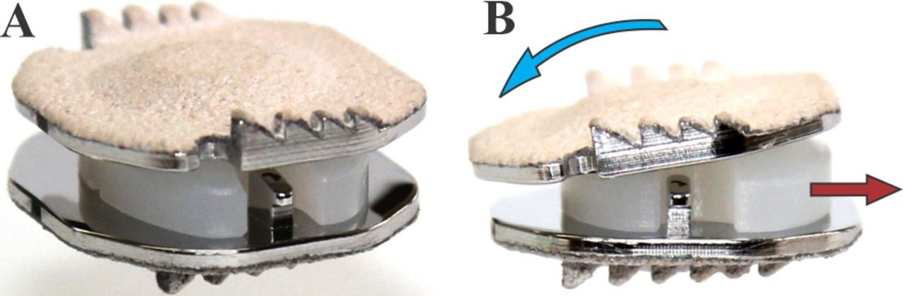
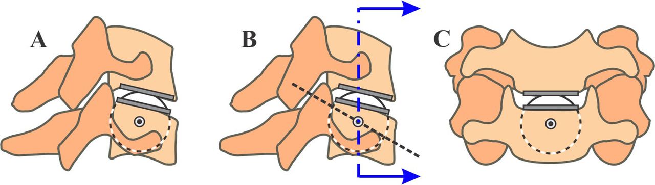
    Another example of a prosthesis with 3 articulating components with 2 bearings is the Mobi-C cervical artificial disc. The bearing (joint) formed by the mobile core with the superior prosthetic endplate is spherical. The core forms a planar bearing with the inferior prosthetic endplate. Endplate angular motion (blue arrow) will result in a corresponding translation of the core (red arrow).

  • Figure 3
    • Download figure
    • Open in new tab
    • Download powerpoint
    Figure 3

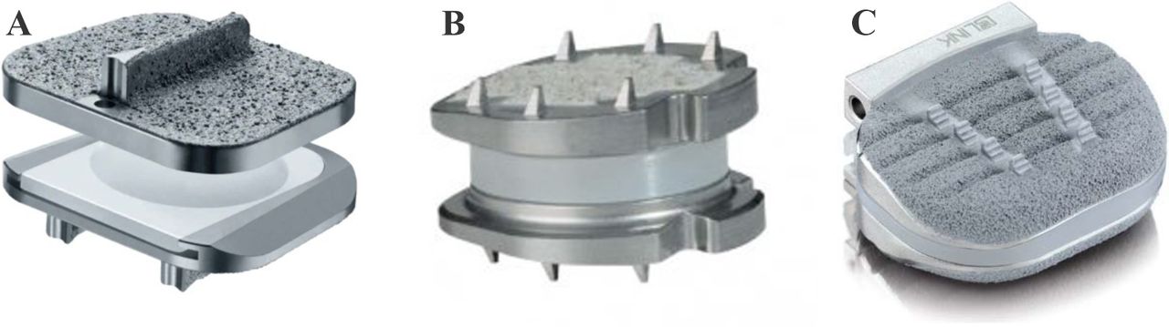
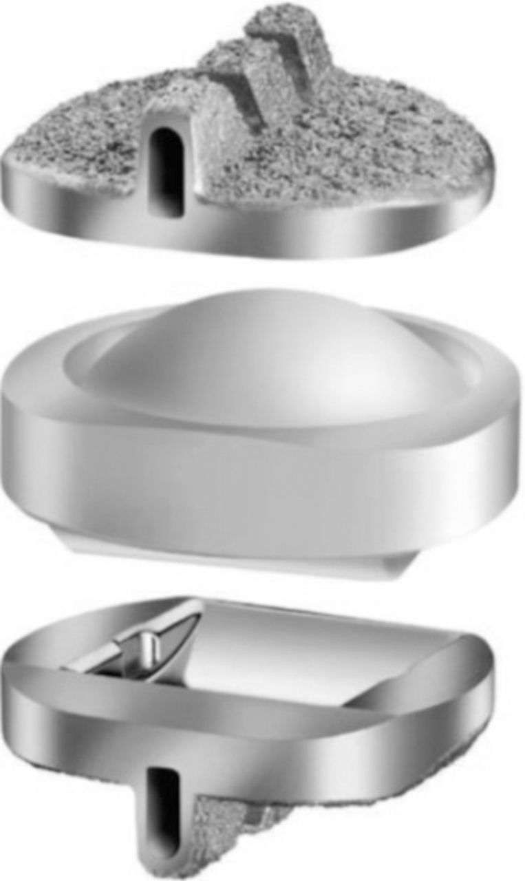
    The Secure-C disc prosthesis has a mobile core that forms a spherical (ball-and-socket) bearing with the superior prosthetic endplate and a cylindrical bearing with the inferior prosthetic endplate with the long axis of the cylinder aligned in the coronal plane (Source: Globus Medical).

  • Figure 4
    • Download figure
    • Open in new tab
    • Download powerpoint
    Figure 4

    Prostheses with 2 components that articulate to form a spherical (ball-and-socket) joint. (A) ProDisc-C (Source: Centinel Spine). (B) Discover (Source: DePuy Synthes Spine). (C) PCM (Source: NuVasive).

  • Figure 5
    • Download figure
    • Open in new tab
    • Download powerpoint
    Figure 5

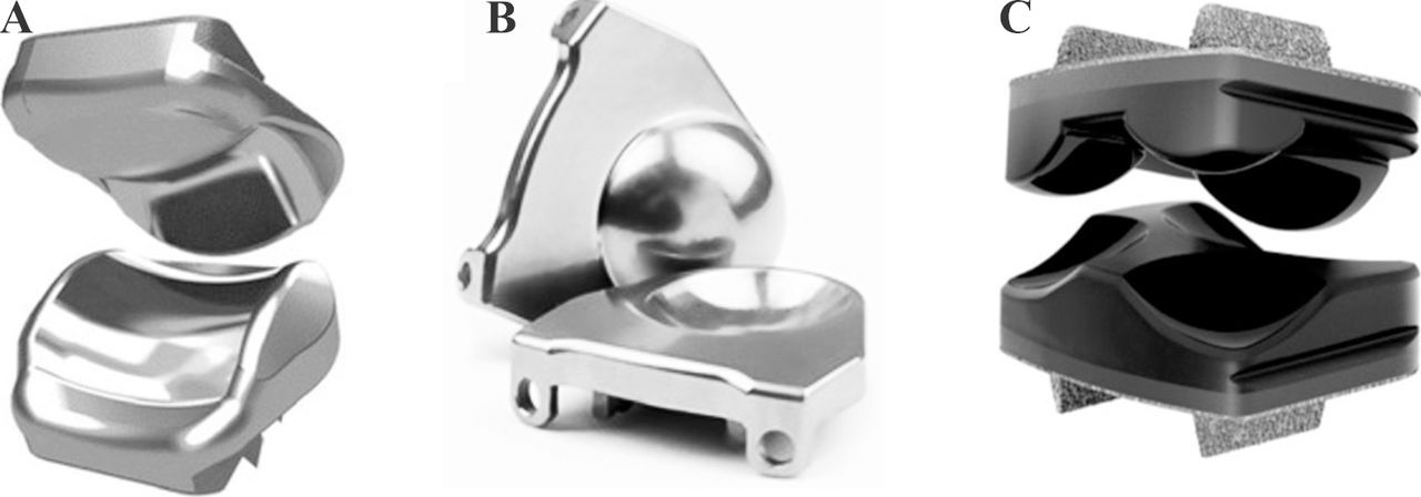
    Prostheses with 2 components that articulate to form (A) a saddle joint (Source: Stryker Spine), (B) a ball-in-trough joint (Source: Medtronic), and (C) 3 noncongruent ball-and-socket joints (Source: Dymicron, Inc).

  • Figure 6
    • Download figure
    • Open in new tab
    • Download powerpoint
    Figure 6

    A class of nonarticulating discs with compressible cores. (A) Bryan disc (Source: Medtronic), (B) M6-C disc (Source: Orthofix Spine), (C) CP-ESP disc (Source: FH Orthopedics Inc.), and (D) Rhine disc (Source: Stryker Spine).

  • Figure 7
    • Download figure
    • Open in new tab
    • Download powerpoint
    Figure 7

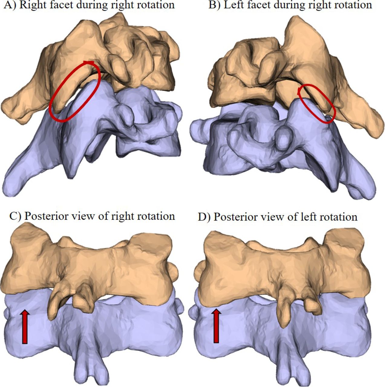
    Axial rotation range of motion test of C5-C6. (A) View of right facet showing full overlap during right rotation, (B) left facet showing minimal facet overlap during right rotation, and (C) right rotation showing coupled right lateral bending. Transparent C5 body shows decreased facet overlap on left (D) left rotation showing coupled left lateral bending. Note increased facet overlap on left.

  • Figure 8
    • Download figure
    • Open in new tab
    • Download powerpoint
    Figure 8

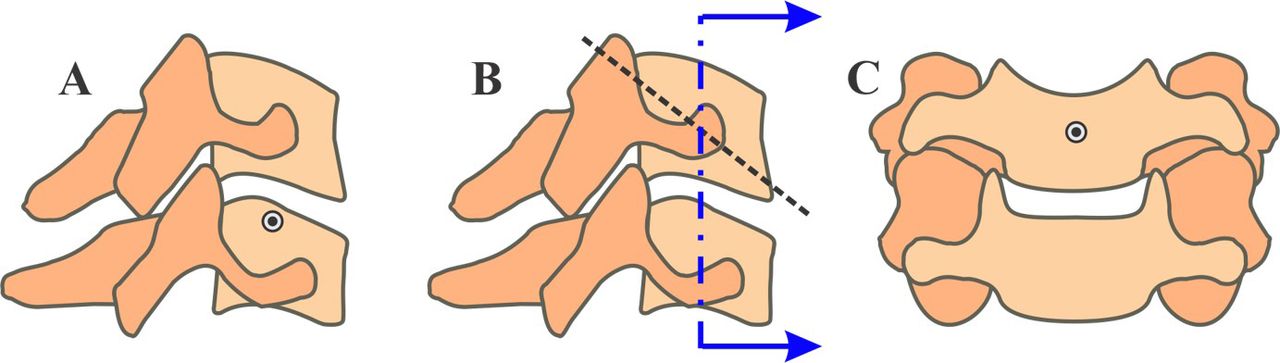
    Axes of rotation in a healthy C5-C6 segment: (A) flexion-extension COR, (B) lateral bending axis of rotation shown in sagittal projection, and (C) intersection of lateral bending axis with the midcoronal plane. COR, center of rotation.

  • Figure 9
    • Download figure
    • Open in new tab
    • Download powerpoint
    Figure 9

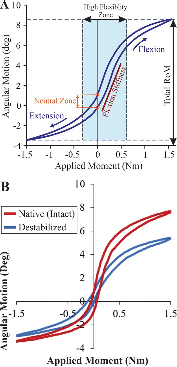
    (A) Kinematic signature of a cervical spine segment in flexion-extension. (B) Effect of decompressive surgery on the kinematic signature showing increased laxity (decreased stiffness) around the neutral posture, resulting in larger range of motion.

  • Figure 10
    • Download figure
    • Open in new tab
    • Download powerpoint
    Figure 10

    Axes of rotation in a reconstructed C5-C6 segment implanted with a single spherical bearing prosthesis: (A) flexion extension COR, (B) lateral bending axis of rotation shown in sagittal projection, and (C) intersection of lateral bending axis with the mid-coronal plane. COR, center of rotation.

  • Figure 11
    • Download figure
    • Open in new tab
    • Download powerpoint
    Figure 11

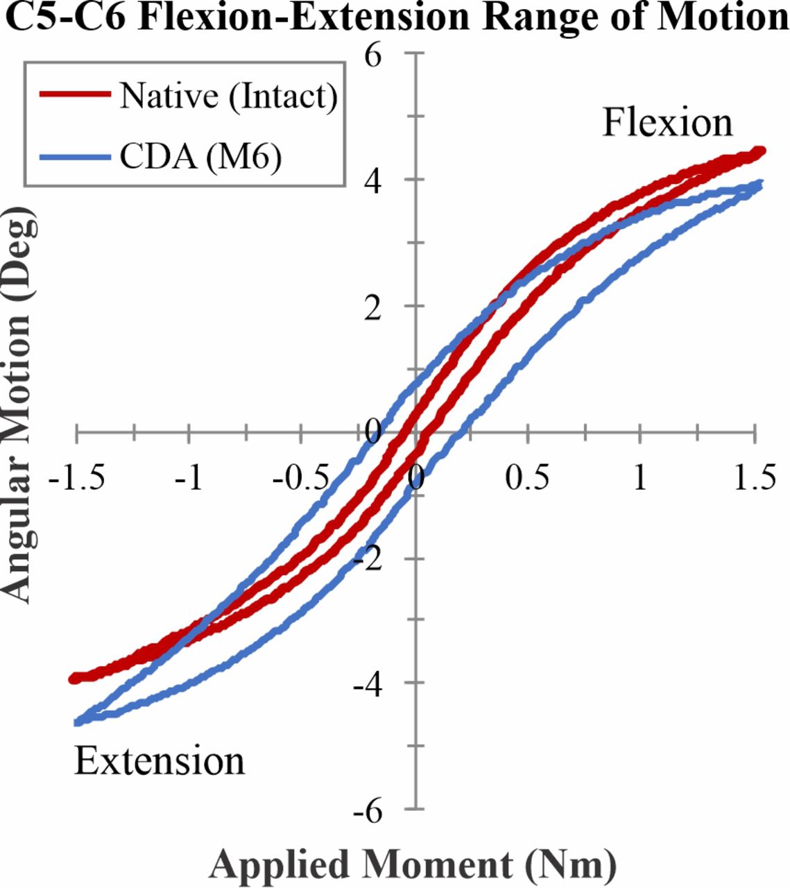
    Examples of kinematic signatures of a cervical spine segment in flexion extension: intact (native) and after CDA using an M6-C disc prosthesis. CDA, cervical disc arthroplasty.

  • Figure 12
    • Download figure
    • Open in new tab
    • Download powerpoint
    Figure 12

    (A, B) Intact cervical segment with the physiologic lateral bending axis of rotation in the cranial body showing normal ROM in lateral bending. (C, D) Implanted cervical segment with nonphysiologic lateral bending axis of rotation in the disc space demonstrating uncinate impingement during lateral bending. ROM, range of motion.

PreviousNext
Back to top

In this issue

International Journal of Spine Surgery: 14 (s2)
International Journal of Spine Surgery
Vol. 14, Issue s2
1 Aug 2020
  • Table of Contents
  • Index by author

Print
Download PDF
Article Alerts
Sign In to Email Alerts with your Email Address
Email Article

Thank you for your interest in spreading the word on International Journal of Spine Surgery.

NOTE: We only request your email address so that the person you are recommending the page to knows that you wanted them to see it, and that it is not junk mail. We do not capture any email address.

Enter multiple addresses on separate lines or separate them with commas.
Biomechanics of Cervical Disc Arthroplasty—A Review of Concepts and Current Technology
(Your Name) has sent you a message from International Journal of Spine Surgery
(Your Name) thought you would like to see the International Journal of Spine Surgery web site.
CAPTCHA
This question is for testing whether or not you are a human visitor and to prevent automated spam submissions.
Citation Tools
Biomechanics of Cervical Disc Arthroplasty—A Review of Concepts and Current Technology
Avinash G. Patwardhan, Robert M. Havey
International Journal of Spine Surgery Aug 2020, 14 (s2) S14-S28; DOI: 10.14444/7087

Citation Manager Formats

  • BibTeX
  • Bookends
  • EasyBib
  • EndNote (tagged)
  • EndNote 8 (xml)
  • Medlars
  • Mendeley
  • Papers
  • RefWorks Tagged
  • Ref Manager
  • RIS
  • Zotero
Share
Biomechanics of Cervical Disc Arthroplasty—A Review of Concepts and Current Technology
Avinash G. Patwardhan, Robert M. Havey
International Journal of Spine Surgery Aug 2020, 14 (s2) S14-S28; DOI: 10.14444/7087
Twitter logo Facebook logo Mendeley logo
  • Tweet Widget
  • Facebook Like
  • Google Plus One

Jump to section

  • Article
    • ABSTRACT
    • INTRODUCTION
    • BIOMECHANICAL FUNCTIONAL GOAL OF CDA
    • CLASSIFICATION OF CERVICAL DISC PROSTHESES DESIGNS
    • KINEMATICS OF HEALTHY CERVICAL SPINE SEGMENTS
    • PROSTHESIS DESIGN AND MOTION AFTER CDA
    • PROSTHESIS DESIGN AND LOAD-SHARING IN THE 3-JOINT COMPLEX
    • INFLUENCE OF SURGICAL FACTORS ON FUNCTIONAL SPINE UNIT KINEMATICS AFTER CDA
    • CONCLUSIONS
    • Footnotes
    • REFERENCES
  • Figures & Data
  • Info & Metrics
  • PDF

Related Articles

  • No related articles found.
  • PubMed
  • Google Scholar

Cited By...

  • No citing articles found.
  • Google Scholar

Similar Articles

Keywords

  • cervical spine
  • cervical disc arthroplasty
  • total disc replacement
  • biomechanics

Content

  • Current Issue
  • Latest Content
  • Archive

More Information

  • About IJSS
  • About ISASS
  • Privacy Policy

More

  • Subscribe
  • Alerts
  • Feedback

Other Services

  • Author Instructions
  • Join ISASS
  • Reprints & Permissions

© 2025 International Journal of Spine Surgery

International Journal of Spine Surgery Online ISSN: 2211-4599

Powered by HighWire