
Main menu

  • Home
  • Content
    • Current Issue
    • Advance Online Publication
    • Archive
  • About Us
    • About ISASS
    • About the Journal
    • Author Instructions
    • Editorial Board
    • Reviewer Guidelines & Publication Criteria
  • More
    • Advertise
    • Subscribe
    • Alerts
    • Feedback
  • Join Us
  • Reprints & Permissions
  • Sponsored Content
  • Other Publications
    • ijss

User menu

  • My alerts

Search

  • Advanced search
International Journal of Spine Surgery
  • My alerts
International Journal of Spine Surgery

Advanced Search

  • Home
  • Content
    • Current Issue
    • Advance Online Publication
    • Archive
  • About Us
    • About ISASS
    • About the Journal
    • Author Instructions
    • Editorial Board
    • Reviewer Guidelines & Publication Criteria
  • More
    • Advertise
    • Subscribe
    • Alerts
    • Feedback
  • Join Us
  • Reprints & Permissions
  • Sponsored Content
  • Follow ijss on Twitter
  • Visit ijss on Facebook
Research ArticleSpecial Issue

Endoscopic Transforaminal Lumbar Interbody Fusion With a Single Oblique PEEK Cage and Posterior Supplemental Fixation

Álvaro Dowling and Kai-Uwe Lewandrowski
International Journal of Spine Surgery December 2020, 14 (s3) S45-S55; DOI: https://doi.org/10.14444/7126
Álvaro Dowling
1Endoscopic Spine Clinic, Santiago, Chile, Department of Orthopaedic Surgery, USP, Ribeirão Preto, Brazil
MD
  • Find this author on Google Scholar
  • Find this author on PubMed
  • Search for this author on this site
Kai-Uwe Lewandrowski
2Center for Advanced Spine Care of Southern Arizona and Surgical Institute of Tucson, Tucson, Arizona, Department Neurosurgery, UNIRIO, Rio de Janeiro, Brazil, Department of Orthopaedics, Fundación Universitaria Sanitas, Bogotá, DC, Colombia
MD
  • Find this author on Google Scholar
  • Find this author on PubMed
  • Search for this author on this site
  • Article
  • Figures & Data
  • Info & Metrics
  • PDF
Loading

Article Figures & Data

Figures

  • Tables
  • Figure 1
    • Download figure
    • Open in new tab
    • Download powerpoint
    Figure 1

    Age distribution of the 43 study patients with the superimposed expected normal distribution (black line). Patients' ages ranged from 35 to 93 years of age and averaged 65.66 years.
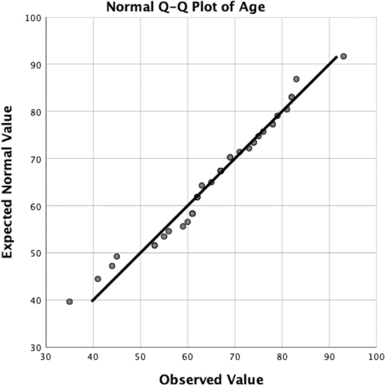
  • Figure 2
    • Download figure
    • Open in new tab
    • Download powerpoint
    Figure 2

    The quantile-quantile plot of the endoscopy patients' age shows normal distribution. The average age was 65.66 ± 11.90 years SD, ranging from 35 to 93 years.

  • Figure 3
    • Download figure
    • Open in new tab
    • Download powerpoint
    Figure 3

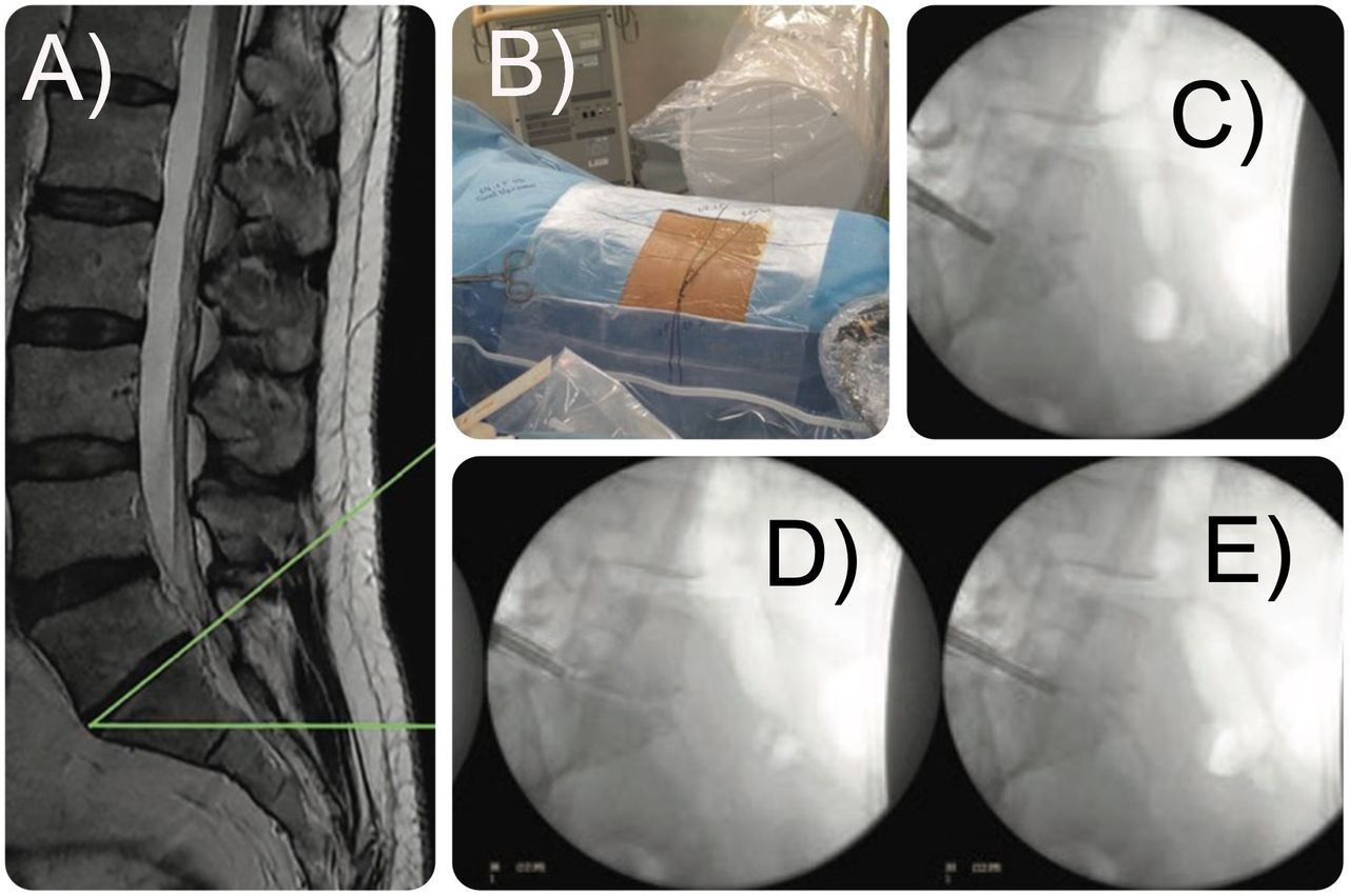
    An exemplary case of a 63-year-old female patient who underwent endoscopically assisted minimally invasive surgery with transforaminal lumbar interbody fusion. Shown is (A) the preoperative sagittal MRI scan showing the sacral slope, (B) positioning and draping of the patient, and (C-E) various steps of the transforaminal interspace preparation with (C) reamers, (D) Kerrison, and (E) chisels.

  • Figure 4
    • Download figure
    • Open in new tab
    • Download powerpoint
    Figure 4

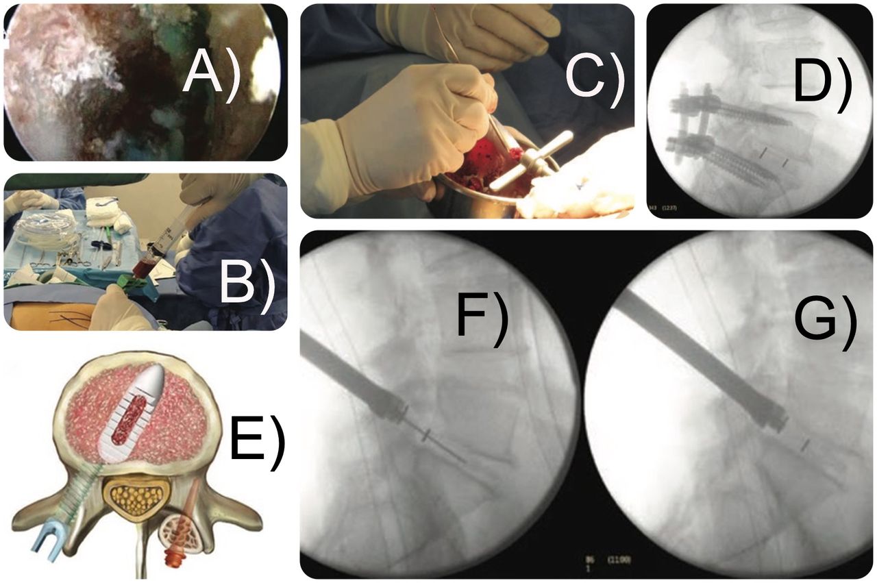
    Intraoperative steps of the fusion surgery of the patient shown in Figure 3 showing the (A) endoscopic view of the decorticated endplates and the empty interspace, (B) the aspiration of bone marrow, (C) the enrichment of allograft chips with bone marrow aspirate and placement through a funnel into the intervertebral disc space, (D) the posterior supplemental percutaneously placed pedicle screw system, (E) the oblique bullet-nosed cannulated interbody fusion cage placed over a nitinol guidewire into the (F) intervertebral disc space into its (G) final position under fluoroscopic control.

  • Figure 5
    • Download figure
    • Open in new tab
    • Download powerpoint
    Figure 5

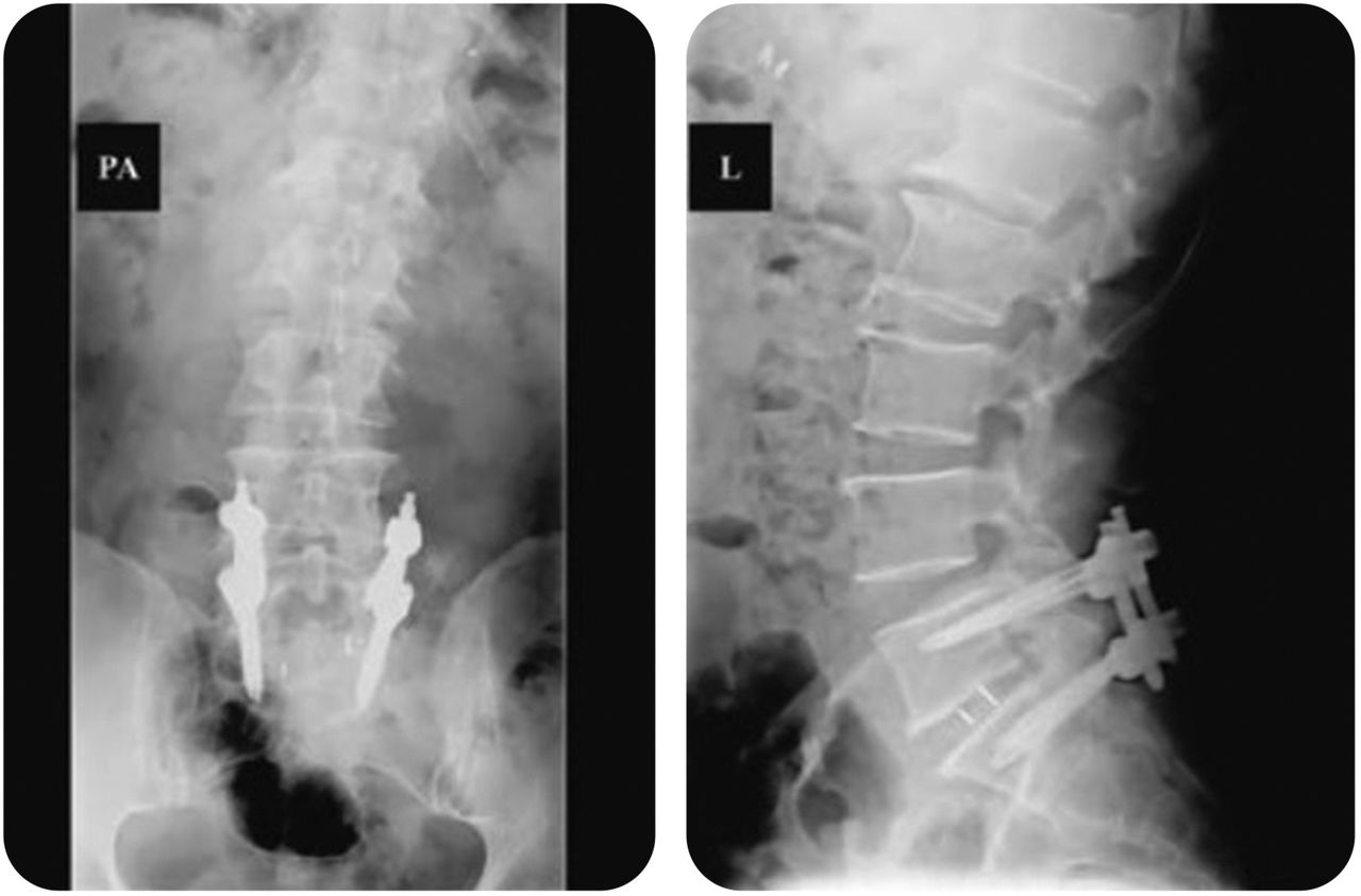
    Postoperative posteroanterior and lateral standing x-rays of the same 63-year-old female patient illustrated in Figures 2 and 3 who underwent endoscopically assisted minimally invasive surgery with transforaminal lumbar interbody fusion are shown. These radiographs were taken at 6 weeks postoperatively.

  • Figure 6
    • Download figure
    • Open in new tab
    • Download powerpoint
    Figure 6

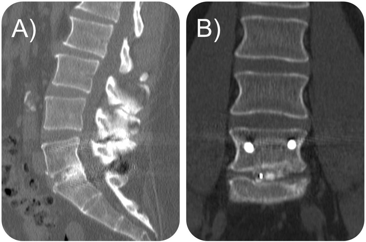
    (A) Sagittal and (B) coronal computed tomography (CT) sections of the same 63-year-old female patient illustrated in Figures 2, 3, and 4 who underwent endoscopically assisted minimally invasive surgery with transforaminal lumbar interbody fusion taken 35 months postoperatively. This CT scan was prompted by recurrent back pain without sciatica that was ultimately attributed to adjacent-level painful L4-5 facet arthropathy which was treated successfully without additional surgery and with medical and interventional supportive care measures.

Tables

  • Figures
  • Table 1
  • Table 2
  • Table 3
  • Table 4
  • Table 5
  • Table 6
  • Table 7
PreviousNext
Back to top

In this issue

International Journal of Spine Surgery
Vol. 14, Issue s3
1 Dec 2020
  • Table of Contents
  • Index by author

Print
Download PDF
Article Alerts
Sign In to Email Alerts with your Email Address
Email Article

Thank you for your interest in spreading the word on International Journal of Spine Surgery.

NOTE: We only request your email address so that the person you are recommending the page to knows that you wanted them to see it, and that it is not junk mail. We do not capture any email address.

Enter multiple addresses on separate lines or separate them with commas.
Endoscopic Transforaminal Lumbar Interbody Fusion With a Single Oblique PEEK Cage and Posterior Supplemental Fixation
(Your Name) has sent you a message from International Journal of Spine Surgery
(Your Name) thought you would like to see the International Journal of Spine Surgery web site.
CAPTCHA
This question is for testing whether or not you are a human visitor and to prevent automated spam submissions.
Citation Tools
Endoscopic Transforaminal Lumbar Interbody Fusion With a Single Oblique PEEK Cage and Posterior Supplemental Fixation
Álvaro Dowling, Kai-Uwe Lewandrowski
International Journal of Spine Surgery Dec 2020, 14 (s3) S45-S55; DOI: 10.14444/7126

Citation Manager Formats

  • BibTeX
  • Bookends
  • EasyBib
  • EndNote (tagged)
  • EndNote 8 (xml)
  • Medlars
  • Mendeley
  • Papers
  • RefWorks Tagged
  • Ref Manager
  • RIS
  • Zotero
Share
Endoscopic Transforaminal Lumbar Interbody Fusion With a Single Oblique PEEK Cage and Posterior Supplemental Fixation
Álvaro Dowling, Kai-Uwe Lewandrowski
International Journal of Spine Surgery Dec 2020, 14 (s3) S45-S55; DOI: 10.14444/7126
Twitter logo Facebook logo Mendeley logo
  • Tweet Widget
  • Facebook Like
  • Google Plus One

Jump to section

  • Article
    • ABSTRACT
    • INTRODUCTION
    • MATERIALS AND METHODS
    • RESULTS
    • DISCUSSION
    • CONCLUSIONS
    • Footnotes
    • REFERENCES
  • Figures & Data
  • Info & Metrics
  • PDF

Related Articles

  • No related articles found.
  • PubMed
  • Google Scholar

Cited By...

  • No citing articles found.
  • Google Scholar

More in this TOC Section

  • Letter to the Editor: Rasch Analysis and High Value Spinal Endoscopy—Another Perspective
  • Real-World Implementation of Artificial Intelligence/Machine Learning for Managing Surgical Spine Patients at 2 Academic Health Care Systems
  • Potential Applications of Artificial Intelligence and Machine Learning in Spine Surgery Across the Continuum of Care
Show more Special Issue

Similar Articles

Keywords

  • lumbar interbody fusion
  • minimally invasive
  • endoscopic
  • direct visualization

Content

  • Current Issue
  • Latest Content
  • Archive

More Information

  • About IJSS
  • About ISASS
  • Privacy Policy

More

  • Subscribe
  • Alerts
  • Feedback

Other Services

  • Author Instructions
  • Join ISASS
  • Reprints & Permissions

© 2025 International Journal of Spine Surgery

International Journal of Spine Surgery Online ISSN: 2211-4599

Powered by HighWire