
Main menu

  • Home
  • Content
    • Current Issue
    • Advance Online Publication
    • Archive
  • About Us
    • About ISASS
    • About the Journal
    • Author Instructions
    • Editorial Board
    • Reviewer Guidelines & Publication Criteria
  • More
    • Advertise
    • Subscribe
    • Alerts
    • Feedback
  • Join Us
  • Reprints & Permissions
  • Sponsored Content
  • Other Publications
    • ijss

User menu

  • My alerts

Search

  • Advanced search
International Journal of Spine Surgery
  • My alerts
International Journal of Spine Surgery

Advanced Search

  • Home
  • Content
    • Current Issue
    • Advance Online Publication
    • Archive
  • About Us
    • About ISASS
    • About the Journal
    • Author Instructions
    • Editorial Board
    • Reviewer Guidelines & Publication Criteria
  • More
    • Advertise
    • Subscribe
    • Alerts
    • Feedback
  • Join Us
  • Reprints & Permissions
  • Sponsored Content
  • Follow ijss on Twitter
  • Visit ijss on Facebook
Research ArticleCervical Spine

A Case Report of Complete Dislocation of Atlantoaxial Joint due to a Traumatic and Pathological Axial Lesion

Xinwei Yuan, Lun Wan, Jiang Hu and Wei Zhang
International Journal of Spine Surgery February 2021, 14 (s4) S5-S9; DOI: https://doi.org/10.14444/7157
Xinwei Yuan
Department of Orthopedics, Sichuan Academy of Medical Sciences and Sichuan Provincial People's Hospital, Chengdu, Sichuan, P.R. China
MD
  • Find this author on Google Scholar
  • Find this author on PubMed
  • Search for this author on this site
Lun Wan
Department of Orthopedics, Sichuan Academy of Medical Sciences and Sichuan Provincial People's Hospital, Chengdu, Sichuan, P.R. China
BD
  • Find this author on Google Scholar
  • Find this author on PubMed
  • Search for this author on this site
Jiang Hu
Department of Orthopedics, Sichuan Academy of Medical Sciences and Sichuan Provincial People's Hospital, Chengdu, Sichuan, P.R. China
MD
  • Find this author on Google Scholar
  • Find this author on PubMed
  • Search for this author on this site
Wei Zhang
Department of Orthopedics, Sichuan Academy of Medical Sciences and Sichuan Provincial People's Hospital, Chengdu, Sichuan, P.R. China
PhD
  • Find this author on Google Scholar
  • Find this author on PubMed
  • Search for this author on this site
  • Article
  • Figures & Data
  • Info & Metrics
  • PDF
Loading

ABSTRACT

Background: Atlantoaxial dislocation usually results in sudden death. The patient had never found any axial lesion before, and the atlantoaxial joint dislocation was caused by rotation of the neck due to discomfort of the neck. The patient was given surgical treatment after the rescue of respiratory and cardiac arrest during transportation, which was extremely rare and rarely reported.

Methods: A 62-year-old male patient presented with limited cervical mobility after a violent rotation of the neck due to neck discomfort. X-ray and computed tomography (CT) scan suggested atlantoaxial dislocation. Sudden respiratory and cardiac arrest during transportation was immediately followed by continuous cranial traction and successful occipital and neck fusion operation.

Results: After the successful rescue of endotracheal intubation, the patient was given continuous cranial traction. After the completion of CT scan, the patient was given occipital neck fusion, and 6 days after the surgery, the patient wore the skull-neck-thorax brace and walked freely.

Conclusion: Continuous cranial traction and posterior occipitocervical fusion are effective methods for treating axial pathological fracture with atlantoaxial dislocation.

Level of Evidence: 5.

  • atlantoaxial dislocation
  • occipitocervical fusion
  • cranial traction

INTRODUCTION

Rheumatic diseases, trauma, and tumor are the common causes of atlantoaxial dislocation.1 Occipitocervical fusion is generally applicable to upper cervical fracture and dislocation where atlantoaxial fusion is not possible.2 It is also one of the effective methods to correct upper cervical instability, and effective bone grafting is also a prerequisite for occipitocervical fusion.3

CASE REPORT

The patient was a 62-year-old male patient who felt neck discomfort 3 days before admission. After being violently attacked by others, he immediately felt neck pain and discomfort with limited movement. Cervical spine computed tomography (CT) scan was performed immediately in the local hospital, indicating that cervical 2 vertebral bone destruction was diagnosed as axial vertebra lesion in the local hospital, and the local hospital suggested to transfer the patient to the hospital for treatment. The family members led the patient to the outpatient clinic of our hospital from a distance of approximately 300 km. In the outpatient examination, the neck was askew to the front and bottom of the right, and the muscle strength of the limbs was normal. American Spinal Cord Injury Association was rated as grade E. X-ray (Figure 1) and CT (Figure 2) examinations of the cervical spine were performed immediately, suggesting pathological axial fracture with atlantoaxial dislocation. The patient was admitted to the Emergency Intensive Care Unit immediately. In the process of transfer, the patient suffered sudden breathing and cardiac arrest, and was immediately given endotracheal intubation and ventilator-assisted treatment. The patient regained spontaneous heart rate and respiration, and sustained cranial traction was given after the condition was stable. Cervical spine CT scan review (Figure 3) indicated good atlantoaxial joint reduction. After the completion of the examination, the patient was given occipitocervical fusion under general anesthesia. Axial vertebral body damage was found during the operation, and local biopsy was taken for examination. Postoperative disease examination suggested that there was a little degenerated bone tissue and necrotic components. Postoperative X-ray showed good internal fixation position (Figure 4).

Figure 1
  • Download figure
  • Open in new tab
  • Download powerpoint
Figure 1

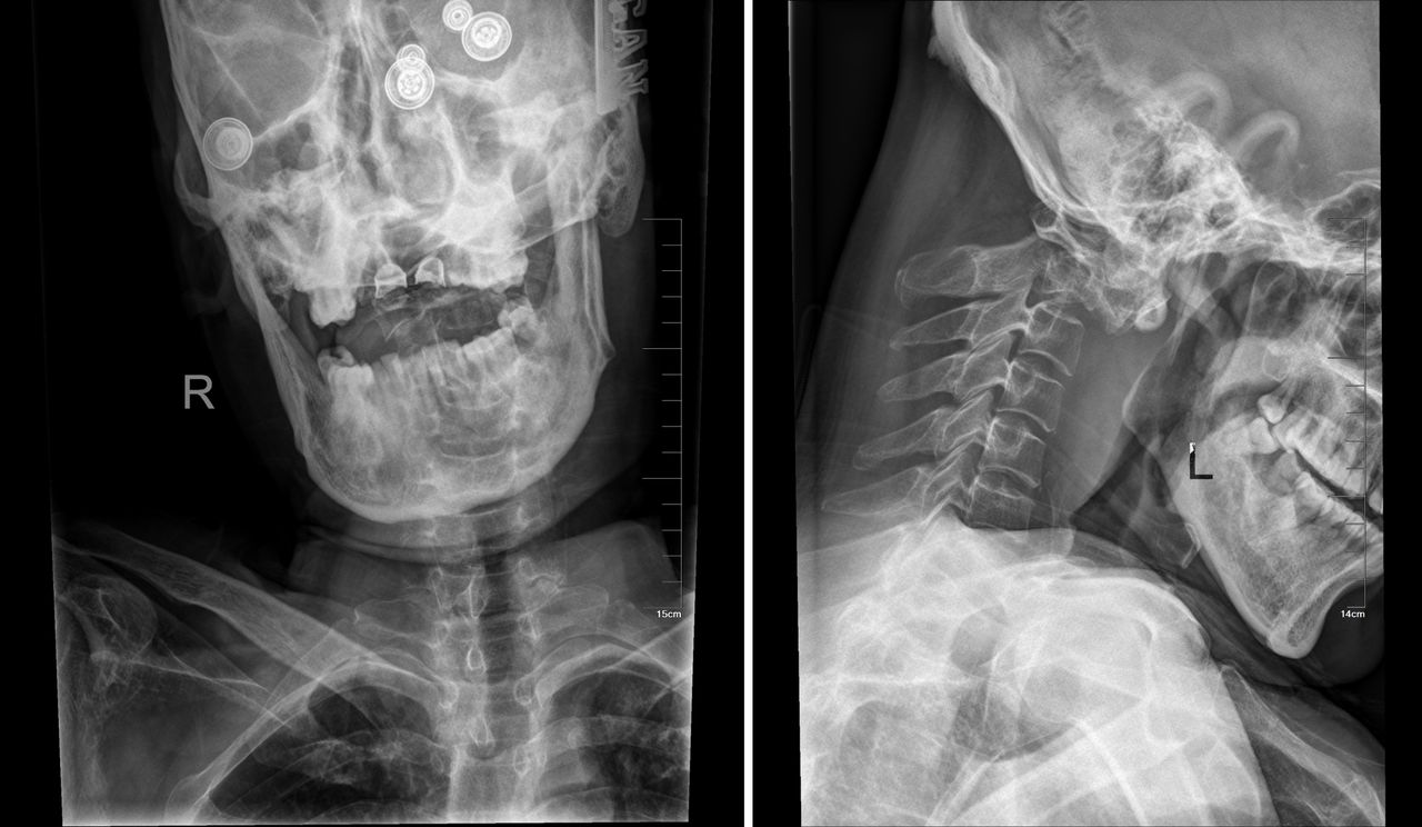
Preoperative anteroposterior x-ray (left) and preoperative lateral x-ray (right). Overlap of the atlas posterior arch with the axial odontoid can be seen on lateral radiographs.

Figure 2
  • Download figure
  • Open in new tab
  • Download powerpoint
Figure 2

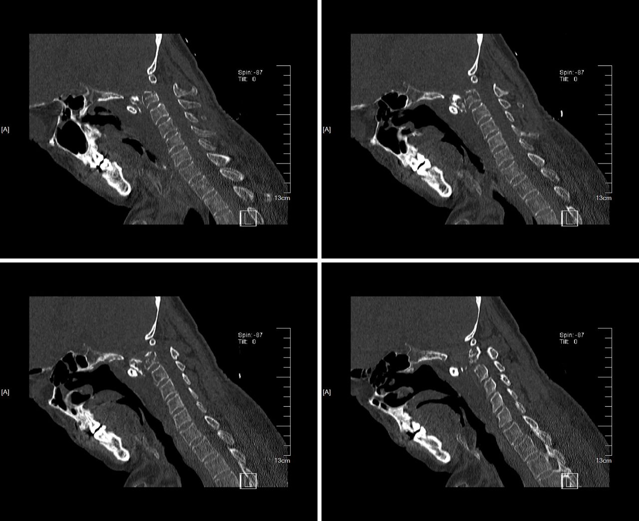
Preoperative computed tomography scan sagittal view showed axial odontoid fracture. The fracture end is located in front of the axial body.

Figure 3
  • Download figure
  • Open in new tab
  • Download powerpoint
Figure 3

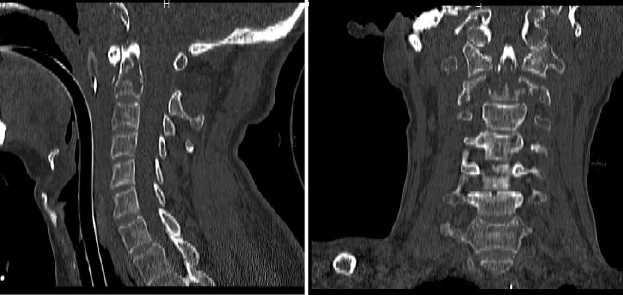
Complete reduction was seen in the sagittal and coronal positions after cranial traction.

Figure 4
  • Download figure
  • Open in new tab
  • Download powerpoint
Figure 4

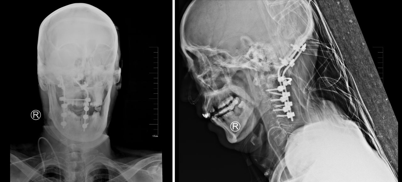
Postoperation anteroposterior x-ray (left) and postoperation lateral x-ray (right).

DISCUSSION

Atlantoaxial dislocation can be divided into partial and complete dislocation. Most patients with complete dislocation die on the spot, although it is rare for the patient to travel to many places without death. The reason may be that dislocation accompanied by local bone destruction leads to slight expansion of spinal canal volume, and no respiratory or cardiac arrest occurs in the mobile position. Tuberculosis of the cervical spine is more rarely reported, accounting for 0.3%–1% of systemic tuberculosis.4,5 Cervical tuberculosis may not be detected at the early stage. Because the patient has no clinical symptoms, the present onset is neck discomfort and posttraumatic atlantoaxial dislocation. Moreover, the role of polymerase chain reaction in the diagnosis of bone tuberculosis is not clear6,7 because it can be rapidly excluded but cannot be diagnosed,8 as shown in this case.

Atlantoaxial tuberculosis often leads to bone destruction of atlantooccipital joint, and local ligament relaxation leads to atlanto-occipital joint instability, so it is particularly important to clear the lesion and reconstruct the stability of upper cervical vertebra.9 Although anterior surgery can achieve the purpose of clearing lesions and decompression of spinal canal, anterior surgery is difficult to reconstruct the stability of atlas and occipital region, and posterior surgery can achieve the purpose of bone grafting and fusion internal fixation,10 but posterior surgery alone is difficult to completely remove lesions, so the selection of surgical methods is particularly important.11 In this case, no local pharyngeal wall abscess was found to form before surgery, so posterior surgery was performed directly after skull traction. However, for other patients with tuberculosis with abscess formation, anterior decompression and posterior fixation and fusion surgery were required, so as to better restore the stability of upper cervical vertebra.

The choice of autologous bone graft or allograft fusion in occipitocervical fusion has always been controversial. Godzik et al12 believed that the success rate of allograft fusion was relatively high, and the occurrence of complications related to body bone was avoided. The thickest part of the occipital region is mostly located in the external carinal process.13 It is still controversial to fix the occipitocervical fusion vertebra to C2 or C3. Pan et al14 found no statistical difference in the rate of fixed C2 or C3 fusion, but there were more complications in the treatment group than in C2. In this case, due to the obvious axial vertebra destruction, we adopted cervical 3 in the operation, and the postoperative effect was good.

CONCLUSIONS

Continuous cranial traction and posterior occipitocervical fusion are effective methods for treating axial pathological fracture with atlantoaxial dislocation.

Footnotes

  • Disclosures and COI: The authors received no funding for this study and report no conflicts of interest.

  • This manuscript is generously published free of charge by ISASS, the International Society for the Advancement of Spine Surgery. Copyright © 2020 ISASS

REFERENCES

  1. 1 .↵
    1. Barbagallo GM,
    2. Certo F,
    3. Visocchi M,
    4. Palmucci S,
    5. Sciacca G,
    6. Albanese V.
    Disappearance of degenerative, non-inflammatory, retro-odontoid pseudotumor following posterior C1-C2 fixation: case series and review of the literature. Eur Spine J. 2013;22(6):S879–S888. doi:10.1007/s00586-013-3004-1
    OpenUrlCrossRef
  2. 2 .↵
    1. Cacciola F,
    2. Boszczyk B,
    3. Perrini P.
    Realignment of basilar invagination by C1-C2 joint distraction: a modified approach to a paradigm shift. Acta Neurochir Suppl. 2019;125:273–277. doi:10.1007/978-3-319-62515-7_39
    OpenUrlCrossRef
  3. 3 .↵
    1. Hussain I,
    2. Schwartz TH,
    Greenfield JP. endoscopic endonasal approach to the upper cervical spine for decompression of the cervicomedullary junction following occipitocervical fusion. Clin Spine Surg. 2018;31(7):285–292. doi:10.1097/BSD.0000000000000620
    OpenUrlCrossRef
  4. 4 .↵
    1. Edwards RJ,
    2. David KM,
    3. Crockard HA.
    Management of tuberculomas of the craniovertebral junction. Br J Neurosurg. 2000;14(1):19–22. doi:10.1080/02688690042852
    OpenUrlCrossRefPubMed
  5. 5 .↵
    1. Teegala R,
    2. Kumar P,
    3. Kale SS, et al.
    Craniovertebral junction tuberculosis: a new comprehensive therapeutic strategy. Neurosurgery. 2008;63(5):946–955. doi:10.1227/01.NEU.0000327696.77814.1E
    OpenUrlCrossRefPubMed
  6. 6 .↵
    1. Raviglione M,
    2. Sulis G.
    Tuberculosis 2015: burden, challenges and strategy for control and elimination. Infect Dis Rep. 2016;8(2):6570. doi:10.4081/idr.2016.6570
    OpenUrlCrossRefPubMed
  7. 7 .↵
    1. Christoforidou A,
    2. Metallidis S,
    3. Kollaras P, et al.
    Tuberculous retropharyngeal abscess as a cause of oropharyngeal dysphagia. Am J Otolaryngol. 2012;33(2):272–274. doi:10.1016/j.amjoto.2011.06.006
    OpenUrlCrossRefPubMed
  8. 8 .↵
    1. Olaru I,
    2. Heyckendorf J,
    3. Grossmann S, et al.
    Time to culture positivity and sputum smear microscopy during tuberculosis therapy. PLoS One. 2014;9(8):e106075. doi:10.1371/journal.pone.0106075
    OpenUrlCrossRef
  9. 9 .↵
    1. Rachdi I,
    2. Fekih Y,
    3. Daoud F.
    Cervical Pott's disease revealed by retropharyngeal abscess. Presse Med. 2018;47(10):918–921. doi:10.1016/j.lpm.2018.08.010
    OpenUrlCrossRef
  10. 10 .↵
    1. Tan LA,
    2. Riew KD,
    3. Traynelis VC.
    Cervical spine deformity—Part 3: posterior techniques, clinical outcome, and complications. Neurosurgery. 2017;81(6):893–898. doi:10.1093/neuros/nyx477
    OpenUrlCrossRef
  11. 11 .↵
    1. Tanaka S,
    2. Aoki M,
    3. Nakanishi T, et al.
    Retrospective case series analysing the clinical data and treatment options of patients with a tubercular abscess of the chest wall. Interact Cardiovasc Thorac Surg. 2012;14(3):249–252. doi:10.1093/icvts/ivr113
    OpenUrlCrossRefPubMed
  12. 12 .↵
    1. Godzik J,
    2. Ravindra VM,
    3. Ray WZ, et al.
    Comparison of structural allograft and traditional autograft technique in occipitocervical fusion: radiological and clinical outcomes from a single institution. J Neurosurg Spine. 2015;23(2):144–152. doi:10.3171/2014.12.SPINE14535
    OpenUrlCrossRef
  13. 13 .↵
    1. Wang YL,
    2. Xu HM,
    3. Wang XY, et al.
    A computed tomographic morphometric study of the pediatric occipital bone thickness: Implications for pediatric occipitocervical fusion. Spine. 2015;40(20):1564–1571. doi:10.1097/BRS.0000000000001091
    OpenUrlCrossRef
  14. 14 .↵
    1. Pan J,
    2. Huang D,
    3. Hao D, et al.
    Occipitocervical fusion: fix to C2 or C3? Clin Neurol Neurosurg. 2014;127:134–139. doi:10.1016/j.clineuro.2014.10.013
    OpenUrlCrossRef
PreviousNext
Back to top

In this issue

International Journal of Spine Surgery
Vol. 14, Issue s4
1 Feb 2021
  • Table of Contents
  • Index by author

Print
Download PDF
Article Alerts
Sign In to Email Alerts with your Email Address
Email Article

Thank you for your interest in spreading the word on International Journal of Spine Surgery.

NOTE: We only request your email address so that the person you are recommending the page to knows that you wanted them to see it, and that it is not junk mail. We do not capture any email address.

Enter multiple addresses on separate lines or separate them with commas.
A Case Report of Complete Dislocation of Atlantoaxial Joint due to a Traumatic and Pathological Axial Lesion
(Your Name) has sent you a message from International Journal of Spine Surgery
(Your Name) thought you would like to see the International Journal of Spine Surgery web site.
CAPTCHA
This question is for testing whether or not you are a human visitor and to prevent automated spam submissions.
Citation Tools
A Case Report of Complete Dislocation of Atlantoaxial Joint due to a Traumatic and Pathological Axial Lesion
Xinwei Yuan, Lun Wan, Jiang Hu, Wei Zhang
International Journal of Spine Surgery Feb 2021, 14 (s4) S5-S9; DOI: 10.14444/7157

Citation Manager Formats

  • BibTeX
  • Bookends
  • EasyBib
  • EndNote (tagged)
  • EndNote 8 (xml)
  • Medlars
  • Mendeley
  • Papers
  • RefWorks Tagged
  • Ref Manager
  • RIS
  • Zotero
Share
A Case Report of Complete Dislocation of Atlantoaxial Joint due to a Traumatic and Pathological Axial Lesion
Xinwei Yuan, Lun Wan, Jiang Hu, Wei Zhang
International Journal of Spine Surgery Feb 2021, 14 (s4) S5-S9; DOI: 10.14444/7157
Twitter logo Facebook logo Mendeley logo
  • Tweet Widget
  • Facebook Like
  • Google Plus One

Jump to section

  • Article
    • ABSTRACT
    • INTRODUCTION
    • CASE REPORT
    • DISCUSSION
    • CONCLUSIONS
    • Footnotes
    • REFERENCES
  • Figures & Data
  • Info & Metrics
  • PDF

Related Articles

  • No related articles found.
  • PubMed
  • Google Scholar

Cited By...

  • No citing articles found.
  • Google Scholar

More in this TOC Section

  • Innovation and Adversity in Spine Surgery: A Retrospective
  • Cervical Myelopathy Secondary to Bilateral Atlantoaxial Pseudoarticulations in Rheumatoid Arthritis: A Case Report
  • Incidence of Traumatic Spinal Injury Following Public Policy Update on Moped Usage in South Carolina
Show more Cervical Spine

Similar Articles

Keywords

  • atlantoaxial dislocation
  • occipitocervical fusion
  • cranial traction

Content

  • Current Issue
  • Latest Content
  • Archive

More Information

  • About IJSS
  • About ISASS
  • Privacy Policy

More

  • Subscribe
  • Alerts
  • Feedback

Other Services

  • Author Instructions
  • Join ISASS
  • Reprints & Permissions

© 2025 International Journal of Spine Surgery

International Journal of Spine Surgery Online ISSN: 2211-4599

Powered by HighWire