
Main menu

  • Home
  • Content
    • Current Issue
    • Advance Online Publication
    • Archive
  • About Us
    • About ISASS
    • About the Journal
    • Author Instructions
    • Editorial Board
    • Reviewer Guidelines & Publication Criteria
  • More
    • Advertise
    • Subscribe
    • Alerts
    • Feedback
  • Join Us
  • Reprints & Permissions
  • Sponsored Content
  • Other Publications
    • ijss

User menu

  • My alerts

Search

  • Advanced search
International Journal of Spine Surgery
  • My alerts
International Journal of Spine Surgery

Advanced Search

  • Home
  • Content
    • Current Issue
    • Advance Online Publication
    • Archive
  • About Us
    • About ISASS
    • About the Journal
    • Author Instructions
    • Editorial Board
    • Reviewer Guidelines & Publication Criteria
  • More
    • Advertise
    • Subscribe
    • Alerts
    • Feedback
  • Join Us
  • Reprints & Permissions
  • Sponsored Content
  • Follow ijss on Twitter
  • Visit ijss on Facebook
Research ArticleOther & Special Categories

Long-Term Reliability of the Two-Segment Fusion Technique in the Treatment of Thoracolumbar Fractures Using Screws in the Fractured Vertebra

Francisco José Martin-Somoza, Jose Miguel Cantero Escribano and Jose Vicente Ramírez-Villaescusa
International Journal of Spine Surgery February 2021, 15 (1) 169-178; DOI: https://doi.org/10.14444/8022
Francisco José Martin-Somoza
1Department of Orthopaedics Surgery and Traumatology, Complejo Hospitalario Universitario de Albacete, Albacete, Spain
MD
  • Find this author on Google Scholar
  • Find this author on PubMed
  • Search for this author on this site
Jose Miguel Cantero Escribano
2Preventive Medicine Unit, La Paz University Hospital, Madrid, Spain
MD
  • Find this author on Google Scholar
  • Find this author on PubMed
  • Search for this author on this site
Jose Vicente Ramírez-Villaescusa
3Department of Orthopaedics Surgery and Traumatology, Spine Unit, Complejo Hospitalario Universitario de Albacete, Albacete, Spain
PHD
  • Find this author on Google Scholar
  • Find this author on PubMed
  • Search for this author on this site
  • Article
  • Figures & Data
  • Info & Metrics
  • PDF
Loading

Article Figures & Data

Figures

  • Tables
  • Figure 1
    • Download figure
    • Open in new tab
    • Download powerpoint
    Figure 1

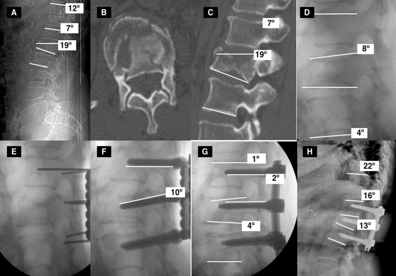
    (A) Measurement technique of vertebral, regional and thoracolumbar kyphosis on x-ray. VK (19°): angle based on a tangential line to the superior and inferior end plate of fractured vertebra. RK (7°): angle based on a tangential line at the superior edge of the superior vertebra and a tangential line at the inferior edge of the inferior vertebra. TLK (12°): angle based on a tangential line at the superior edge of T10 and a tangential line at the inferior edge of L2. (B, C) Axial and sagittal CT images that suggest a type A2 burst fracture according to AO classification. We can also perform the measurement technique on sagittal CT images. (D) Indirect initial reduction with patient positioned in prone decubitus. (VK 8°, RK 4°). (E) Fractured vertebra instrumented with cannulated screws on both pedicles toward the lower vertebral plate. (F, G) The placement of the slightly molded bars in lordosis and its union to the screw allowed for an adequate correction with indirect reduction of its deformity (VK 4°, RK 1°). (H) X-ray image at the end of follow-up (8 years after surgery). A long-term progression of VK, RK, and TLK is observed (TLK 22°, RK 16°, and VK 13°). VK indicates vertebral kyphosis; RK, regional kyphosis; TLK, thoracolumbar kyphosis; CT, computed tomography.

  • Figure 2
    • Download figure
    • Open in new tab
    • Download powerpoint
    Figure 2

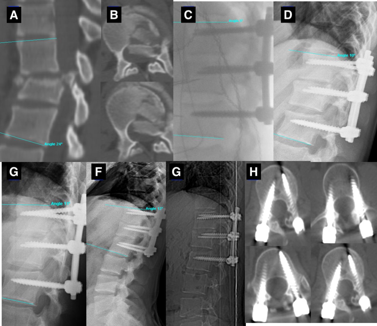
    (A) Sagittal CT view of 25-year-old man with an L1 A3 burst fracture and preoperative T12–L2 kyphosis of 23°. (B) Axial CT views, where we can see a significant posterior fragment compressing the spinal cord that is neurologically intact. (C) Sagittal intraoperative view following polyaxial screw instrumentation with a TLK of 6°. (D–F) Sagittal view at 1 month (10°), 3 months postoperative (11°), and 5-year follow-up (12°) with a slight increase of TLK kyphosis. (G, H) Sagittal and axial CT images. The pedicle screws were well placed at upper and lower levels, and canal clearance was observed at the L1 level. CT indicates computed tomography; TLK, thoracolumbar kyphosis.

  • Figure 3
    • Download figure
    • Open in new tab
    • Download powerpoint
    Figure 3

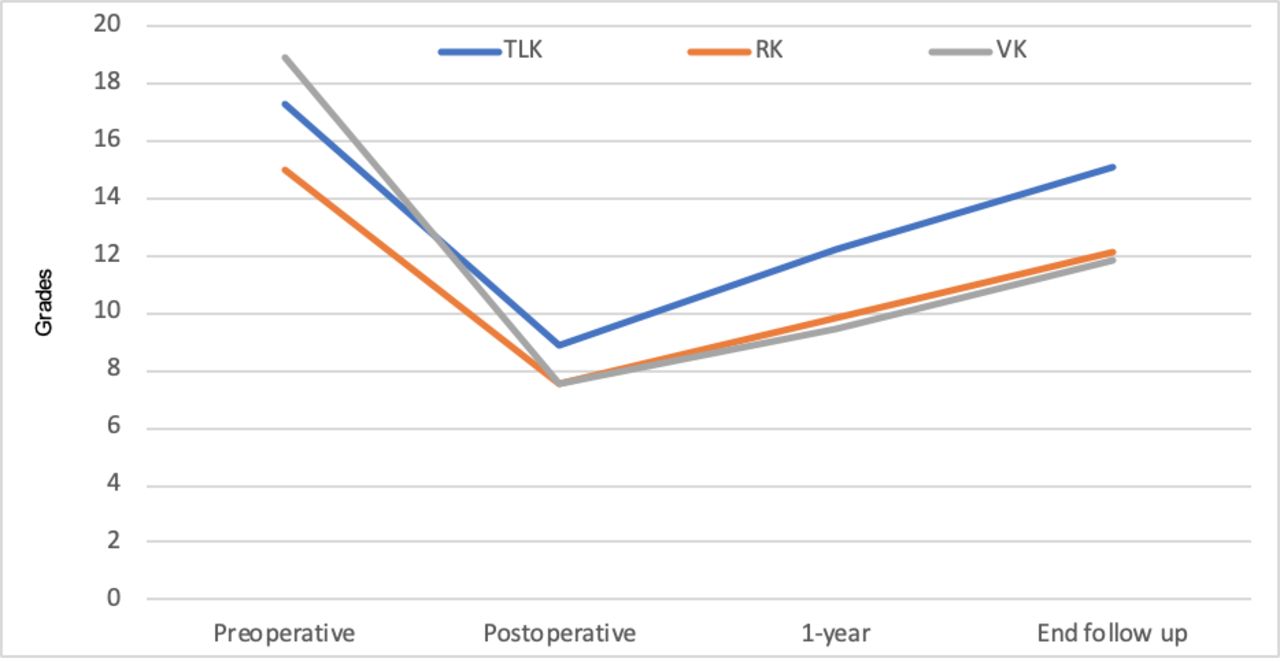
    Evolution of the different measurements of preoperative kyphosis until the end of treatment (mean treatment 8 years).

  • Figure 4
    • Download figure
    • Open in new tab
    • Download powerpoint
    Figure 4

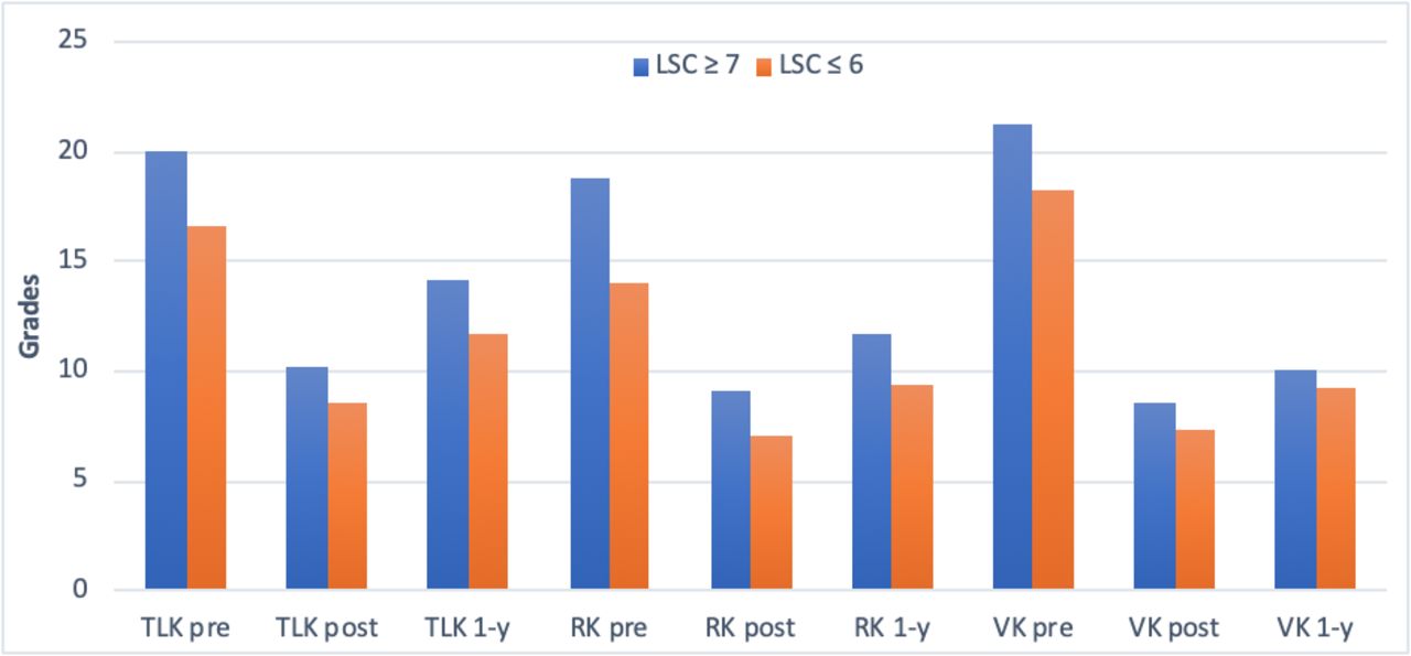
    Representation of thoracolumbar, regional, and vertebral kyphosis evolution during the first tracking year according to LSC ≤6 to ≥7. LSC indicates load-sharing classification.

  • Figure 5
    • Download figure
    • Open in new tab
    • Download powerpoint
    Figure 5

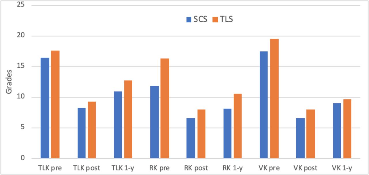
    Regional, vertebral, and thoracolumbar kyphosis evolution depending on the type of instrumentation used (side-connecting screws or top-loading screws) during the first year of tracking time.

  • Figure 6
    • Download figure
    • Open in new tab
    • Download powerpoint
    Figure 6

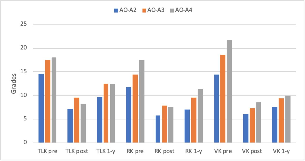
    Thoracolumbar, vertebral, and regional kyphosis evolution depending on the type of lesion according to the AO classification (A2–A4) during the first tracking year.

Tables

  • Figures
  • Table 1
  • Table 2
  • Table 3
  • Table 4
  • Table 5
PreviousNext
Back to top

In this issue

International Journal of Spine Surgery
Vol. 15, Issue 1
1 Feb 2021
  • Table of Contents
  • Index by author

Print
Download PDF
Article Alerts
Sign In to Email Alerts with your Email Address
Email Article

Thank you for your interest in spreading the word on International Journal of Spine Surgery.

NOTE: We only request your email address so that the person you are recommending the page to knows that you wanted them to see it, and that it is not junk mail. We do not capture any email address.

Enter multiple addresses on separate lines or separate them with commas.
Long-Term Reliability of the Two-Segment Fusion Technique in the Treatment of Thoracolumbar Fractures Using Screws in the Fractured Vertebra
(Your Name) has sent you a message from International Journal of Spine Surgery
(Your Name) thought you would like to see the International Journal of Spine Surgery web site.
CAPTCHA
This question is for testing whether or not you are a human visitor and to prevent automated spam submissions.
Citation Tools
Long-Term Reliability of the Two-Segment Fusion Technique in the Treatment of Thoracolumbar Fractures Using Screws in the Fractured Vertebra
Francisco José Martin-Somoza, Jose Miguel Cantero Escribano, Jose Vicente Ramírez-Villaescusa
International Journal of Spine Surgery Feb 2021, 15 (1) 169-178; DOI: 10.14444/8022

Citation Manager Formats

  • BibTeX
  • Bookends
  • EasyBib
  • EndNote (tagged)
  • EndNote 8 (xml)
  • Medlars
  • Mendeley
  • Papers
  • RefWorks Tagged
  • Ref Manager
  • RIS
  • Zotero
Share
Long-Term Reliability of the Two-Segment Fusion Technique in the Treatment of Thoracolumbar Fractures Using Screws in the Fractured Vertebra
Francisco José Martin-Somoza, Jose Miguel Cantero Escribano, Jose Vicente Ramírez-Villaescusa
International Journal of Spine Surgery Feb 2021, 15 (1) 169-178; DOI: 10.14444/8022
Twitter logo Facebook logo Mendeley logo
  • Tweet Widget
  • Facebook Like
  • Google Plus One

Jump to section

  • Article
    • ABSTRACT
    • INTRODUCTION
    • MATERIALS AND METHODS
    • RESULTS
    • DISCUSSION
    • ACKNOWLEDGMENTS
    • Footnotes
    • REFERENCES
  • Figures & Data
  • Info & Metrics
  • PDF

Related Articles

  • No related articles found.
  • PubMed
  • Google Scholar

Cited By...

  • No citing articles found.
  • Google Scholar

More in this TOC Section

  • Hemivertebra Resection and Spinal Arthrodesis by Single-Stage Posterior Approach in Congenital Scoliosis and Kyphoscoliosis: Results at 9.6 Years Mean Follow-up
  • Prevalence and Significance of Vitamin D Deficiency in Patients Undergoing Corrective Surgery for Adolescent Idiopathic Scoliosis
  • Adult Congenital Spine Deformity: Clinical Features and Motivations for Surgical Treatment
Show more Other & Special Categories

Similar Articles

Keywords

  • spinal cord injury
  • surgical treatment
  • thoracolumbar burst fractures

Content

  • Current Issue
  • Latest Content
  • Archive

More Information

  • About IJSS
  • About ISASS
  • Privacy Policy

More

  • Subscribe
  • Alerts
  • Feedback

Other Services

  • Author Instructions
  • Join ISASS
  • Reprints & Permissions

© 2025 International Journal of Spine Surgery

International Journal of Spine Surgery Online ISSN: 2211-4599

Powered by HighWire