


Article Figures & Data
Figures
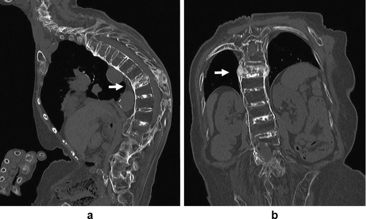
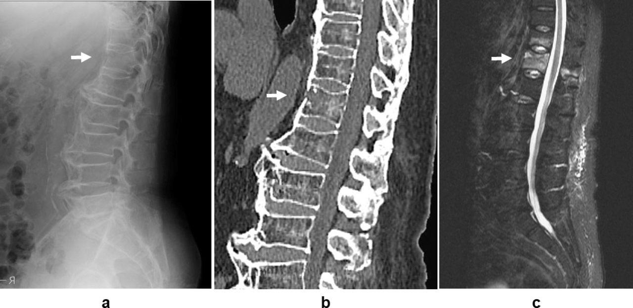
- Figure 1
Case 1: Pretherapeutic x-ray, computed tomography (CT), magnetic resonance imaging (MRI). (a) X-ray lateral view. No obvious vertebral collapse was observed; although, thoracic hyperkyphosis with the ossification of anterior ligament of the whole spine was identified. (b) CT sagittal view. Ossification of anterior longitudinal ligament was discontinued, and the posterior wall of the vertebral body was distended at the T10 vertebral level. (c) MRI STIR (short T1 inversion recovery) sagittal view. High-intensity changes of the T10 vertebral body and the posterior elements were confirmed.
- Figure 2
Case 1: Computed tomography (CT) at 2 years, 1 month after injury. (a) Sagittal view. Bone union of both the T10 vertebral body and the spinous process was achieved. (b) Coronal view. Callus formation was recognized at not only the central part but also the lateral sides of the T10 vertebral body.
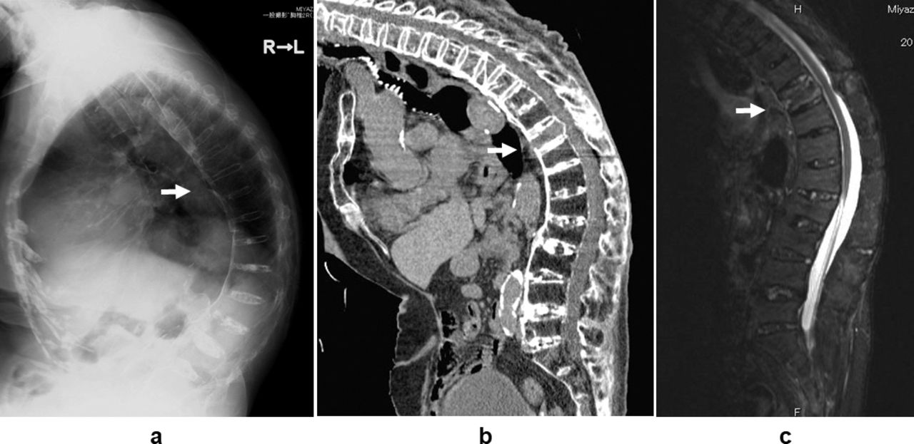
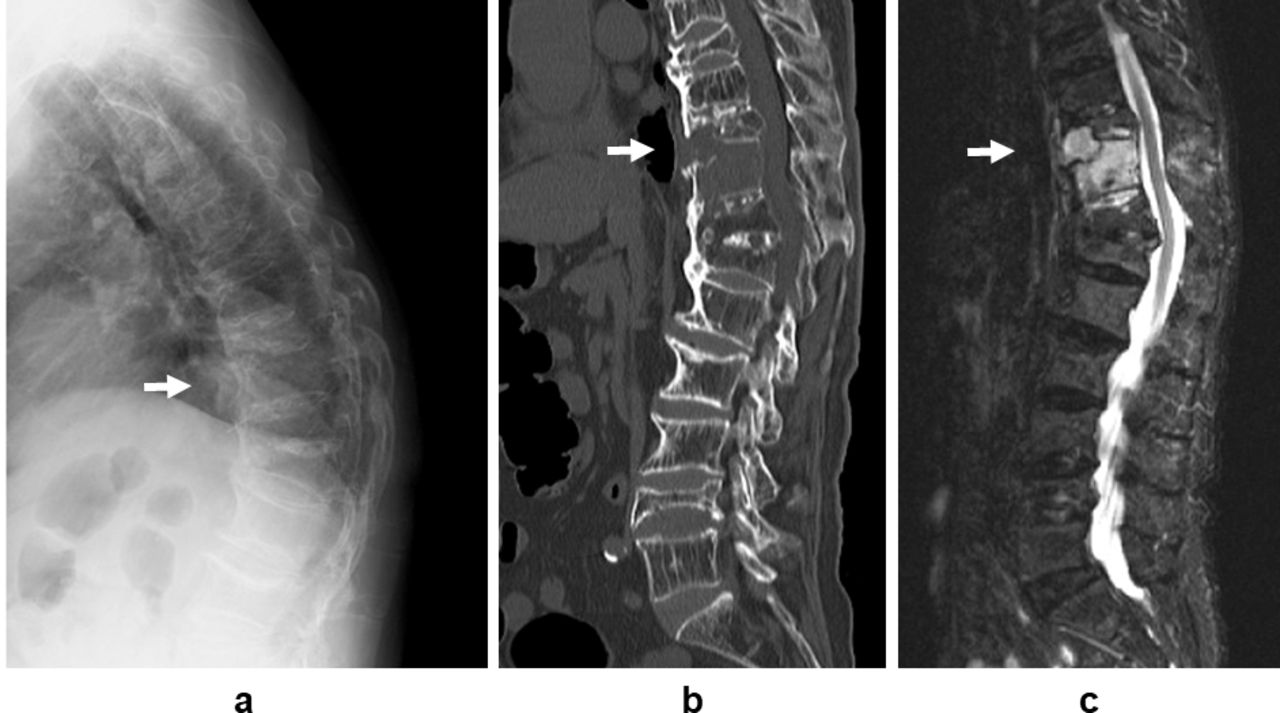
- Figure 3
Case 2: Pretherapeutic x-ray, computed tomography (CT), magnetic resonance imaging (MRI). (a) X-ray lateral view. Massive osteophyte formation of the anterior portion of the spine was observed; although, neither abnormality of spinal alignment nor collapse of vertebral bodies existed. (b) CT sagittal view. Ossification of anterior longitudinal ligament was discontinued at the thoracolumbar level. (c) MRI STIR (short T1 inversion recovery) sagittal view. High-intensity changes of the T12 vertebral body and the posterior elements were confirmed.
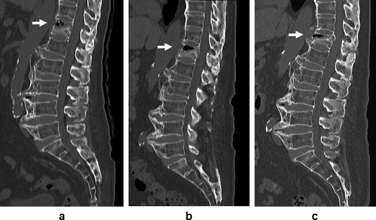
- Figure 4
Case 2: Sequential changes of computed tomography (CT). (a) 3 months after injury. The cavity in the anterior part of the T12 vertebral body was recognized. (b) 5 months after injury. The cavity in the anterior part of the T12 vertebral body was enlarged. (c) 1 year after injury. In the T12 vertebral body, collapse has progressed, but the cavity was diminished in size.
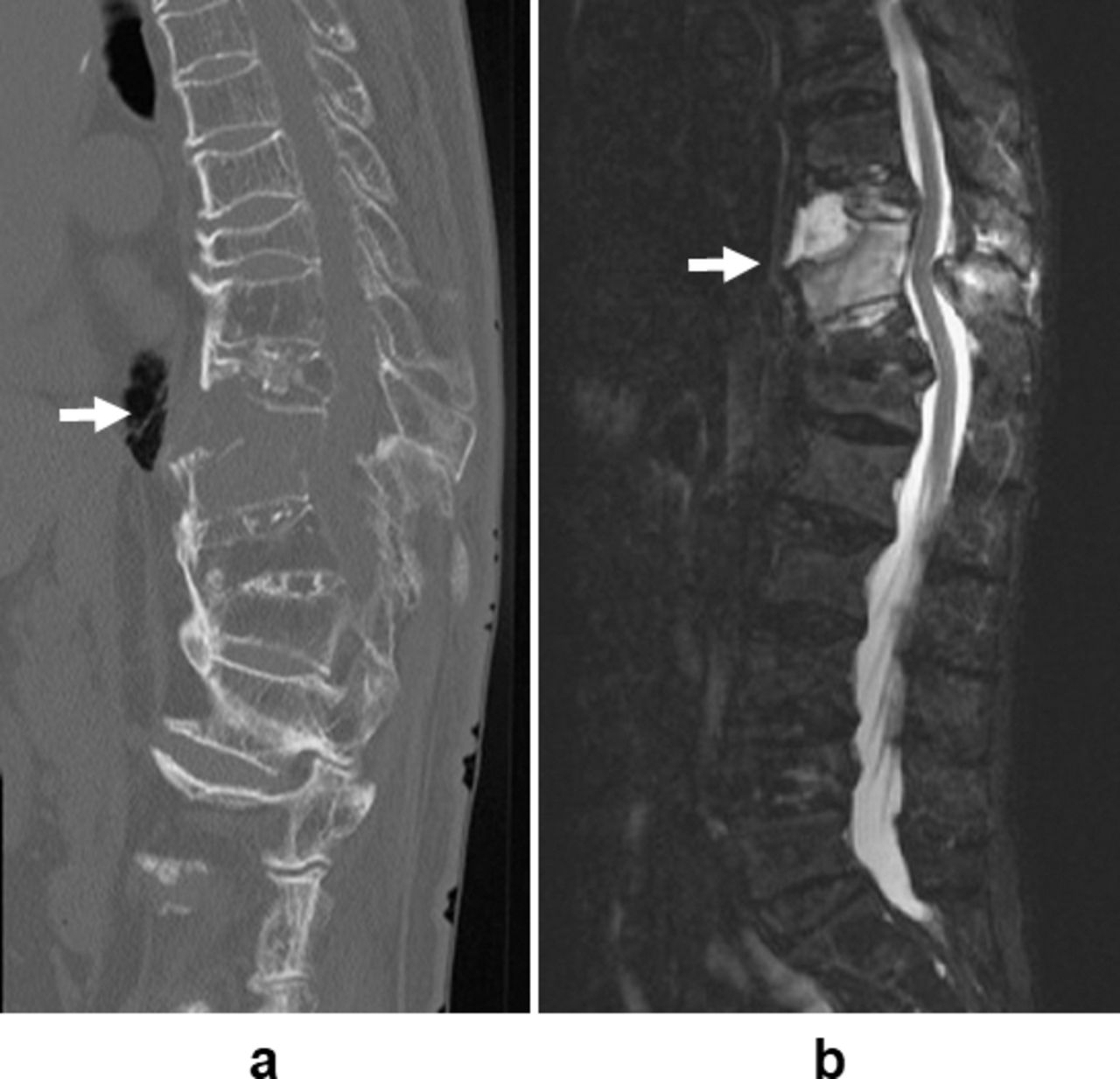
- Figure 5
Case 3: Pretherapeutic x-ray, computed tomography (CT), magnetic resonance imaging (MRI). (a) X-ray lateral view. Multiple-vertebral collapse with thoracic hyperkyphosis was recognized. (b) CT sagittal view. Ossification of anterior longitudinal ligament was discontinued at the T10 vertebral level, and the spinous processes of T9, 10, and T11 were spontaneously fused. (c) MRI STIR (short T1 inversion recovery) sagittal view. High-intensity changes of the T10 vertebral body and the posterior elements were confirmed.
- Figure 6
Case 3: Computed tomography (CT) and magnetic resonance imaging (MRI) at the advent of paralysis. (a) CT sagittal view. Angular displacement of fracture has been progressed, and the cavity of the T10 vertebral body was spread. (b) MRI STIR (short T1 inversion recovery) sagittal view. Mild compression and meandering of the spinal cord due to spinal canal stenosis created by advanced angular displacement of fracture was identified.
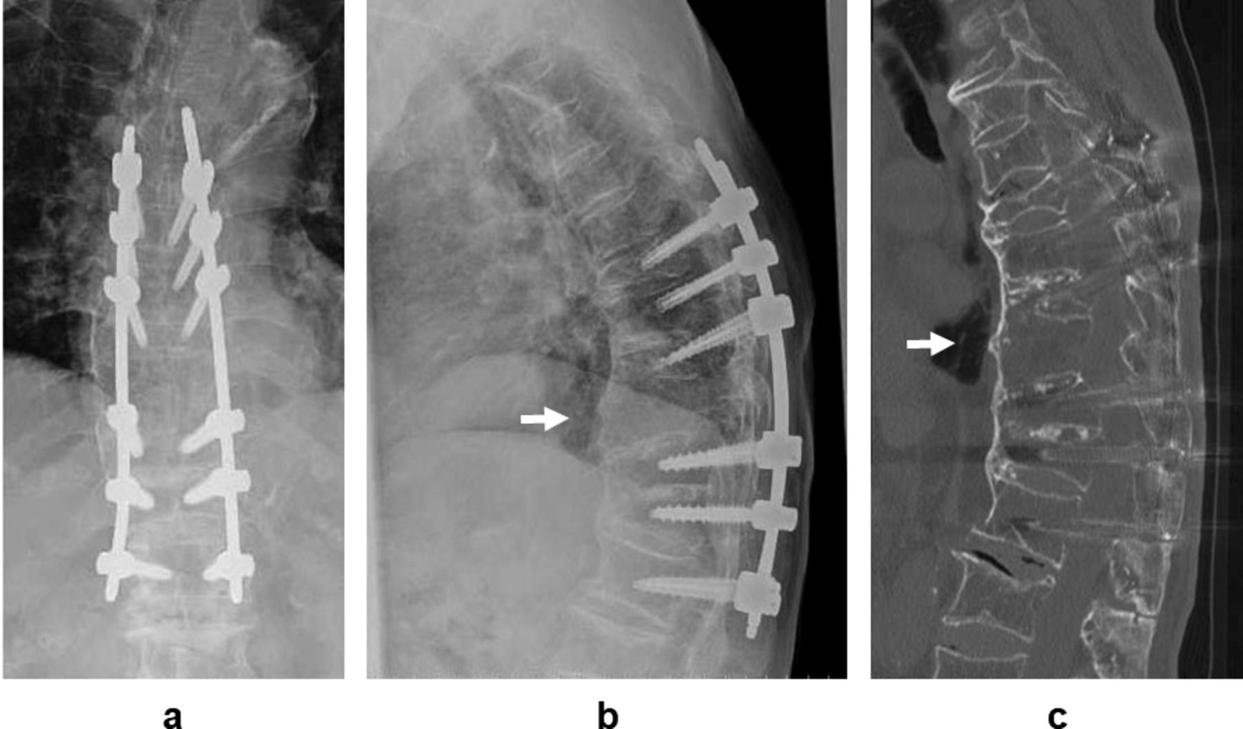
- Figure 7
Case 3: X-ray and computed tomography (CT) at 1 year, 4 months after surgery. (a) X-ray anterior-posterior view. (b) X-ray lateral view. (c) CT sagittal view. Bone union of the T10 vertebral body with proper sagittal spinal alignment was confirmed.


Tables
In this issue


Jump to section
Related Articles
Cited By...
- No citing articles found.