
Main menu

  • Home
  • Content
    • Current Issue
    • Advance Online Publication
    • Archive
  • About Us
    • About ISASS
    • About the Journal
    • Author Instructions
    • Editorial Board
    • Reviewer Guidelines & Publication Criteria
  • More
    • Advertise
    • Subscribe
    • Alerts
    • Feedback
  • Join Us
  • Reprints & Permissions
  • Sponsored Content
  • Other Publications
    • ijss

User menu

  • My alerts

Search

  • Advanced search
International Journal of Spine Surgery
  • My alerts
International Journal of Spine Surgery

Advanced Search

  • Home
  • Content
    • Current Issue
    • Advance Online Publication
    • Archive
  • About Us
    • About ISASS
    • About the Journal
    • Author Instructions
    • Editorial Board
    • Reviewer Guidelines & Publication Criteria
  • More
    • Advertise
    • Subscribe
    • Alerts
    • Feedback
  • Join Us
  • Reprints & Permissions
  • Sponsored Content
  • Follow ijss on Twitter
  • Visit ijss on Facebook
Research ArticleLumbar Spine

Anatomical Variations in the Origin of the Lumbar Artery: A Cadaveric Study Augmented by CT Findings

Thomas Mombo Amuti, Ibsen Henric Ongidi, Innocent Peter Ouko, Dennis Nick, Joseph Mageto, Laura Ocholla, Beda Olabu and Julius Alexander Ogeng'o
International Journal of Spine Surgery October 2021, 15 (5) 906-914; DOI: https://doi.org/10.14444/8118
Thomas Mombo Amuti
Department of Human Anatomy, School of Medicine, University of Nairobi, Nairobi, Kenya
BSC
  • Find this author on Google Scholar
  • Find this author on PubMed
  • Search for this author on this site
Ibsen Henric Ongidi
Department of Human Anatomy, School of Medicine, University of Nairobi, Nairobi, Kenya
BSC
  • Find this author on Google Scholar
  • Find this author on PubMed
  • Search for this author on this site
Innocent Peter Ouko
Department of Human Anatomy, School of Medicine, University of Nairobi, Nairobi, Kenya
BSC
  • Find this author on Google Scholar
  • Find this author on PubMed
  • Search for this author on this site
Dennis Nick
Department of Human Anatomy, School of Medicine, University of Nairobi, Nairobi, Kenya
  • Find this author on Google Scholar
  • Find this author on PubMed
  • Search for this author on this site
Joseph Mageto
Department of Human Anatomy, School of Medicine, University of Nairobi, Nairobi, Kenya
  • Find this author on Google Scholar
  • Find this author on PubMed
  • Search for this author on this site
Laura Ocholla
Department of Human Anatomy, School of Medicine, University of Nairobi, Nairobi, Kenya
  • Find this author on Google Scholar
  • Find this author on PubMed
  • Search for this author on this site
Beda Olabu
Department of Human Anatomy, School of Medicine, University of Nairobi, Nairobi, Kenya
BSC, MBCHB, MSC, MMED
  • Find this author on Google Scholar
  • Find this author on PubMed
  • Search for this author on this site
Julius Alexander Ogeng'o
Department of Human Anatomy, School of Medicine, University of Nairobi, Nairobi, Kenya
BSC, MBCHB, MD, PHD, DSC
  • Find this author on Google Scholar
  • Find this author on PubMed
  • Search for this author on this site
  • Article
  • Figures & Data
  • Info & Metrics
  • PDF
Loading

Article Figures & Data

Figures

  • Tables
  • Figure 1
    • Download figure
    • Open in new tab
    • Download powerpoint
    Figure 1

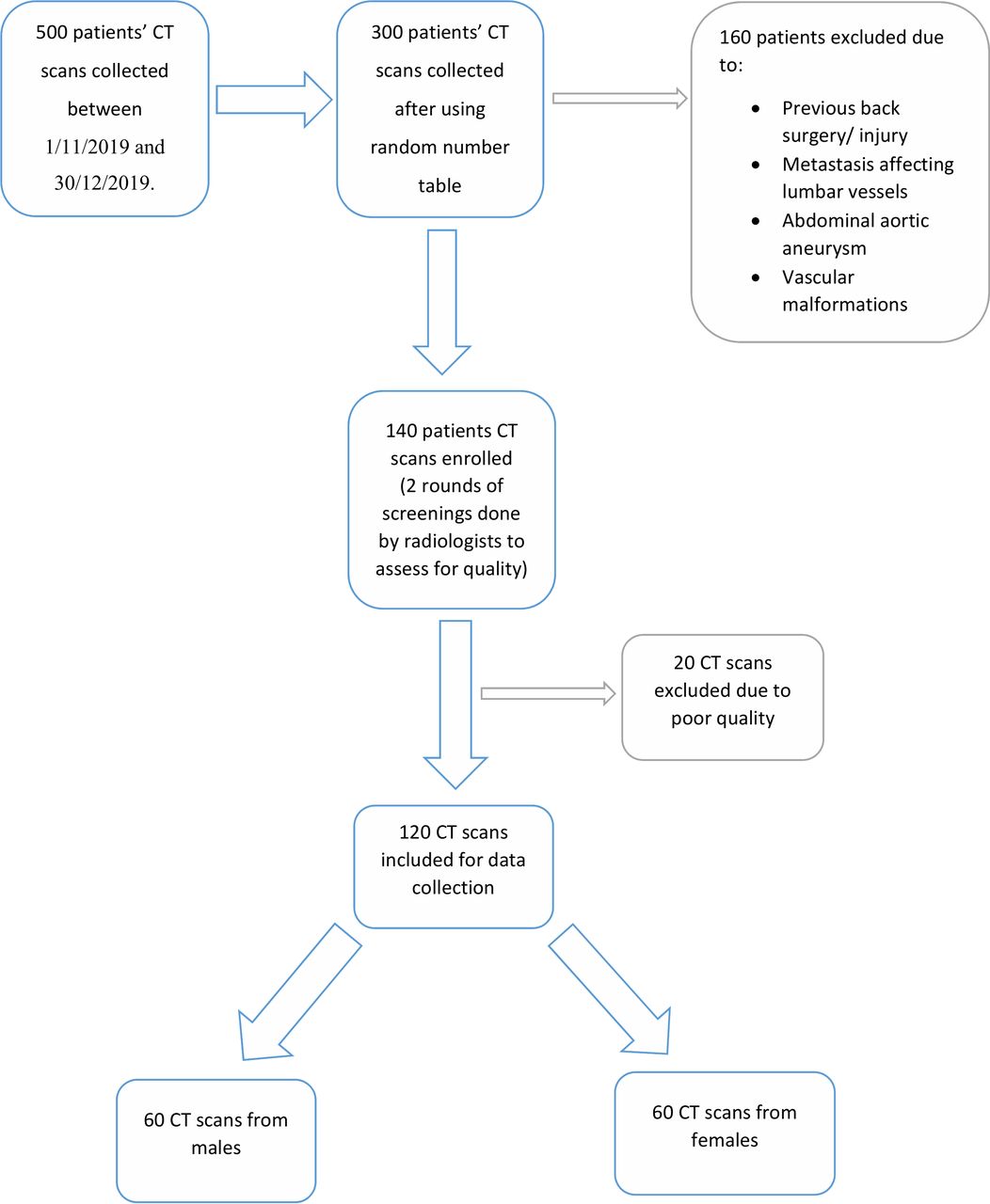
    Flow chart showing the selection process of CT scans obtained from the Kenyatta National Hospital Radiology Department.
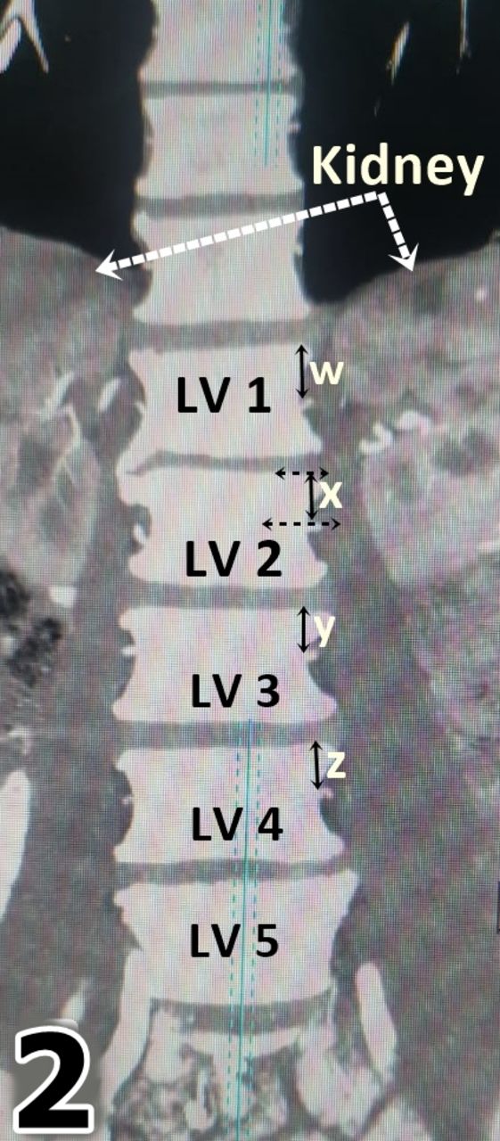
  • Figure 2
    • Download figure
    • Open in new tab
    • Download powerpoint
    Figure 2

    CT showing the measurement of the distances (W, X, Y, Z) between the origin of the lumbar arteries and the top of their respective vertebra. LV, lumbar vertebra.

  • Figure 3
    • Download figure
    • Open in new tab
    • Download powerpoint
    Figure 3

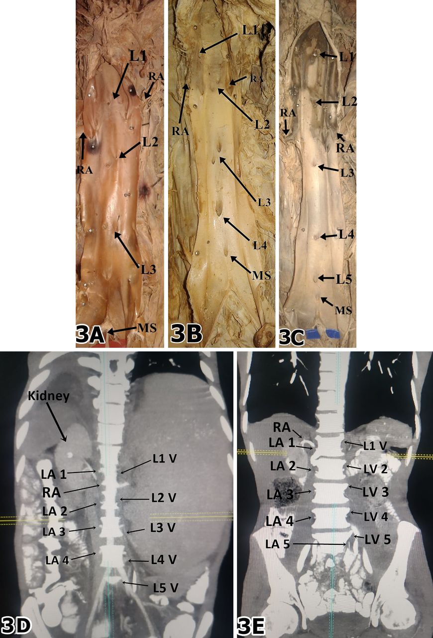
    (A) Dissected aorta showing ostia for 3 pairs of lumbar arteries (L1, L2, and L3). (B) Dissected aorta showing ostia for 4 pairs of lumbar arteries (L1, L2, L3, L4). (C) Dissected aorta showing ostia for 5 pairs of lumbar arteries (L1, L2, L3, L4, L5), with the L2 originating below the renal artery. (D) CT scan showing 4 pairs of lumbar arteries (LA 1, LA 2, LA 3, LA 4). (E) CT scan showing 5 pairs of lumbar arteries (LA 1, LA 2, LA 3, LA 4, LA 5). LV, lumbar vertebra; MS, middle sacral artery; RA, renal artery.

Tables

  • Figures
  • Table 1
  • Table 2
  • Table 3
  • Table 4
  • Table 5
  • Table 6
PreviousNext
Back to top

In this issue

International Journal of Spine Surgery
Vol. 15, Issue 5
1 Oct 2021
  • Table of Contents
  • Index by author

Print
Download PDF
Article Alerts
Sign In to Email Alerts with your Email Address
Email Article

Thank you for your interest in spreading the word on International Journal of Spine Surgery.

NOTE: We only request your email address so that the person you are recommending the page to knows that you wanted them to see it, and that it is not junk mail. We do not capture any email address.

Enter multiple addresses on separate lines or separate them with commas.
Anatomical Variations in the Origin of the Lumbar Artery: A Cadaveric Study Augmented by CT Findings
(Your Name) has sent you a message from International Journal of Spine Surgery
(Your Name) thought you would like to see the International Journal of Spine Surgery web site.
CAPTCHA
This question is for testing whether or not you are a human visitor and to prevent automated spam submissions.
Citation Tools
Anatomical Variations in the Origin of the Lumbar Artery: A Cadaveric Study Augmented by CT Findings
Thomas Mombo Amuti, Ibsen Henric Ongidi, Innocent Peter Ouko, Dennis Nick, Joseph Mageto, Laura Ocholla, Beda Olabu, Julius Alexander Ogeng'o
International Journal of Spine Surgery Oct 2021, 15 (5) 906-914; DOI: 10.14444/8118

Citation Manager Formats

  • BibTeX
  • Bookends
  • EasyBib
  • EndNote (tagged)
  • EndNote 8 (xml)
  • Medlars
  • Mendeley
  • Papers
  • RefWorks Tagged
  • Ref Manager
  • RIS
  • Zotero
Share
Anatomical Variations in the Origin of the Lumbar Artery: A Cadaveric Study Augmented by CT Findings
Thomas Mombo Amuti, Ibsen Henric Ongidi, Innocent Peter Ouko, Dennis Nick, Joseph Mageto, Laura Ocholla, Beda Olabu, Julius Alexander Ogeng'o
International Journal of Spine Surgery Oct 2021, 15 (5) 906-914; DOI: 10.14444/8118
Twitter logo Facebook logo Mendeley logo
  • Tweet Widget
  • Facebook Like
  • Google Plus One

Jump to section

  • Article
    • ABSTRACT
    • INTRODUCTION
    • MATERIALS AND METHODS
    • RESULTS
    • DISCUSSION
    • CONCLUSIONS
    • ACKNOWLEDGMENTS
    • Footnotes
    • REFERENCES
  • Figures & Data
  • Info & Metrics
  • PDF

Related Articles

  • No related articles found.
  • PubMed
  • Google Scholar

Cited By...

  • No citing articles found.
  • Google Scholar

More in this TOC Section

  • Sexual Dysfunction and Urinary Incontinence in Female Patients Following Primary Anterior Lumbar Interbody Fusion: A Survey of 84 Patients
  • Comparison of Stand-Alone Anterior Lumbar Interbody Fusion, 360° Anterior Lumbar Interbody Fusion, and Arthroplasty for Recurrent Lumbar Disc Herniation: Focus on Nerve Decompression and Painful Spinal Instability Resolution
  • Recovery Trajectories After Lumbar Fusion Stratified by Baseline Patient-Reported Outcomes Measurement Information System Physical Function Disability Levels
Show more Lumbar Spine

Similar Articles

Keywords

  • lumbar arteries
  • anatomic variation
  • computed tomography
  • aorta
  • spine
  • surgery

Content

  • Current Issue
  • Latest Content
  • Archive

More Information

  • About IJSS
  • About ISASS
  • Privacy Policy

More

  • Subscribe
  • Alerts
  • Feedback

Other Services

  • Author Instructions
  • Join ISASS
  • Reprints & Permissions

© 2025 International Journal of Spine Surgery

International Journal of Spine Surgery Online ISSN: 2211-4599

Powered by HighWire