
Main menu

  • Home
  • Content
    • Current Issue
    • Advance Online Publication
    • Archive
  • About Us
    • About ISASS
    • About the Journal
    • Author Instructions
    • Editorial Board
    • Reviewer Guidelines & Publication Criteria
  • More
    • Advertise
    • Subscribe
    • Alerts
    • Feedback
  • Join Us
  • Reprints & Permissions
  • Sponsored Content
  • Other Publications
    • ijss

User menu

  • My alerts

Search

  • Advanced search
International Journal of Spine Surgery
  • My alerts
International Journal of Spine Surgery

Advanced Search

  • Home
  • Content
    • Current Issue
    • Advance Online Publication
    • Archive
  • About Us
    • About ISASS
    • About the Journal
    • Author Instructions
    • Editorial Board
    • Reviewer Guidelines & Publication Criteria
  • More
    • Advertise
    • Subscribe
    • Alerts
    • Feedback
  • Join Us
  • Reprints & Permissions
  • Sponsored Content
  • Follow ijss on Twitter
  • Visit ijss on Facebook
Research ArticleEndoscopic Minimally Invasive Surgery

Full Endoscopic Thoracic Discectomy: Is the Interlaminar Approach an Alternative to the Transforaminal Approach? A Technical Note

Marcelo Campos Moraes Amato, Bruno César Aprile, Luiz Adriano Esteves, Vinicius Marques Carneiro and Ricardo Santos de Oliveira
International Journal of Spine Surgery April 2022, 16 (2) 309-317; DOI: https://doi.org/10.14444/8209
Marcelo Campos Moraes Amato
1 Neurosurgeon, Amato – Instituto de Medicina Avançada, São Paulo, Brazil
MD, PhD
  • Find this author on Google Scholar
  • Find this author on PubMed
  • Search for this author on this site
Bruno César Aprile
2 Orthopedic surgeon, Clínica SOU, São Paulo, Brazil
MD
  • Find this author on Google Scholar
  • Find this author on PubMed
  • Search for this author on this site
Luiz Adriano Esteves
3 Neurosurgeon, Universidade Estadual de Campinas, Campinas, Brazil
MD, PhD
  • Find this author on Google Scholar
  • Find this author on PubMed
  • Search for this author on this site
Vinicius Marques Carneiro
4 Neurosurgeon, Faculdade de Medicina de Ribeirão Preto, Universidade de São Paulo, São Paulo, Brazil
MD, PhD
  • Find this author on Google Scholar
  • Find this author on PubMed
  • Search for this author on this site
Ricardo Santos de Oliveira
4 Neurosurgeon, Faculdade de Medicina de Ribeirão Preto, Universidade de São Paulo, São Paulo, Brazil
MD, PhD
  • Find this author on Google Scholar
  • Find this author on PubMed
  • Search for this author on this site
  • Article
  • Figures & Data
  • Info & Metrics
  • PDF
Loading

Article Figures & Data

Figures

  • Tables
  • Figure 1
    • Download figure
    • Open in new tab
    • Download powerpoint
    Figure 1

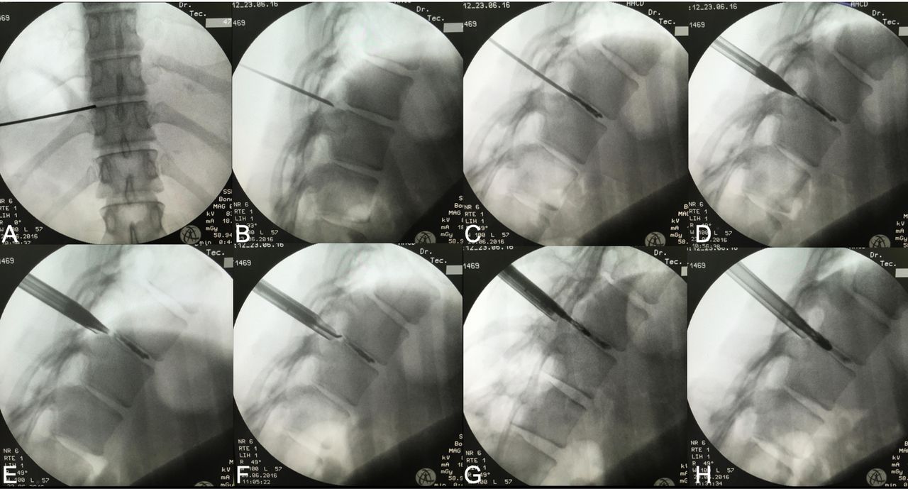
    Fluoroscopic views of the transforaminal approach of patient 1. (A) Insertion of the needle at T10-T11 level at anteroposterior (AP) view until needle tip reaches the midpedicular line. (B) After reaching the disc at the AP view, the C-arm is turned to lateral position, the needle tip is observed a few millimeters behind the posterior intervertebral disc line due to the disc protrusion. (C) The needle is now observed inside the disc space, and intraoperative discography is performed by injecting a mixture of radiopaque dye and methylene blue. (D) A guide wire is inserted through the needle and the dilator followed over the guide wire, note the tip of the dilator at the same point where the needle tip had touched the disc extrusion. (E) The guide wire is removed and the operating sheath is inserted over the dilator. (F) Through the endoscope, a dissector touches the disc herniation. (G and H) Progressive reamers are inserted inside the disc to assure nucleotomy.

  • Figure 2
    • Download figure
    • Open in new tab
    • Download powerpoint
    Figure 2

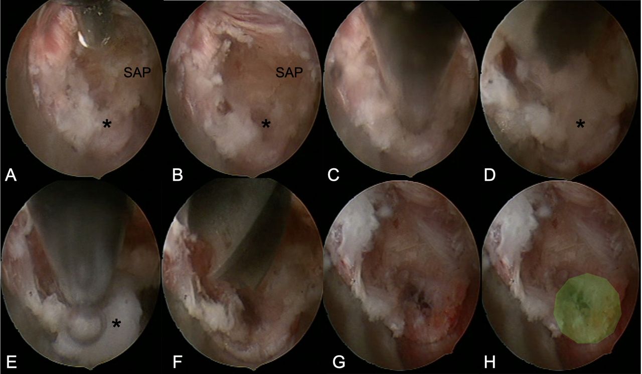
    (A and B) After insertion of the transforaminal endoscope, it is possible to identify the superior articular process (SAP) and the disc herniation (*). The radiofrequency probe is used to palpate and clean the SAP. (C) A trephine is used to open de annulus. (D and E) Further disc material is resected using the Lowe punch. (F) A burr is used to complete discectomy. (G and H) At the end of the surgery, it is possible to notice the widened foramen (green area).

  • Figure 3
    • Download figure
    • Open in new tab
    • Download powerpoint
    Figure 3

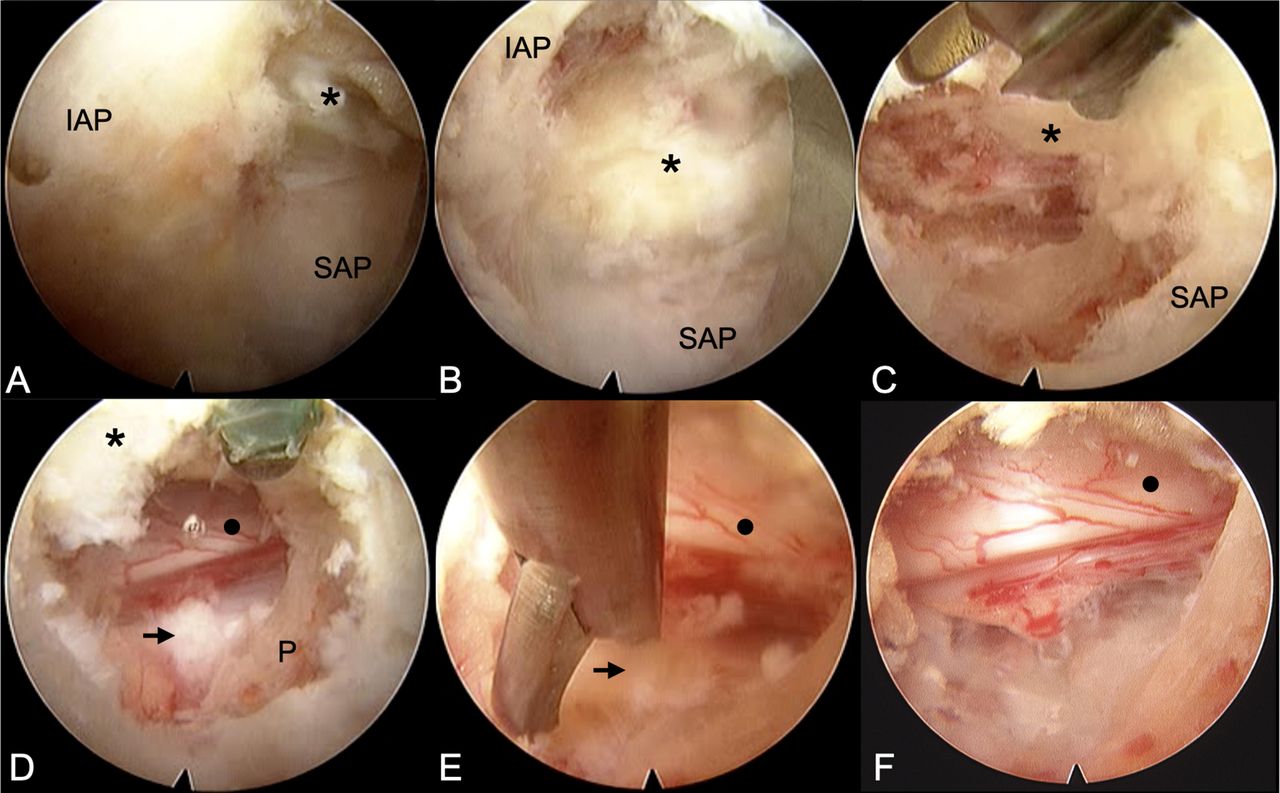
    Endoscopic view of the interlaminar approach. (A) After preparing the operating field it is possible to identify the inferior articular process (IAP), the superior articular process (SAP), and the interlaminar window with the flavum ligament (*). (B) After drilling the IAP and enlarging the interlaminar window, the SAP is ready to be drilled. (C) It is possible to see the burr drilling the inferior laminae and the SAP, lateral to the flavum ligament (*) it is possible to identify the opening of the spinal canal. (D) Above the pedicle (P), it is possible to identify the extruded disc herniation (arrow) and the partially compressed dural sac (circle). (E) Lowe punch resecting the disc herniation. (F) Dural sac free at the end of the surgery.

  • Figure 4
    • Download figure
    • Open in new tab
    • Download powerpoint
    Figure 4

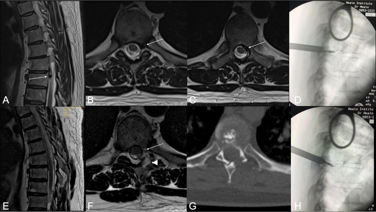
    (A) Sagittal T2-weighted magnetic resonance imaging (MRI) showing the extruded disc herniation with caudal migration (arrow). (B and C) Axial T2-weighted MRI showing the paramedian disc herniation with migration medial to pedicle. (E and F) Postoperative images showing good resection of the disc herniation. It is possible to see the partial facetectomy (arrow head), with complete preservation of the spinal muscles. (G) Computed tomographic image showing partial facetectomy. (D and H) Intraoperative images showing access to the intervertebral disc and manipulation of the instrument medial to the pedicle.

  • Figure 5
    • Download figure
    • Open in new tab
    • Download powerpoint
    Figure 5

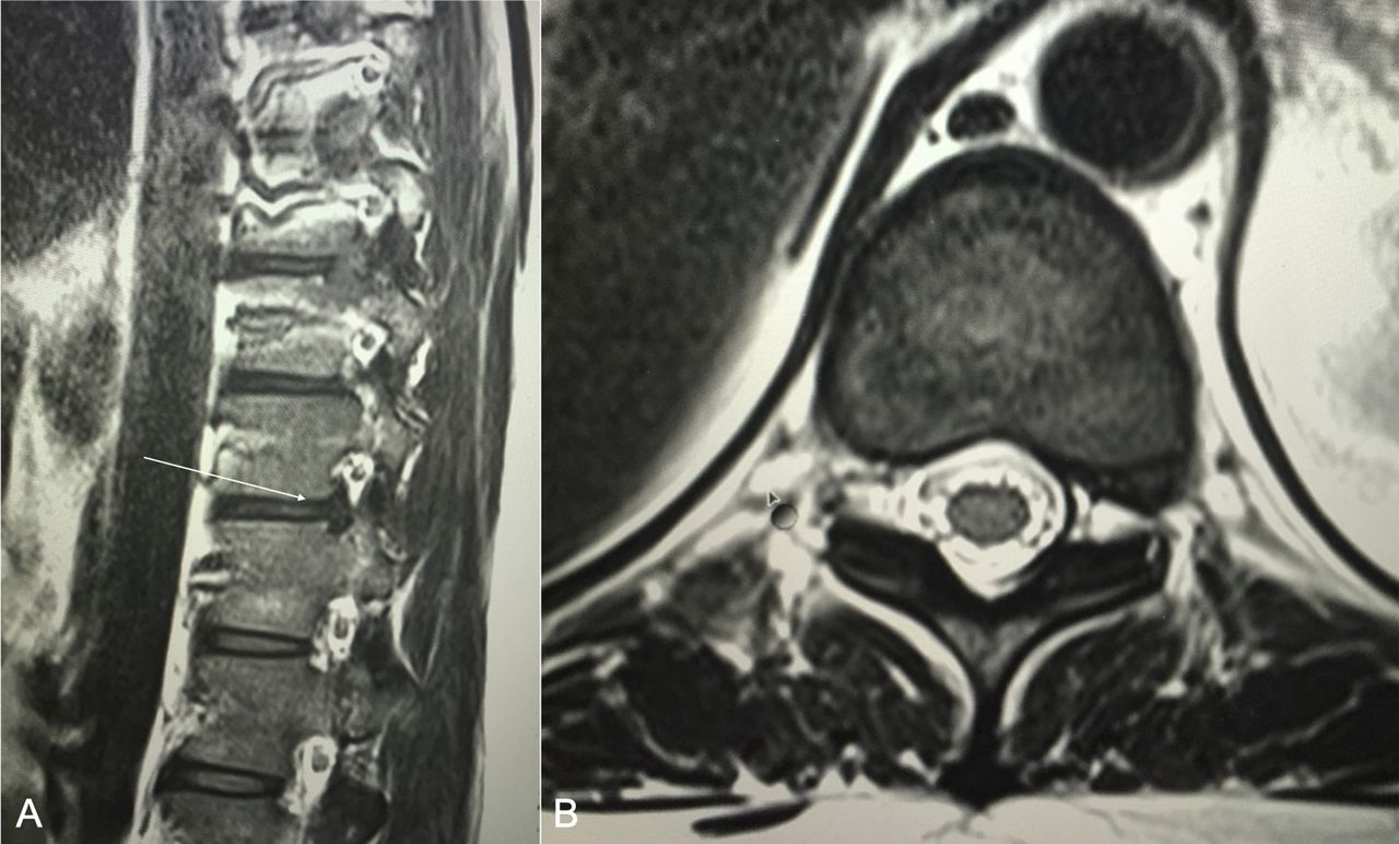
    (A and B) Sagittal and axial T2-weighted magnetic resonance images of patient 1 showing a T10-T11 foraminal disc herniation.

  • Figure 6
    • Download figure
    • Open in new tab
    • Download powerpoint
    Figure 6

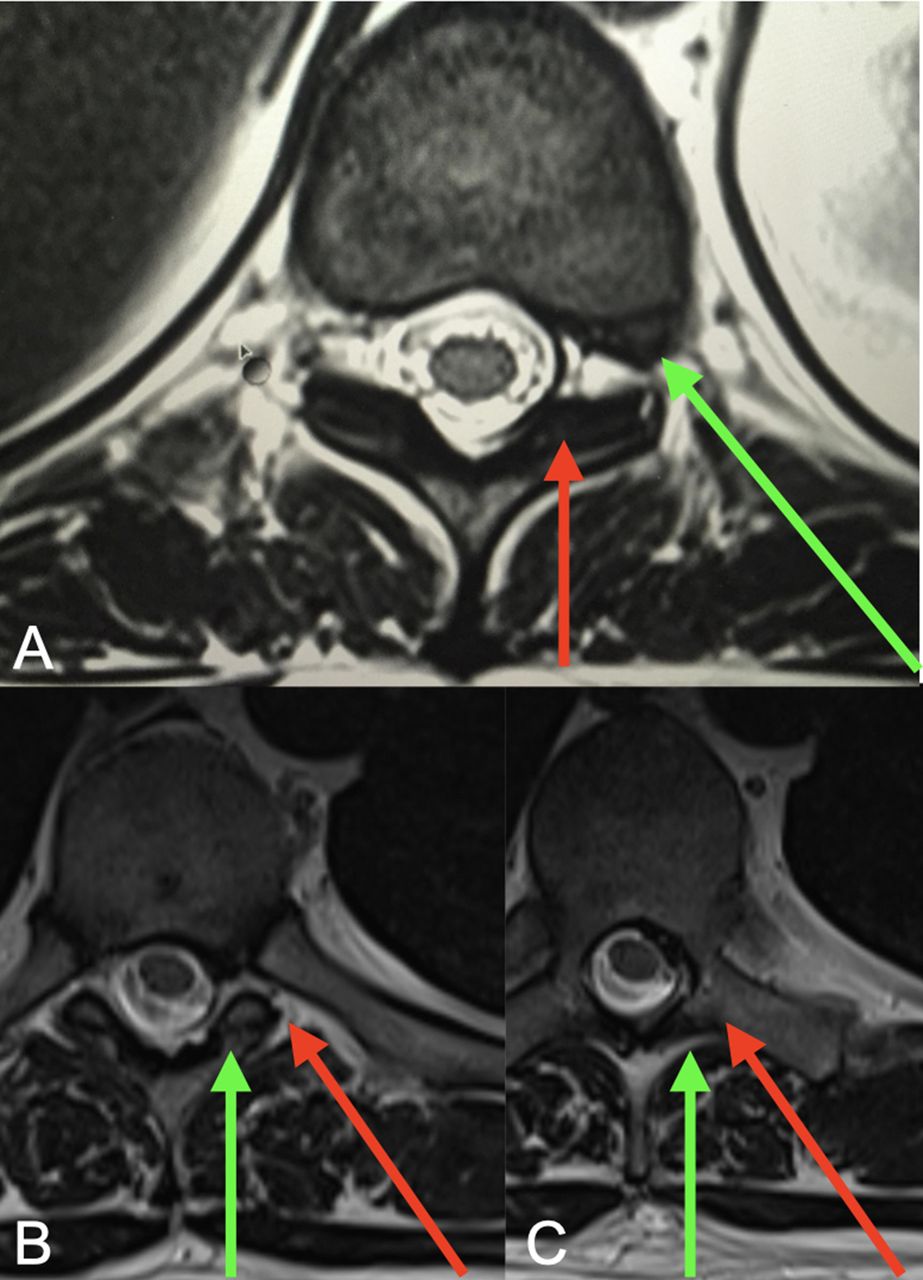
    (A) Patient 1 axial magnetic resonance image (MRI) showing a T10-T11 foramina disc herniation; the green arrow shows the selected transforaminal approach, which is a straight forward direction to the disease, avoiding bone resection. (B and C) Patient 2 axial MRI showing a T7-T8 paramedian disc herniation; the green arrow shows the selected interlaminar approach, the need of medial facetectomy is more adequate than a lateral facetectomy that would be needed in a transforaminal approach (red arrow), because it allows navigation inside the spinal canal to access the migrated material, sparing the pedicle.

Tables

  • Figures
    • View popup
    Table 1

    Published patients series of interlaminar full endoscopic spine surgery for thoracic disc herniations.

    Patient SeriesPathologyN (M:F)Age, y (range)Operative Time, minLength of Stay, dFollow-Up, moPostoperative CourseComplications
    Ruetten, 20187 T1-T2 (6), T4-T5 (1), T7-T8 (2), T9-T10 (4), T10-T11 (5), T11-T12 (2)20 (8:12)53 (23–71)95 (35–135)3 (2–5)18All patients with radiculopathy showed symptom regression. Preoperative thoracic spine pain was reduced, but not significantly.Epidural hematoma ×1 (revision), transient intercostal neuralgia, deterioration of myelopathy
    Hur, 201915 T10-T111 (1:0)6595Not reported1.5Paresthesia immediately subsided, motor grade improved from G2-G3 to G3-G4None
    Liu L, 202016 T10-T11 and ossification of ligamentum flavum1 (0:1)5811036Motor grade improved from G4 to G4+ bilaterally. The VAS score improved from 8 to 5.None
    • All patients received general anesthesia.

    • F, female; M, male; VAS, visual analog score.

    • View popup
    Table 2

    Patients’ results.

    PatientLevelApproachAnesthesiaTotal Time, minIrrigation Time, minHospital Stay, hComplications
    Patient 1: female, 38 yT10–T11TransforaminalLocal and sedation50326None
    Patient 2: male, 39 yT7–T8InterlaminarGeneral80703None
PreviousNext
Back to top

In this issue

International Journal of Spine Surgery
Vol. 16, Issue 2
1 Apr 2022
  • Table of Contents
  • Index by author

Print
Download PDF
Article Alerts
Sign In to Email Alerts with your Email Address
Email Article

Thank you for your interest in spreading the word on International Journal of Spine Surgery.

NOTE: We only request your email address so that the person you are recommending the page to knows that you wanted them to see it, and that it is not junk mail. We do not capture any email address.

Enter multiple addresses on separate lines or separate them with commas.
Full Endoscopic Thoracic Discectomy: Is the Interlaminar Approach an Alternative to the Transforaminal Approach? A Technical Note
(Your Name) has sent you a message from International Journal of Spine Surgery
(Your Name) thought you would like to see the International Journal of Spine Surgery web site.
CAPTCHA
This question is for testing whether or not you are a human visitor and to prevent automated spam submissions.
Citation Tools
Full Endoscopic Thoracic Discectomy: Is the Interlaminar Approach an Alternative to the Transforaminal Approach? A Technical Note
Marcelo Campos Moraes Amato, Bruno César Aprile, Luiz Adriano Esteves, Vinicius Marques Carneiro, Ricardo Santos de Oliveira
International Journal of Spine Surgery Apr 2022, 16 (2) 309-317; DOI: 10.14444/8209

Citation Manager Formats

  • BibTeX
  • Bookends
  • EasyBib
  • EndNote (tagged)
  • EndNote 8 (xml)
  • Medlars
  • Mendeley
  • Papers
  • RefWorks Tagged
  • Ref Manager
  • RIS
  • Zotero
Share
Full Endoscopic Thoracic Discectomy: Is the Interlaminar Approach an Alternative to the Transforaminal Approach? A Technical Note
Marcelo Campos Moraes Amato, Bruno César Aprile, Luiz Adriano Esteves, Vinicius Marques Carneiro, Ricardo Santos de Oliveira
International Journal of Spine Surgery Apr 2022, 16 (2) 309-317; DOI: 10.14444/8209
Twitter logo Facebook logo Mendeley logo
  • Tweet Widget
  • Facebook Like
  • Google Plus One

Jump to section

  • Article
    • Abstract
    • INTRODUCTION
    • METHODS
    • RESULTS
    • DISCUSSION
    • Footnotes
    • References
  • Figures & Data
  • Info & Metrics
  • PDF

Related Articles

  • No related articles found.
  • PubMed
  • Google Scholar

Cited By...

  • No citing articles found.
  • Google Scholar

More in this TOC Section

  • Effective Biportal Endoscopic Spine Surgery Technique With Better Facet Joint Preserving for Lumbar Lateral Recess Stenosis
  • Risk Analysis of Neurological Deterioration Associated With Fluid Insufflation in Uniportal Spine Endoscopy: A Case Series and Literature Review
  • Comparison of Pain and Functional Outcomes Among Geriatric and Nongeriatric Adults Following Full Endoscopic Spine Surgery for Degenerative Lumbar Pathology
Show more Endoscopic Minimally Invasive Surgery

Similar Articles

Keywords

  • Intervertebral disc displacement
  • percutaneous discectomy
  • endoscopic surgical procedures
  • thoracic vertebrae

Content

  • Current Issue
  • Latest Content
  • Archive

More Information

  • About IJSS
  • About ISASS
  • Privacy Policy

More

  • Subscribe
  • Alerts
  • Feedback

Other Services

  • Author Instructions
  • Join ISASS
  • Reprints & Permissions

© 2025 International Journal of Spine Surgery

International Journal of Spine Surgery Online ISSN: 2211-4599

Powered by HighWire