
Main menu

  • Home
  • Content
    • Current Issue
    • Advance Online Publication
    • Archive
  • About Us
    • About ISASS
    • About the Journal
    • Author Instructions
    • Editorial Board
    • Reviewer Guidelines & Publication Criteria
  • More
    • Subscribe
    • Alerts
    • Feedback
  • Join Us
  • Reprints & Permissions
  • Sponsored Content
  • Other Publications
    • ijss

User menu

  • My alerts

Search

  • Advanced search
International Journal of Spine Surgery
  • My alerts
International Journal of Spine Surgery

Advanced Search

  • Home
  • Content
    • Current Issue
    • Advance Online Publication
    • Archive
  • About Us
    • About ISASS
    • About the Journal
    • Author Instructions
    • Editorial Board
    • Reviewer Guidelines & Publication Criteria
  • More
    • Subscribe
    • Alerts
    • Feedback
  • Join Us
  • Reprints & Permissions
  • Sponsored Content
  • Follow ijss on Twitter
  • Visit ijss on Facebook
LetterBiomechanics

Response Letter

Andrew L. Freeman, Joan E. Bechtold and David W. Polly
International Journal of Spine Surgery February 2023, 17 (1) 164-165; DOI: https://doi.org/10.14444/8361
Andrew L. Freeman
1 Department of Biomedical Engineering, University of Minnesota, Minneapolis, MN, USA
  • Find this author on Google Scholar
  • Find this author on PubMed
  • Search for this author on this site
  • For correspondence: freem085@umn.edu
Joan E. Bechtold
1 Department of Biomedical Engineering, University of Minnesota, Minneapolis, MN, USA
2 Department of Orthopaedic Surgery, University of Minnesota, Minneapolis, MN, USA
  • Find this author on Google Scholar
  • Find this author on PubMed
  • Search for this author on this site
David W. Polly
2 Department of Orthopaedic Surgery, University of Minnesota, Minneapolis, MN, USA
3 Minneapolis Medical Research Foundation and Excelen Center for Bone & Joint Research and Education, Minneapolis, MN, USA
  • Find this author on Google Scholar
  • Find this author on PubMed
  • Search for this author on this site
  • Article
  • Figures & Data
  • Info & Metrics
  • PDF
Loading
  • sacroiliac joint
  • low back pain
  • fusion
  • range of motion

We thank Kampkuiper et al for their insightful comments. In general, we agree with their comments.

Placing the sacroiliac fusion devices so that they are fully contained within the bone with no neural impingement is the first constraint in construct design. Implant malposition is the most common reason for revision surgery with Cher et al1 reporting it to be about 1%. This appears to have improved slightly from 2010 to 2014 (1%–0.9%). In a follow-up study, Cher et al2 reported an overall revision rate of 3% for cases performed between 2015 to 2018. Out of 14,210 cases, 435 underwent revisions. Of these, 54% were for symptomatic implant malposition, which implies a 1.6% implant malposition rate.

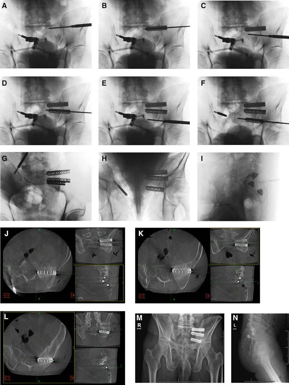
The senior author has done all of his cases using intraoperative navigation. It is also routine to do a postimplant placement check scan prior to leaving the operating room. This began in 2010. Having done >270 primaries (average 2.8 implants per case) and >60 revision cases, there have been well over 1000 implants placed. Of those implants, 2 needed repositioning. This corresponds with the Kampkuiper et al’s comments citing Cleveland.

The key to being able to achieve the desired implant placement is the individual bony anatomy. Women typically only have S1 and S2 articulations, whereas men typically have S1-S3 articulations. In addition, the rate of occurrence of dysmorphic sacra (Figure 1) is reported by Matson et al to be between 3.9% and 35.6% of the population.3

Figure 1
  • Download figure
  • Open in new tab
  • Download powerpoint
Figure 1

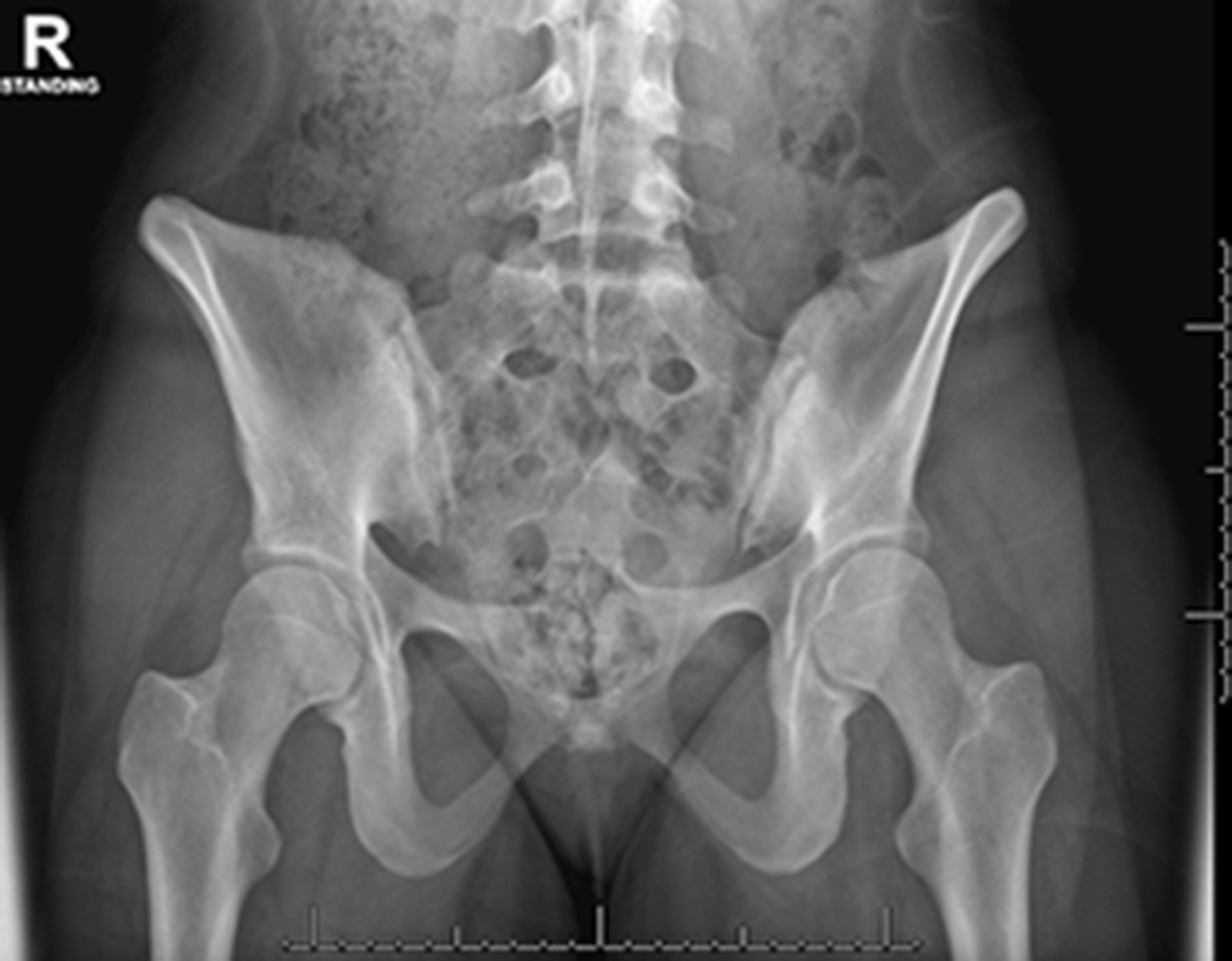
Dysmorphic sacrum as indicated by the upsloped sacral ala.

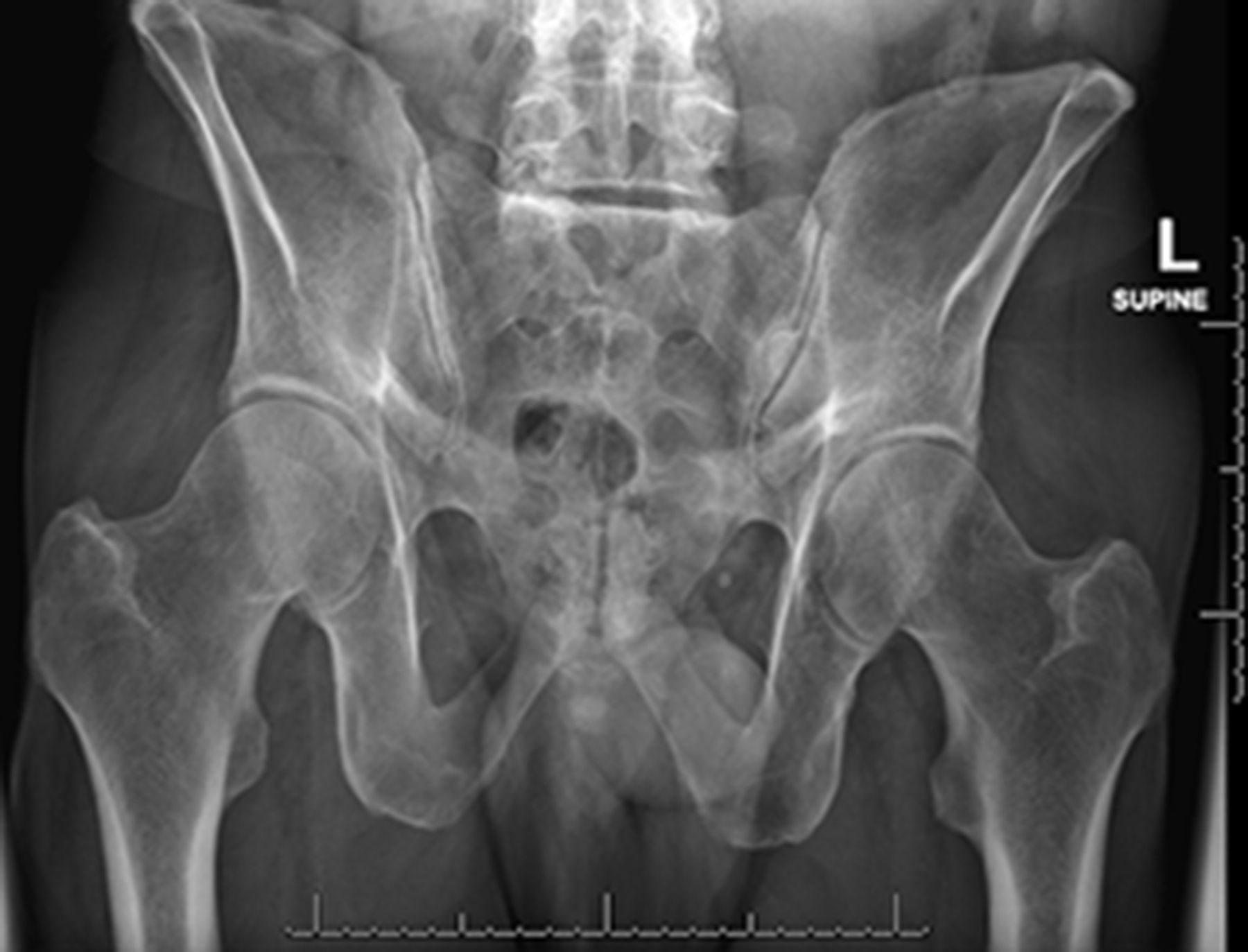
In normal morphology pelves (Figure 2), it is the senior author’s experience that the cephalad S1 implant is positioned parallel to the sacral ala (Figure 3). The caudal S1 implant is then angled parallel to the ventral sacral cortex. The S2 implant is then placed in what would be a through-and-through trajectory of the S2 corridor. This allows for inadvertent pin advancement without the risk of neural injury (Figure 3). We then routinely do 2-dimensional and 3-dimensional check images to ensure optimal placement. This case probably represents the maximum achievable between device angulation and distance. This is in agreement with the diagram and thoughts from Kampkuiper et al.

Figure 2
  • Download figure
  • Open in new tab
  • Download powerpoint
Figure 2

Normal morphology sacrum.

Figure 3
  • Download figure
  • Open in new tab
  • Download powerpoint
Figure 3

Intraoperative imaging and device placement with 2-dimensional and 3-dimensional imaging.

Acknowledgments

The authors acknowledge SI Bone for donating the implants and financial support for the original study.

Footnotes

  • Funding The authors received no financial support for the research, authorship, and/or publication of this letter.

  • Declaration of Conflicting Interests David Polly reports consulting for SI Bone and Globus Medical; royalties from SI Bone and Springer; and research support (paid to institutino) from Medtronic and Mizuho OSI. The remaining authors have nothing to report.

  • This manuscript is generously published free of charge by ISASS, the International Society for the Advancement of Spine Surgery. Copyright © 2023 ISASS. To see more or order reprints or permissions, see http://ijssurgery.com.

References

  1. 1.↵
    1. Cher DJ ,
    2. Reckling WC ,
    3. Capobianco RA
    . Implant survivorship analysis after minimally invasive sacroiliac joint fusion using the ifuse implant system(®). Med Devices (Auckl). 2015;8:485–492. doi:10.2147/MDER.S94885
    OpenUrlCrossRef
  2. 2.↵
    1. Cher D ,
    2. Wroe K ,
    3. Reckling WC ,
    4. Yerby S
    . Postmarket surveillance of 3D-printed implants for sacroiliac joint fusion. Med Devices (Auckl). 2018;11:337–343. doi:10.2147/MDER.S180958
    OpenUrlCrossRef
  3. 3.↵
    1. Matson DM ,
    2. Maccormick LM ,
    3. Sembrano JN ,
    4. Polly DW
    . Sacral dysmorphism and lumbosacral transitional vertebrae (LSTV) review. Int J Spine Surg. 2020;14(Suppl 1):14–19. doi:10.14444/6075
    OpenUrlCrossRef
Previous
Back to top

In this issue

International Journal of Spine Surgery
Vol. 17, Issue 1
1 Feb 2023
  • Table of Contents
  • Index by author
Print
Download PDF
Article Alerts
Sign In to Email Alerts with your Email Address
Email Article

Thank you for your interest in spreading the word on International Journal of Spine Surgery.

NOTE: We only request your email address so that the person you are recommending the page to knows that you wanted them to see it, and that it is not junk mail. We do not capture any email address.

Enter multiple addresses on separate lines or separate them with commas.
Response Letter
(Your Name) has sent you a message from International Journal of Spine Surgery
(Your Name) thought you would like to see the International Journal of Spine Surgery web site.
CAPTCHA
This question is for testing whether or not you are a human visitor and to prevent automated spam submissions.
Citation Tools
Response Letter
Andrew L. Freeman, Joan E. Bechtold, David W. Polly
International Journal of Spine Surgery Feb 2023, 17 (1) 164-165; DOI: 10.14444/8361

Citation Manager Formats

  • BibTeX
  • Bookends
  • EasyBib
  • EndNote (tagged)
  • EndNote 8 (xml)
  • Medlars
  • Mendeley
  • Papers
  • RefWorks Tagged
  • Ref Manager
  • RIS
  • Zotero
Share
Response Letter
Andrew L. Freeman, Joan E. Bechtold, David W. Polly
International Journal of Spine Surgery Feb 2023, 17 (1) 164-165; DOI: 10.14444/8361
Reddit logo Twitter logo Facebook logo Mendeley logo
  • Tweet Widget
  • Facebook Like
  • Google Plus One

Jump to section

  • Article
    • Acknowledgments
    • Footnotes
    • References
  • Figures & Data
  • Info & Metrics
  • PDF

Related Articles

  • No related articles found.
  • PubMed
  • Google Scholar

Cited By...

  • No citing articles found.
  • Google Scholar

More in this TOC Section

  • Predicting Pedicle Screw Pullout and Fatigue Performance: Comparing Lateral Dual-Energy X-Ray Absorptiometry, Anterior to Posterior Dual-Energy X-Ray Absorptiometry, and Computed Tomography Hounsfield Units
  • Effect of Sacropelvic Hardware on Axis and Center of Rotation of the Sacroiliac Joint: A Finite Element Study
  • 5.5-mm Cobalt-Chrome vs 6-mm Titanium Alloy Rods in Surgical Treatment of Lenke 1 Adolescent Idiopathic Scoliosis With High-Density Pedicle Screws and Direct Vertebral Rotation on Differently Shaped Rods: A Retrospective Comparative Cohort Study
Show more Biomechanics

Similar Articles

Keywords

  • sacroiliac joint
  • low back pain
  • fusion
  • range of motion

Content

  • Current Issue
  • Latest Content
  • Archive

More Information

  • About IJSS
  • About ISASS
  • Privacy Policy

More

  • Subscribe
  • Alerts
  • Feedback

Other Services

  • Author Instructions
  • Join ISASS
  • Reprints & Permissions

© 2023 International Journal of Spine Surgery

International Journal of Spine Surgery Online ISSN: 2211-4599

Powered by HighWire