

Fig. 1

Fig. 1
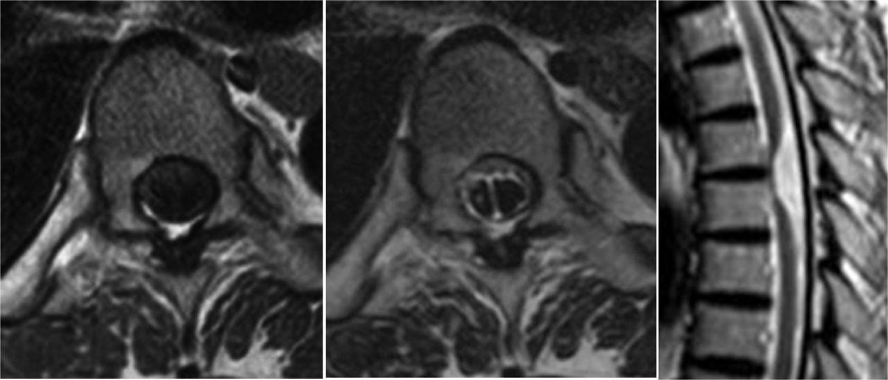
Axial T1, T2-weighted MRI (left, center) shows significant right ventral shift of the spinal cord and a dorsal midline subarachnoid septum at T5–T6 disc level. The midline septum is observed only at this level. Sagittal T2-weighted MRI (right) shows ventral displacement of the spinal cord at T5–T6 disc level. Focal ventral kinking and adhesion of the spinal cord is apparent with an enlarged dorsal subarachnoid space.
In this issue


Related Articles
Cited By...
- No citing articles found.