


Article Figures & Data
Figures
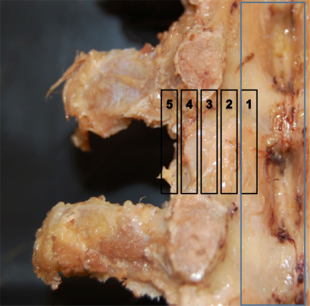
- Fig. 1
DRG region. Zone 1 and 2: intraspinal; Zone 3: medial neuroforamina; Zone 4: lateral neuroforamina; Zone 5: extraforaminal space.
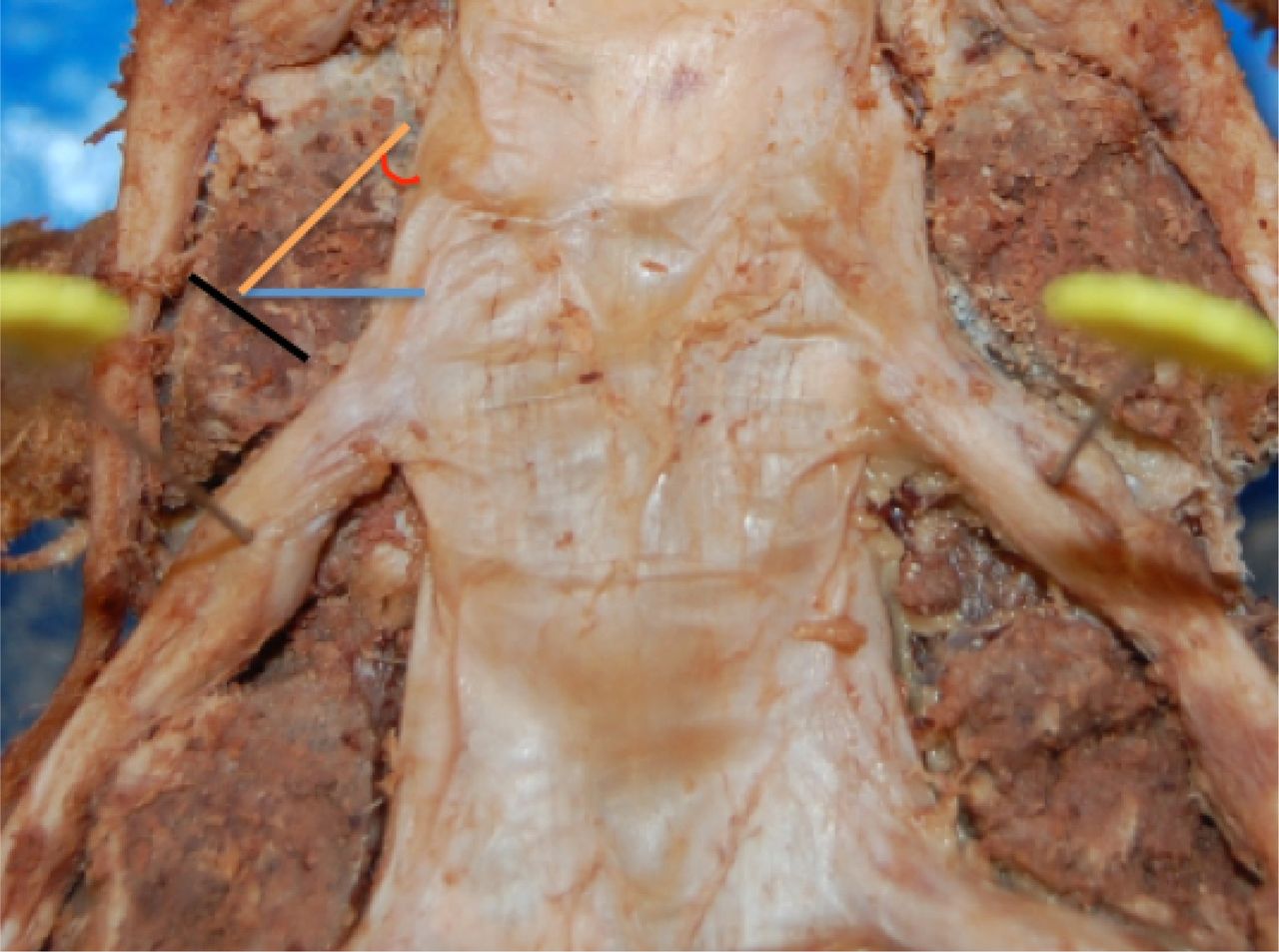
- Fig. 2
DRG Size (mm). Width is the width of DRG at the pin (maximal width of DRG). The nerve root angle is determined from sin(O) calculated by the right/diagonal lines relative to the width.

Tables
DRG Size (mm) Left Right L1 L2 L3 L4 L5 L1 L2 L3 L4 L5 Width: mean 3.2 4.3 4.9 6.1 6.5 3.2 4.3 4.9 6.1 6.5 Width: sem 0.7 0.4 0.3 0.4 0.5 0.7 0.4 0.3 0.4 0.5 DRG Region Left Right L1 L2 L3 L4 L5 L1 L2 L3 L4 L5 Zone: mean 3.0 3.0 3.5 3.9 4.3 3.0 3.1 3.5 3.8 4.2 Zone: sem 0.3 0.1 0.1 0.1 0.1 0.3 0.2 0.1 0.1 0.1 Intervertebral Foramina Size (mm) Left Right L1 L2 L3 L4 L5 L1 L2 L3 L4 L5 AP: mean 6.6 7.4 7.6 8.4 8.7 6.8 7.2 7.4 8.5 8.6 AP: sem 0.2 0.2 0.3 0.4 0.4 0.4 0.2 0.3 0.2 0.4 PP: mean 7.2 8.1 9.4 10.1 10.1 7.3 8.5 9.4 10.4 10.3 PP: sem 1.8 0.7 0.8 0.8 0.7 1.8 0.7 0.8 0.8 0.7 Pedicle width (medial to lateral) (mm) Left Right L1 L2 L3 L4 L5 L1 L2 L3 L4 L5 Width: mean 7.0 7.0 7.8 8.7 9.7 6.7 7.1 7.9 8.9 9.7 Width: sem 0.3 0.3 0.2 0.3 0.4 0.2 0.3 0.2 0.4 0.4 Nerve Root Angle Left Right L1 L2 L3 L4 L5 L1 L2 L3 L4 L5 Angle (rounded to nearest 10th) 50.5 52.2 55.2 57.4 58.8 50.5 52.2 55.2 57.2 56.3 Key: DRG Region zone 1 and 2: intraspinal; zone 3: medial neuroforamina; zone 4: lateral neuroforamina; zone 5: extraforaminal space. Intervertebral Foraminal Size. Anteriorposterior (AP): maximal distance from posterior body/disk to facet joint. Pedicle to pedicle (PP) distance: inferior border of upper to superior border of lower pedicle. SEM: standard error of the mean. Angle determined via sin(O) = right/diagonal.
- Table 2
Raw Values Model-Adjusted Values Level Proportion Agree % Agree % Agree (95% Confidence Interval) 1 8/12 66.7 ---- 2 22/29 75.9 77.4 (58.0, 89.5) 3 25/32 78.1 79.4 (61.2, 90.4) 4 28/32 87.5 88.6 (71.9, 95.9) 5 30/32 93.8 94.4 (79.0, 98.7)
In this issue


Jump to section
Related Articles
Cited By...
- No citing articles found.