
Main menu

  • Home
  • Content
    • Current Issue
    • Advance Online Publication
    • Archive
  • About Us
    • About ISASS
    • About the Journal
    • Author Instructions
    • Editorial Board
    • Reviewer Guidelines & Publication Criteria
  • More
    • Advertise
    • Subscribe
    • Alerts
    • Feedback
  • Join Us
  • Reprints & Permissions
  • Sponsored Content
  • Other Publications
    • ijss

User menu

  • My alerts

Search

  • Advanced search
International Journal of Spine Surgery
  • My alerts
International Journal of Spine Surgery

Advanced Search

  • Home
  • Content
    • Current Issue
    • Advance Online Publication
    • Archive
  • About Us
    • About ISASS
    • About the Journal
    • Author Instructions
    • Editorial Board
    • Reviewer Guidelines & Publication Criteria
  • More
    • Advertise
    • Subscribe
    • Alerts
    • Feedback
  • Join Us
  • Reprints & Permissions
  • Sponsored Content
  • Follow ijss on Twitter
  • Visit ijss on Facebook
Research ArticleArticles

Assessment of Magnetic Resonance Imaging Artifact Following Cervical Total Disc Arthroplasty

Amir H. Fayyazi, Jennifer Taormina, David Svach, Jeff Stein and Nathaniel R. Ordway
International Journal of Spine Surgery January 2015, 9 30; DOI: https://doi.org/10.14444/2030
Amir H. Fayyazi
1OAA Orthopaedic Specialists, Allentown, PA
MD
  • Find this author on Google Scholar
  • Find this author on PubMed
  • Search for this author on this site
Jennifer Taormina
2SUNY Upstate Medical University, Syracuse, NY
MS
  • Find this author on Google Scholar
  • Find this author on PubMed
  • Search for this author on this site
David Svach
3Research & Testing, Depuy Synthes, West Chester, PA
MS
  • Find this author on Google Scholar
  • Find this author on PubMed
  • Search for this author on this site
Jeff Stein
4Clinical Biostatistics & Data Management, DePuy Synthes, West Chester, PA
PhD
  • Find this author on Google Scholar
  • Find this author on PubMed
  • Search for this author on this site
Nathaniel R. Ordway
2SUNY Upstate Medical University, Syracuse, NY
MS
  • Find this author on Google Scholar
  • Find this author on PubMed
  • Search for this author on this site
  • Article
  • Figures & Data
  • Info & Metrics
  • PDF
Loading

Article Figures & Data

Figures

  • Tables
  • Fig. 1
    • Download figure
    • Open in new tab
    • Download powerpoint
    Fig. 1

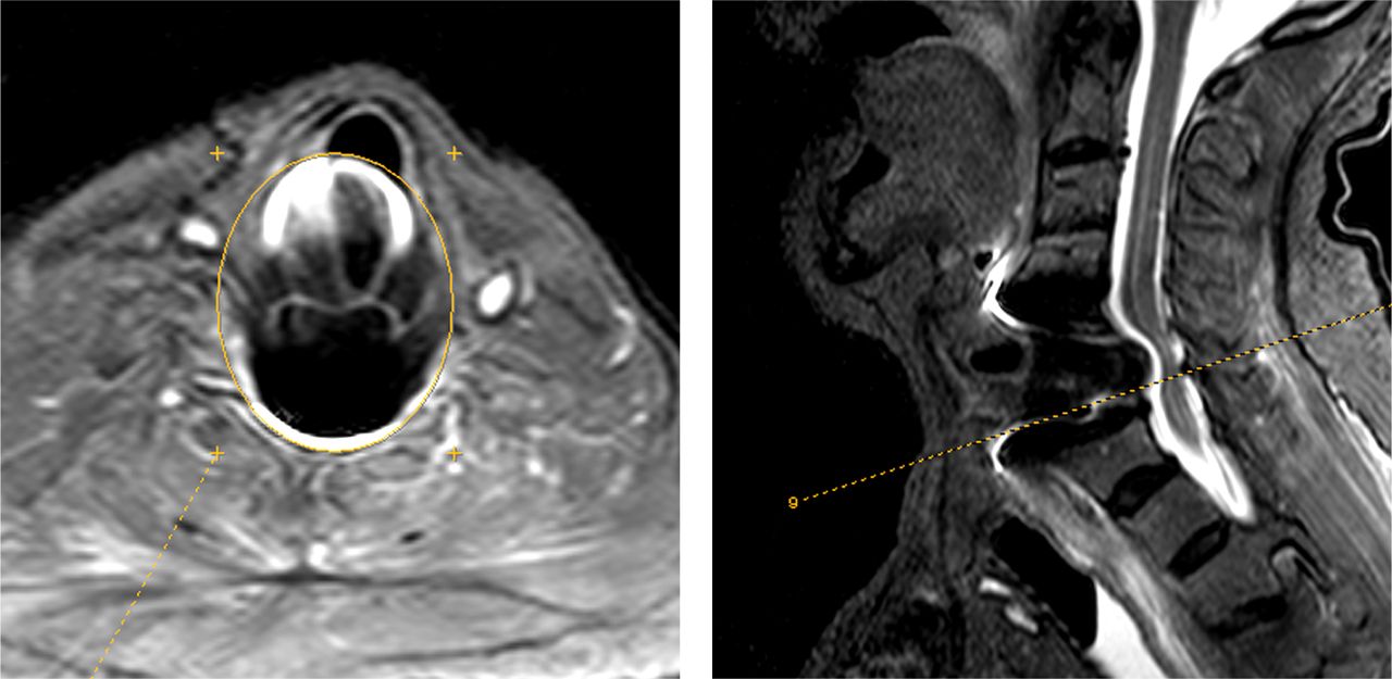
    The area of the artifact was measured on each axial image using eFilm Lite software as demonstrated.
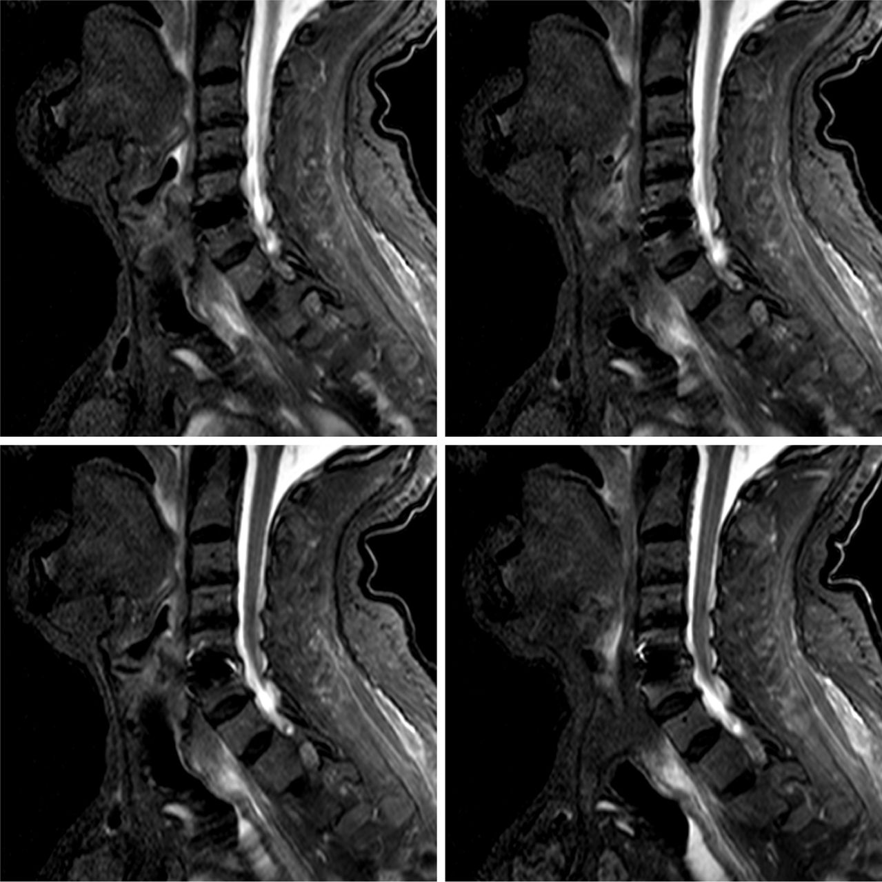
  • Fig. 2
    • Download figure
    • Open in new tab
    • Download powerpoint
    Fig. 2

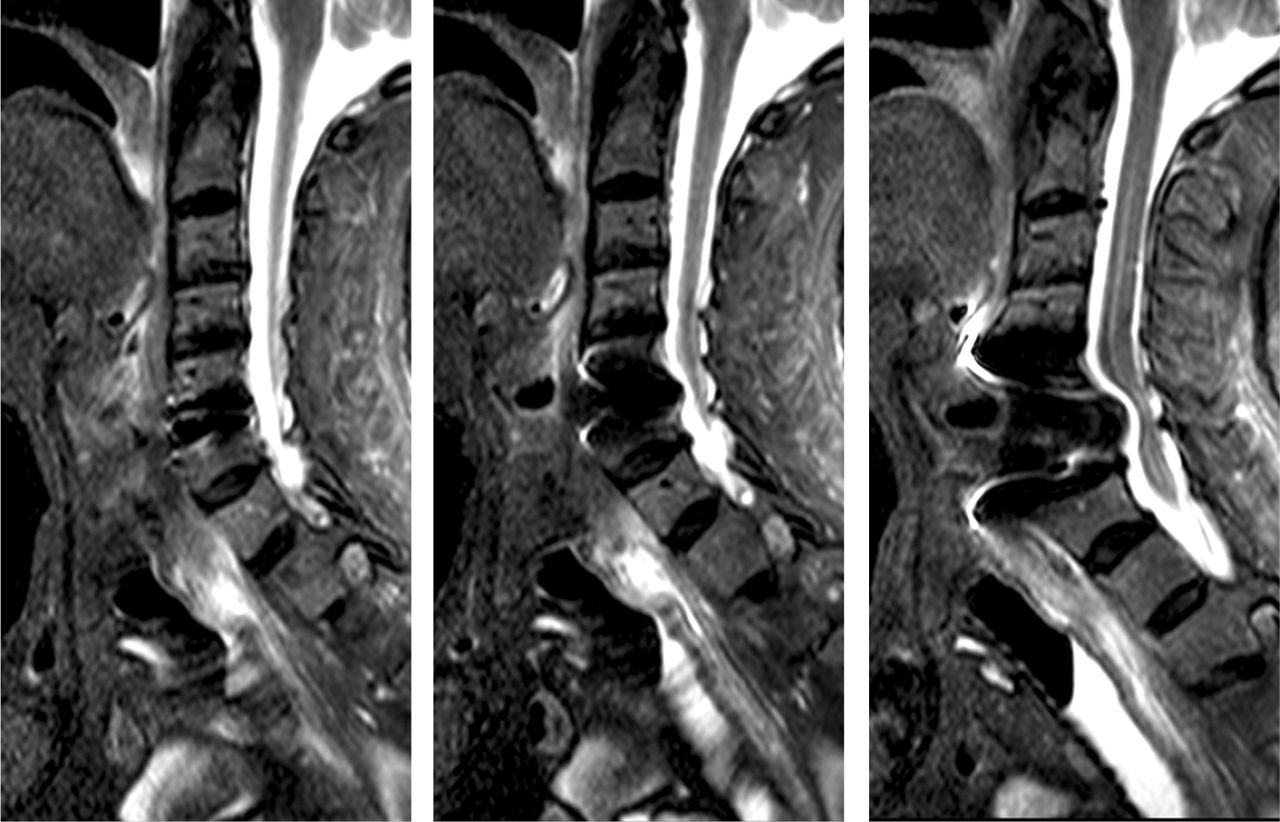
    Corresponding T2 weighted sagittal and axial images of Visualized, Partially Distorted, and Fully Distorted implanted segments.

  • Fig. 3
    • Download figure
    • Open in new tab
    • Download powerpoint
    Fig. 3

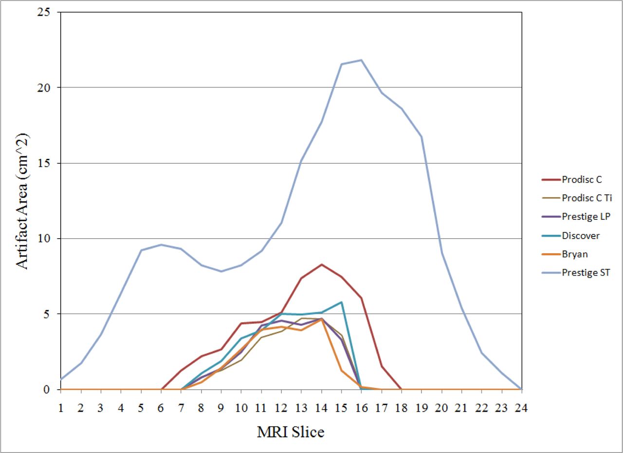
    The cross-sectional area of the artifact measures in each slice was dependent on the shape and material property of the device.

  • Fig. 4
    • Download figure
    • Open in new tab
    • Download powerpoint
    Fig. 4

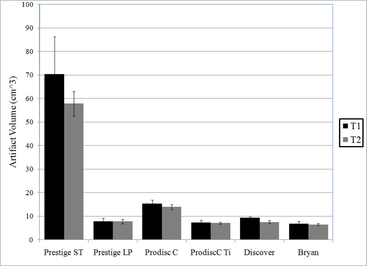
    The volume of the artifact measured was significantly larger following implantation of the Prestige ST implant.

  • Fig. 5
    • Download figure
    • Open in new tab
    • Download powerpoint
    Fig. 5

    Minimal artifact was seen following the implantation of the Titanium Devices; Bryan (Top Left), Discover (Top Right), Prestige LP (Lower Left), and Prodisc C Ti (Lower Right).

  • Fig. 6
    • Download figure
    • Open in new tab
    • Download powerpoint
    Fig. 6

    Significant distortion was noted following implantation of the stainless steel Prestige ST implant; neither the index nor the adjacent levels could be assessed following implantation.

Tables

  • Figures
    • View popup
    Table 1

    MRI imaging sequences

    T1T2
    SagittalTR: 400ms
    TE: 7.4ms
    TR: 3500ms
    TE: 120ms
    AxialTR: 1089ms
    TE: 7.8ms
    TR: 3500ms
    TE: 80ms
    • View popup
    Table 2

    List of cervical implants imaged as well as the size and weight ranges.

    Implant MaterialsSize Used (mm) (deep x width x height)Size Range (mm)Weight Range (grams)
    Prodisc-CCoCrMo endplates with plasma-sprayed titanium surface, UHMWPE inlay12 x 15 x 512 – 18 D
    15 – 19 W
    5 – 7 H
    4.43 – 9.26
    Prodisc-C Ti (prototype)TAN Endplates, superior CoCrMo insert, inferior UHMWPE inlay12 x 15 x 512 – 18 D
    15 – 19 W
    5 – 7 H
    2.73 – 5.64
    Prestige LPTitanium Alloy/Titanium Carbide Composite, plasma-sprayed titanium coating12 x 17.8 x 612 – 18 D
    17.8 W
    6 – 8 H
    3.65 – 7.67
    DiscoverTitanium Alloy Endplates, Polyethylene Core15.2 x 18.7 x 813.7 – 16.7 D
    14.2 – 18.7 W
    5 – 9 H
    2.14 – 5.54
    BryanTitanium shells, Polyurethane nucleus and sheath14 (diameter) x 614 – 18 D
    6 H
    Prestige STStainless steel14 x 17.8 x 712 – 18 D
    17.8 W
    6 – 7 H
    10.41 – 14.74
    • D: depth; W: width; H: height.

    • View popup
    Table 3

    Qualitative scoring method used for classifying image artifact.

    IVisualizedNo distinguishable artifact in either sagittal or axial images of the spinal canal or bilateral neural foramina.
    IIPartially DistortedDistortion of either neural foramina or the spinal canal that allows partial visualization of the neural structures.
    IIIFully DistortedInability to visualize the neural structures in bilateral neural foramina and the canal.
    • View popup
    Table 4

    Pearson Product Moment Correlation Coefficients.

    TI (All)T1 Without Prestige STT2 (All)T2 Without Prestige ST
    Overall0.9990.9880.9970.990
    Observer A0.9990.9970.9990.996
    Observer B0.9980.9991.0000.999
    Observer C0.9990.9920.9990.974
    • View popup
    Table 5

    Concordance Correlation Coefficients.

    T1T2
    First Measurement (All)0.8980.974
    Repeat Measurement (All)0.9110.992
    First Measurement Without Prestige ST0.860.913
    Repeat Measurement Without Prestige ST0.8610.924
    • View popup
    Table 6

    Qualitative measurements by level and implant.

    LevelProDisc-CProDisc-C TiPrestige LPDiscoverBryanPrestige ST
    IndexPartially DistortedFully VisualizedFully VisualizedFully VisualizedFully VisualizedFully Distorted
    AdjacentFully VisualizedFully VisualizedFully VisualizedFully VisualizedFully VisualizedFully Distorted
PreviousNext
Back to top

In this issue

International Journal of Spine Surgery
Vol. 9
1 Jan 2015
  • Table of Contents
  • Index by author

Print
Download PDF
Article Alerts
Sign In to Email Alerts with your Email Address
Email Article

Thank you for your interest in spreading the word on International Journal of Spine Surgery.

NOTE: We only request your email address so that the person you are recommending the page to knows that you wanted them to see it, and that it is not junk mail. We do not capture any email address.

Enter multiple addresses on separate lines or separate them with commas.
Assessment of Magnetic Resonance Imaging Artifact Following Cervical Total Disc Arthroplasty
(Your Name) has sent you a message from International Journal of Spine Surgery
(Your Name) thought you would like to see the International Journal of Spine Surgery web site.
CAPTCHA
This question is for testing whether or not you are a human visitor and to prevent automated spam submissions.
Citation Tools
Assessment of Magnetic Resonance Imaging Artifact Following Cervical Total Disc Arthroplasty
Amir H. Fayyazi, Jennifer Taormina, David Svach, Jeff Stein, Nathaniel R. Ordway
International Journal of Spine Surgery Jan 2015, 9 30; DOI: 10.14444/2030

Citation Manager Formats

  • BibTeX
  • Bookends
  • EasyBib
  • EndNote (tagged)
  • EndNote 8 (xml)
  • Medlars
  • Mendeley
  • Papers
  • RefWorks Tagged
  • Ref Manager
  • RIS
  • Zotero
Share
Assessment of Magnetic Resonance Imaging Artifact Following Cervical Total Disc Arthroplasty
Amir H. Fayyazi, Jennifer Taormina, David Svach, Jeff Stein, Nathaniel R. Ordway
International Journal of Spine Surgery Jan 2015, 9 30; DOI: 10.14444/2030
Twitter logo Facebook logo Mendeley logo
  • Tweet Widget
  • Facebook Like
  • Google Plus One

Jump to section

  • Article
    • Abstract
    • Introduction
    • Material And Methods
    • Results
    • Discussion
    • Conclusion
    • Disclosures
    • References
  • Figures & Data
  • Info & Metrics
  • PDF

Related Articles

  • No related articles found.
  • Google Scholar

Cited By...

  • No citing articles found.
  • Google Scholar

More in this TOC Section

  • Validity and reliability of a novel patient reported outcome tool to evaluate post-operative dysphagia, odynophagia, and voice (DOV) disability after anterior cervical procedures
  • Surgical Revision after Sacroiliac Joint Fixation or Fusion
  • A Rare Case of T1-2 Thoracic Disc Herniation Mimicking Cervical Radiculopathy
Show more Articles

Similar Articles

Keywords

  • cervical spine
  • total disc arthroplasty
  • MRI
  • Artifact
  • Quantitative Measurement
  • Qualitative Measurement

Content

  • Current Issue
  • Latest Content
  • Archive

More Information

  • About IJSS
  • About ISASS
  • Privacy Policy

More

  • Subscribe
  • Alerts
  • Feedback

Other Services

  • Author Instructions
  • Join ISASS
  • Reprints & Permissions

© 2025 International Journal of Spine Surgery

International Journal of Spine Surgery Online ISSN: 2211-4599

Powered by HighWire