


Article Figures & Data
Figures
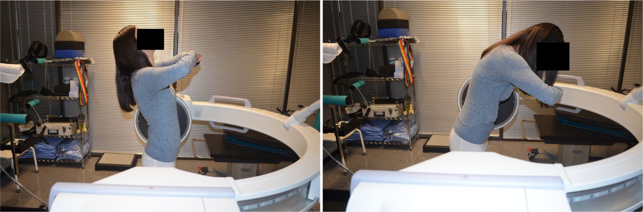
- Fig. 1
VMA standing flexion/extension position.
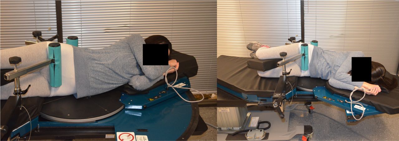
- Fig. 2
VMA recumbent flexion/extension position.
- Fig. 3
Flexion extension radiographs (FE) at a self-selected pace and position.

Tables
- Table 1
Bending posture and bending method classifications for classifying studies reviewed as part of the review of medical literature.
Bending Posture Bending Method A. Standing uncontrolled Patient stands uprights and bends on their own without grabbing anything or being bolstered to anything. I. Max Voluntary Patient bends as far as they can move them-selves without using their hands to pull into flexion. B. Hips flexed In flexion, bending can occur in the hips and lumbar spine. Knees may or may not be flexed. II. Patient pulling Patient pulls on a fixed object to go further into flexion than the patient could otherwise go. C. Standing pelvis bolster Upright standing with a bolster to minimize pelvis motion. Knees kept in full extension. III. Rad Tech Pulling A technician pushes and/or pulls the patient into a maximal spine bend. D. Side lying Lying in lateral decubitus position and bending forward and backward. IV. Stan-dard End Range IVT and IVR are measured at a standardized overall trunk bending angle, short of the maximal end range position. E. Traction & compression Using external forces to add load (compression) or to distract (traction) the spine while in an upright standing posture - Table 2
Average angles and translations measured for both VMA and FE imaging radiographs (N = 2239 intervertebral levels).
Panel A. IVR measured in degrees (°) FE VMA (Standing) VMA (Lying) Δ VMA (Standing)-FE Δ VMA (Lying)-FE Level Mean Std Dev Mean Std Dev Mean Std Dev Mean Std Dev Mean Std Dev L1-L2 5.9 2.3 7.6 4.3 8.6 3.6 1.7** 2.0 2.7** 1.3 L2-L3 6.8 2.4 11.2 4.8 10.1 3.7 4.4*** 2.4 3.3*** 1.3 L3-L4 7.7 2.6 10.1 5.1 9.2 4.1 2.4** 2.5 1.5* 1.5 L4-L5 9.1 3.3 11.9 7.1 8.3 4.4 2.8** 3.8 0.8 1.1 L5-S1 8.5 4.2 8.1 6.2 10.3 4.6 0.4 2.0 1.8* 0.4 Average 7.6 3.0 9.8 5.5 9.3 4.1 2.3** 2.5 2.0** 1.1 Panel B. IVT in percentage vertebral body depth (%) FE VMA (Standing) VMA (Lying) Δ VMA (Standing)-FE Δ VMA (Lying)-FE Level Mean Std Dev Mean Std Dev Mean Std Dev Mean Std Dev Mean Std Dev L1-L2 4.4 1.4 5.7 3.8 5.4 2.3 1.3* 2.4 1.0 0.9 L2-L3 5.2 1.3 6.1 4.0 7.9 2.1 0.9 2.7 2.7** 0.8 L3-L4 5.9 1.5 6.6 4.5 9.0 2.7 0.7 3.0 3.1** 1.2 L4-L5 7.7 2.1 5.8 5.2 8.3 2.0 1.9* 3.1 0.6 0.1 L5-S1 6.9 3.3 6.4 3.7 2.8 1.1 0.5 0.5 4.1*** 2.2 Average 6.0 1.9 6.1 4.2 6.7 2.1 1.04* 2.3 2.3** 1.1 Level n Contribution L1-L2 446 11 L2-L3 438 16 L3-L4 471 27 L4-L5 452 23 L5-S1 432 23 Total 2239 - Table 4
Specificity and NPV of the VMA and FE lumbar imaging radiographs in detecting radiographic instability across levels, and the prevalence of VMA- and FE-detectable radiographic instability.
IVR IVT Method Specificity NPV Prevalence Specificity NPV Prevalence VMA 99.4% 90.7% 12.3% 99.1% 87.4% 11.9% FE 98.3% 72.4% 6.1% 98.2% 71.2% 5.4% Difference 1.1% 18.3% 6.2% 0.9% 16.2% 6.5% - Table 5
Average angles and translations measured for both VMA and FE imaging radiographs in the agreement dataset.
Panel A. IVR measured in degrees (°) FE VMA (Standing) VMA (Lying) Δ VMA (Standing)-FE Δ VMA (Lying)-FE Level Mean Std Dev Mean Std Dev Mean Std Dev Mean Std Dev Mean Std Dev L1-L2 6.3 1.8 9.1 4.2 10.3 3.3 2.8** 2.4 4.0*** 1.5 L2-L3 7.1 2.2 12.2 5.1 9.8 3.5 5 1*** 2.9 2 7** 1.3 L3-L4 7.9 2.1 11.6 3.8 10.2 3.6 3.7** 1.7 2 3** 1.5 L4-L5 10.2 2.7 12.3 4.6 9.7 3.8 2.1** 1.9 0.5 1.1 L5-S1 9.4 3.8 10.2 5.1 11.1 3.9 0.8 1.3 1.7* 0.1 Average 8.2 2.5 11.1 4.6 10.2 3.6 2.9** 2.0 2.2** 1.1 Panel B. IVT in percentage vertebral body depth (%) FE VMA (Standing) VMA (Lying) Δ VMA (Standing)-FE Δ VMA (Lying)-FE Level Mean Std Dev Mean Std Dev Mean Std Dev Mean Std Dev Mean Std Dev L1-L2 5.1 1.5 6.2 2.9 5.9 1.9 1.1* 1.4 0.8 0.4 L2-L3 5.8 1.1 6.4 3.7 7.4 2.2 0.6 2.6 1.6* 1.1 L3-L4 6.2 1.4 7.5 4.1 8.7 2.8 1.3* 2.7 2.5** 1.4 L4-L5 7.1 1.8 6.4 5.3 8.9 3.1 0.7 3.5 1.8* 1.3 L5-S1 7.8 2.8 6.9 3.2 3.2 2.2 0.9* 0.4 4.60*** 0.6 Average 6.4 1.7 6.7 3.8 6.8 2.4 0.9* 2.1 2.3** 1.0 Intra/Inter Observer Intra Subject Level n Contribution n Contribution L1-2 446 11 - - L2-3 438 16 74 14 L3-4 471 27 71 23 L4-5 452 23 72 27 L5-S1 432 23 70 28 Total 705 287 - Table 7
Intra and inter observer variability and intra subject variability in VMA lumbar imaging radiographs.
Measurement Mean in Differences CI Upper LOA CI Lower LOA CI CR ICC (3,1) CI SEM Observer Variability Rotation 0.02 0.03 to 0.09 2.52 2.68 to 2.36 -2.49 -2.25 to -2.73 2.49 0.974 0.978 to 0.97 0.13 Intra-Observer Variability Translation (%) 0.06 0.05 to 0.13 2.69 2.91 to 2.47 -2.62 2.44 to -2.81 2.62 0.926 0.936 to 0.916 0.18 Inter-Observer Variability Rotation 0.05 0.06 to 0.19 1.97 2.16 to 1.78 -2.03 -1.81 to -2.25 1.99 0.939 0.946 to 0.932 0.23 Inter-Observer Variability Translation (%) 0.03 0.04 to 0.21 2.84 3.07 to 2.61 -2.91 -2.60 to -3.22 2.81 0.946 0.957 to 0.935 0.24 Intra-Subject Variability Rotation -0.14 -0.15 to -0.26 2.46 2.71 to 2.22 -2.52 2.25 to -2.79 2.49 0.919 0.928 to 0.911 0.27 Intra-Subject Variability Translation (%) -0.11 -0.09 to -0.21 2.67 2.87 to 2.34 -2.69 -2.34 to -2.96 2.57 0.923 09.38 to 0.913 0.25 This table reports the confidence intervals (CI), mean absolute error, upper and lower limits of agreement (LOA), interclass correlation (ICC), and standard error of the mean (SEM).
In this issue


Jump to section
Related Articles
Cited By...
- No citing articles found.