


Article Figures & Data
Figures
- Fig. 1
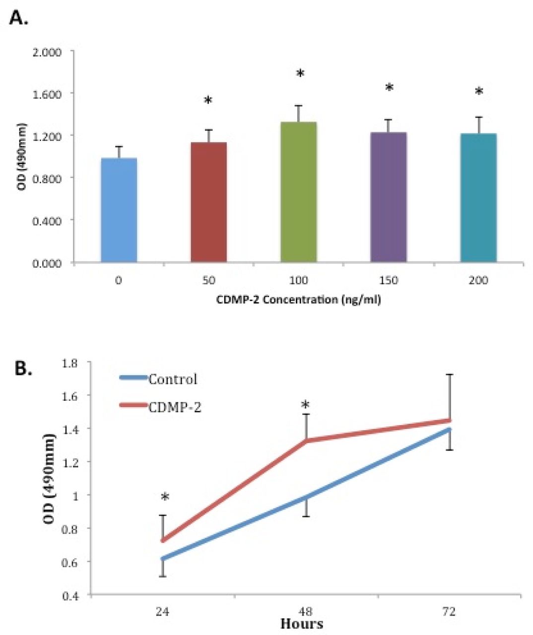
A) Effect of CDMP-2 on cellular proliferation of C28/I2 cells at varying doses for 48 hours. Absorbance was significantly higher (p < 0.05) in all groups treated with CDMP-2 compared to control. Each bar represents combined data from three individual experiments performed in quintuplicate (mean±SD). * indicates significant difference compared with control group (p < 0.05). B) Effect of CDMP-2 (100ng/ml) on cellular proliferation of C28/I2 cells at varying times. At 24 and 48 hours, there was significantly higher absorbance (p< 0.05) in groups treated with CDMP-2. Data is presented as mean ± SD and results are derived from three individual experiments each performed in quintuplicate. * indicates significant difference compared with the control group of the same time point (p < 0.05).
- Fig. 2
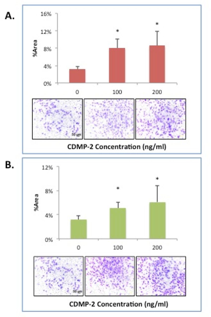
A) Effect of CDMP-2 on C28/CI2 cell migration capacity. The presence of CDMP-2 in the upper chamber of a transwell plate significantly increased cell migration as observed with Boyden Chamber assay with varying doses at 45 hours, compared to cultured alone. (Top panel) Graphical representation of Crystal Violet –stained cells after migration; percentage of area staining was calculated with Image-J software. (Bottom panel) Representative microscopic images of Boyden Chamber filters mounted on slides. Varying concentrations of CDMP-2 were used. All groups with CDMP-2 as a stimulant had significantly higher %area of staining compared to control (p < 0.05). Data is presented as mean ± standard deviation (SD). * indicates significant difference compared with control (p < 0.05). B) Effect of CDMP-2 on C28/CI2 cell migration capacity. The presence of CDMP-2 in the bottom chamber of a transwell plate significantly increased cell migration as observed with Boyden Chamber assay with varying doses at 45 hours, compared to cultured alone. (Tope panel) Graphical representation of Crystal Violet –stained cells after migration; percentage of area staining was calculated with Image-J software. (Bottom panel) Representative microscopic images of Boyden Chamber filters mounted on slides. Varying concentrations of CDMP-2 were used. As an attractant, doses of 200ng/ml and 300ng/ml of CDMP-2 resulted in significantly higher % area of staining compared to control (p < 0.05). Data is presented as mean ± standard deviation (SD). * indicates significant difference compared with control (p < 0.05).
- Fig. 3
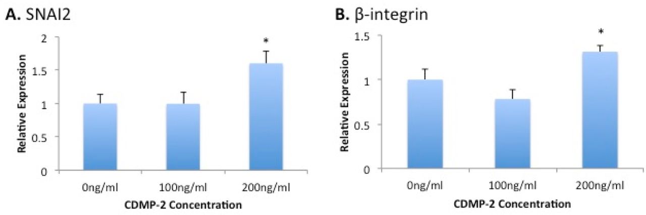
Gene expression analysis of migration markers in C28/I2 cells under CDMP-2 stimulation. Represented as CDMP-2 concentration (x-axis) against normalised relative expression (y-axis). Relative expression was calculated using RT-PCR analysis, with 3 reference genes (GAPDH, B2M, HPRT1). Values were normalised to the control (0ng/ml). Each reaction was repeated 4-times, values represented as mean + SEM. * indicates significant difference compared to control group (p < 0.05). A) SNAI2; B) β-integrin.
- Fig. 4
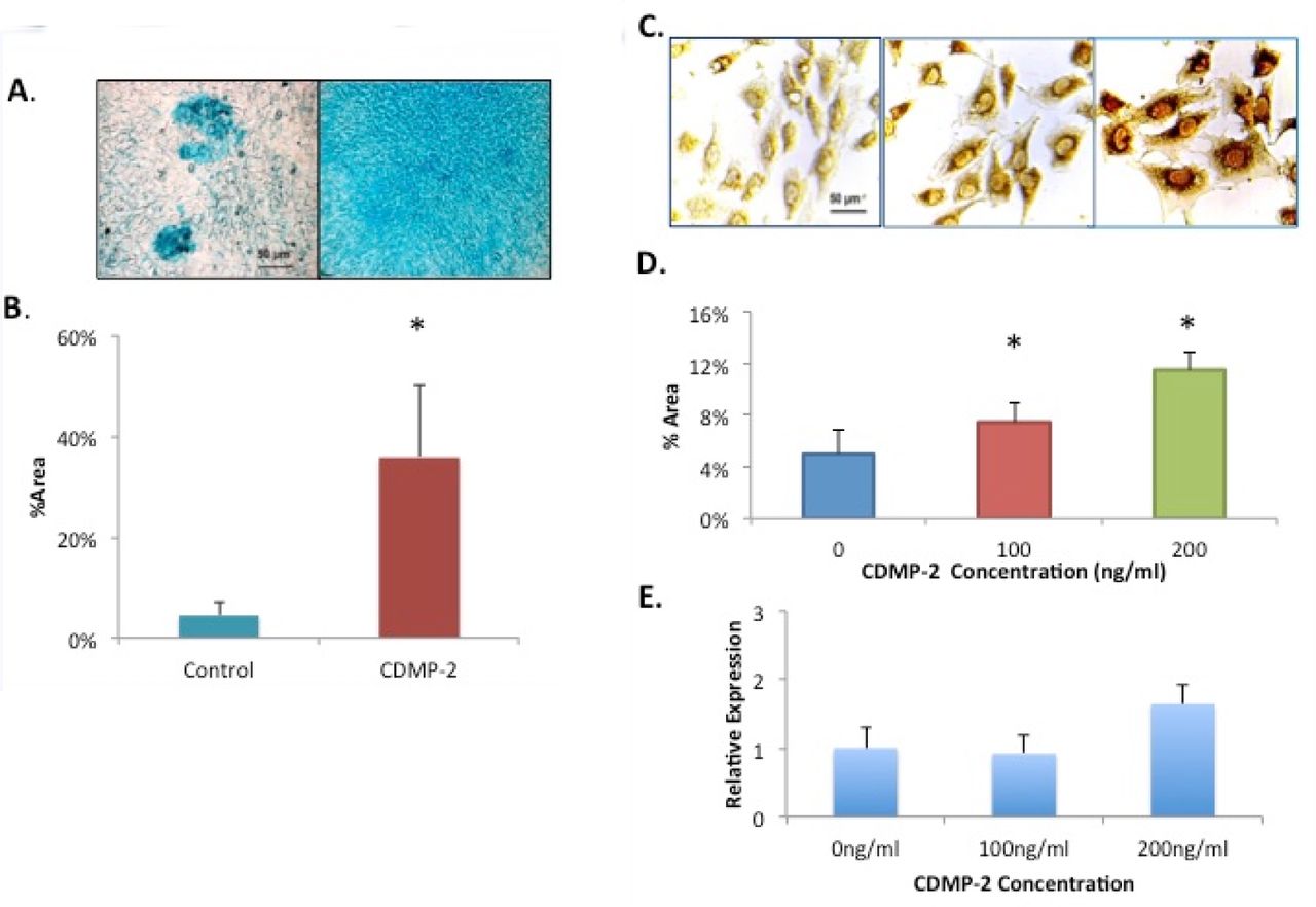
A) Microscopic view of C28/I2 cells with Alcian blue staining. Stained cells with GAG production is observed to be more prominent in the group treated with CDMP-2 (right) than the control group (left). B) Effect of CDMP-2 (100ng/ml) on proteoglycan production as observed with Alcian blue staining assay and ImageJ analysis. Mean %area of staining is 7.8 times more in the CDMP-2 treated group than the control group (p = 0.048). Data is presented as mean ± standard deviation (SD) with each bar representing the combined data from three individual experiments. C) Microscopic view of C28/I2 cells of immuno-staining with primary antibody for aggrecan. Strong brown staining is observed in cells stimulated with CDMP-2 of 100ng/ml (middle panel) and 200ng/ml (right panel) than with control (left panel). The figure is a representative of cultured from three independent samples (original magnification: X400). D) Mean % area of staining is used to quantify aggrecan immuno-staining of C28/I2 cells. Under the influence of CDMP-2 of varying concentrations, cells increased functional production of aggrecans by 50% at 100ng/ml and 130% at 200ng/ml compared to the control (p < 0.05). Data is presented as mean ± standard deviation (SD). E) Gene expression analysis of aggrecan in C28/ I2 cells under CDMP-2 stimulation. Represented as CDMP-2 concentration (x-axis) against normalised relative expression (y-axis). Relative expression was calculated using RT-PCR analysis, with 3 reference genes (GAPDH, B2M and HPRT1). Values were normalised to the control (0ng/ml). Each reaction was repeated 4-times, values represented as mean + SEM. Results were not statistically significant (p < 0.05). * indicates significant difference compared with the control group (p < 0.05).
- Fig. 5
Effect of CDMP-2 (100ng/ml) on total collagen production measured with [3H]-proline incorporation assay. Total collagen synthesis is 3 times more in the CDMP-2 treated group than the control group (p < 0.05). Data is presented as mean ± standard deviation (SD) with each bar representing combined data from three individual experiments.
- Fig. 6
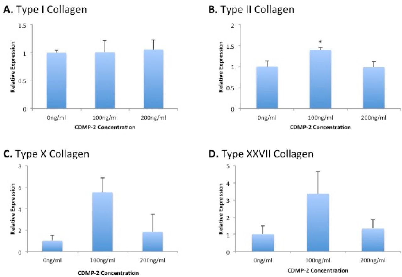
Gene expression analysis of chondrocytic markers and receptors in C28/I2 cells under CDMP-2 stimulation (0ng/ml, 100ng/ml, 200ng/ml) for 48 hours. Represented as CDMP-2 concentration (x-axis) against normalised relative expression (y-axis). Relative expression was calculated using RT-PCR analysis, with 3 reference genes. Values were normalised to the control (0ng/ml). Each reaction was repeated 4-times, values represented as mean + SEM. * indicates significant difference compared with the control group (p < 0.05). A) Type I Collagen; B) Type II Collagen; C) Type X Collagen; D) Type XXVII Collagen.
- Fig. 7
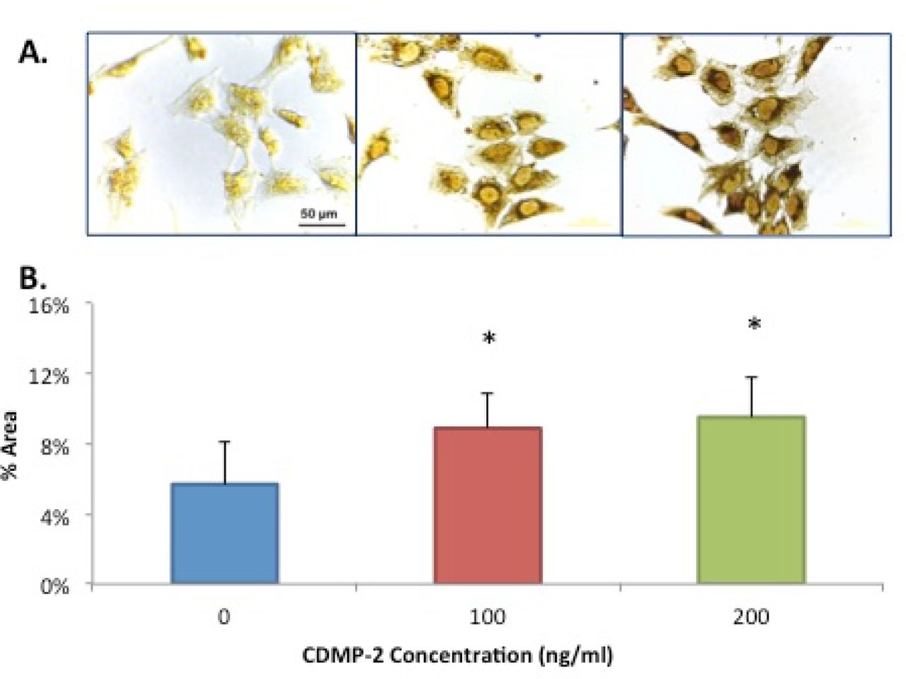
A) Microscopic view (x40 magnification) of C28/I2 cells of immuno-staining for type II collagen. Strong brown staining is observed in cells stimulated with CDMP-2 of 200ng/ml (right panel) and 100ng/ml (middle panel) than with control (left panel). The figure is a representative of culture from three independent samples. B) Protein expression of type II collagen production of C28/I2 cells under CDMP-2 influence, demonstrated using immunocytochemistry and ImageJ analysis. Data presented as mean ± standard deviation (SD). N = 5. * indicates significant difference compared with control (p < 0.05).
- Fig. 8
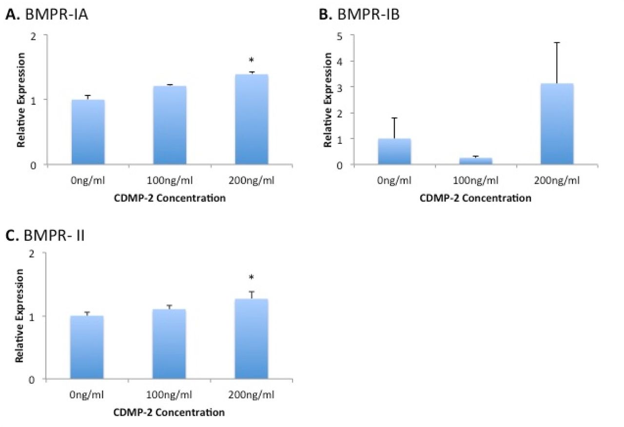
Gene expression analysis of BMP receptors in C28/I2 cells under CDMP-2 stimulation. Represented as CDMP-2 concentration (x-axis) against normalised relative expression (y-axis). Relative expression was calculated using RT-PCR analysis, with 3 reference genes. Values were normalised to the control (0ng/ml). Each reaction was repeated 4-times, values represented as mean + SEM. * indicates significant difference compared to control group (p < 0.05). A) BMPR-IA; B) BMPR-IB; C) BMPR- II.


In this issue


Jump to section
Related Articles
Cited By...
- No citing articles found.