
Main menu

  • Home
  • Content
    • Current Issue
    • Advance Online Publication
    • Archive
  • About Us
    • About ISASS
    • About the Journal
    • Author Instructions
    • Editorial Board
    • Reviewer Guidelines & Publication Criteria
  • More
    • Advertise
    • Subscribe
    • Alerts
    • Feedback
  • Join Us
  • Reprints & Permissions
  • Sponsored Content
  • Other Publications
    • ijss

User menu

  • My alerts

Search

  • Advanced search
International Journal of Spine Surgery
  • My alerts
International Journal of Spine Surgery

Advanced Search

  • Home
  • Content
    • Current Issue
    • Advance Online Publication
    • Archive
  • About Us
    • About ISASS
    • About the Journal
    • Author Instructions
    • Editorial Board
    • Reviewer Guidelines & Publication Criteria
  • More
    • Advertise
    • Subscribe
    • Alerts
    • Feedback
  • Join Us
  • Reprints & Permissions
  • Sponsored Content
  • Follow ijss on Twitter
  • Visit ijss on Facebook
Research ArticleBiomechanics

Assessment of Cervical and Lumbar Kinematics in Simulated Open and Closed Kinetic Chain

Farid Amirouche, Martine Dolan, Sonia Pradhan, Mark Mikhael and Jad Bou Monsef
International Journal of Spine Surgery October 2022, 8382; DOI: https://doi.org/10.14444/8382
Farid Amirouche
1 Department of Orthopaedic Surgery, Northshore University HealthSystem an Affiliate of University of Chicago Pritzker School of Medicine, Evanston, IL, USA
2 Department of Orthopaedic Surgery, University of Illinois College of Medicine at Chicago, Chicago, IL, USA
PʜD
  • Find this author on Google Scholar
  • Find this author on PubMed
  • Search for this author on this site
Martine Dolan
2 Department of Orthopaedic Surgery, University of Illinois College of Medicine at Chicago, Chicago, IL, USA
BS
  • Find this author on Google Scholar
  • Find this author on PubMed
  • Search for this author on this site
Sonia Pradhan
2 Department of Orthopaedic Surgery, University of Illinois College of Medicine at Chicago, Chicago, IL, USA
MPT
  • Find this author on Google Scholar
  • Find this author on PubMed
  • Search for this author on this site
Mark Mikhael
1 Department of Orthopaedic Surgery, Northshore University HealthSystem an Affiliate of University of Chicago Pritzker School of Medicine, Evanston, IL, USA
3 Orthopaedic Spine Surgery, Illinois Bone and Joint Institute, Glenview, IL, USA
MD
  • Find this author on Google Scholar
  • Find this author on PubMed
  • Search for this author on this site
Jad Bou Monsef
4 Department of Orthopaedic Surgery and Rehabilitation Medicine, SUNY Downstate Health Sciences University, Brooklyn, NY, USA
MD
  • Find this author on Google Scholar
  • Find this author on PubMed
  • Search for this author on this site
  • Article
  • Figures & Data
  • Info & Metrics
  • PDF
Loading

Article Figures & Data

Figures

  • Figure 1
    • Download figure
    • Open in new tab
    • Download powerpoint
    Figure 1

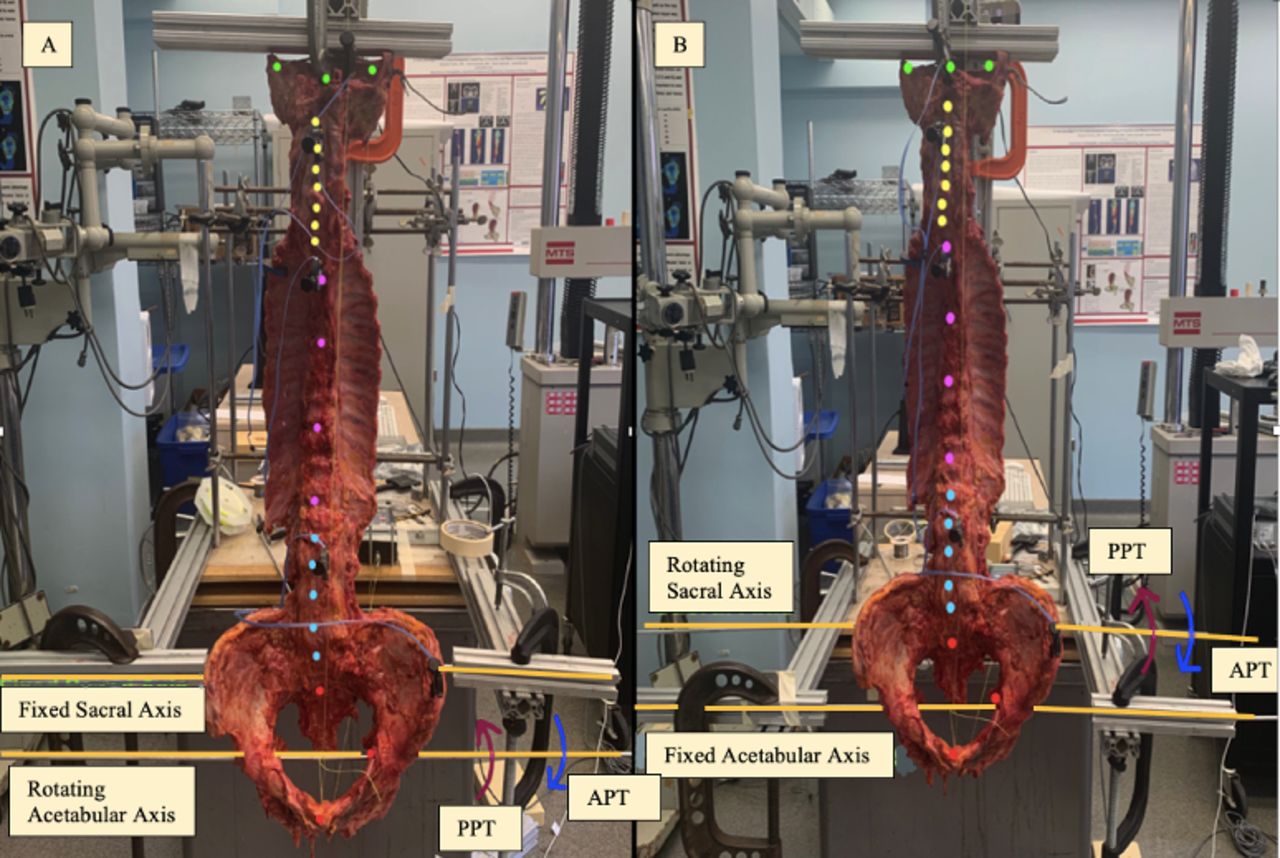
    Experimental setup: (A) coronal/front view with sacral axis fixed and (B) coronal/front view with acetabular axis fixed. The dots represent the digitized points taken for the Optotrak: 3 green dots for the skull, 7 yellow dots for the cervical spine vertebral body (VB), 4 pink dots for the thoracic spine VB, 5 blue dots for the lumbar spine VB, and 5 red dots for the sacrum and pelvis (only 3 seen in the frontal plane). APT, anterior pelvic tilt; PPT, posterior pelvic tilt.

  • Figure 2
    • Download figure
    • Open in new tab
    • Download powerpoint
    Figure 2

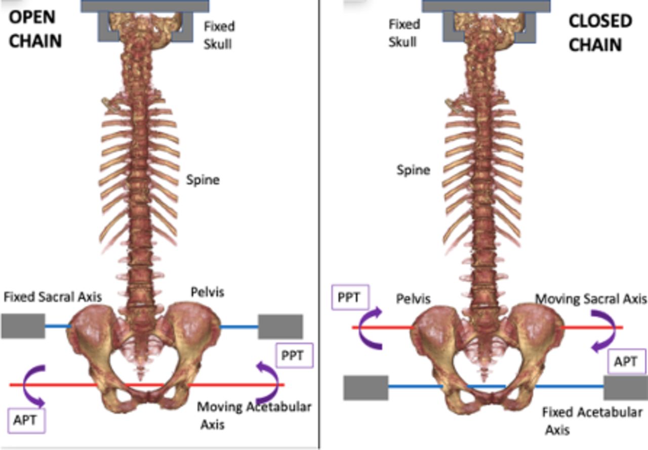
    Simulated open- vs closed-chain movement. In open-chain movements (left image), the sacral axis is fixed, and the pelvis rotates using the acetabular axis. In closed-chain movements (right image), the acetabular axis is fixed, and the pelvis rotates using the sacral axis. APT, anterior pelvic tilt; PPT, posterior pelvic tilt.

  • Figure 3
    • Download figure
    • Open in new tab
    • Download powerpoint
    Figure 3

    Visualization of how pelvic incidence (PI), pelvic tilt (PT), and sacral slope (SS) were determined. The red dots represent the digitized Optotrak points used to calculate these values.

  • Figure 4
    • Download figure
    • Open in new tab
    • Download powerpoint
    Figure 4

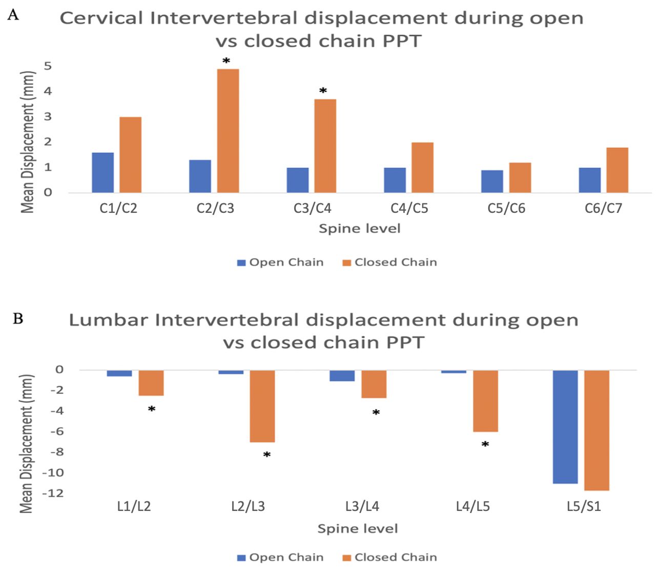
    Cervical (graph A) and lumbar (graph B) intervertebral displacement in an open-chain and closed-chain pelvic tilt. Asterisk indicates significant differences between open- and closed-chain posterior pelvic tilt (PPT) at a specific spinal level. (A) Intervertebral decompression during closed-chain PPT was significantly greater than open-chain PPT at the cervical spine levels C2/C3 (4.85 vs 1.20 mm) and C3/C4 (3.48 vs 1.39 mm) (P < 0.05). (B) Intervertebral compression during closed-chain PPT was significantly greater than open-chain PPT at L1/L2 (−2.54 vs −0.69 mm), L2/L3 (−6.99 vs −0.43 mm), L3/L4 (−2.78 vs −1.17 mm), and L4/L5 (−6.10 vs −0.40 mm) (P < 0.05 for all).

  • Figure 5
    • Download figure
    • Open in new tab
    • Download powerpoint
    Figure 5

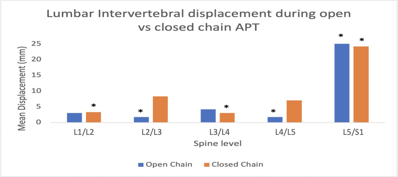
    Lumbar intervertebral displacement in open- and closed-chain anterior pelvic tilt (APT). Since there were no significant differences between open and closed chains, asterisks indicate significant differences between spinal levels within either open or closed chains. In closed-chain APT, significant differences in relative intervertebral decompression displacement were noted between spinal levels L1/L2 (2.87 mm) and L5/S1 (24.48 mm) and between L3/L4 (2.94 mm) and L5/S1 (24.48 mm) (P < 0.05 for both). In open-chain APT, significant differences in relative intervertebral decompression displacement existed between spinal levels L4/L5 (1.53 mm) and L5/S1 (25.14 mm) and between L2/L3 (1.68 mm) and L5/S1 (25.14 mm) (P < 0.05 for both).
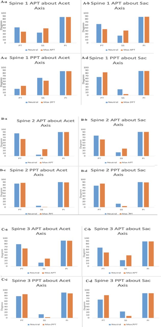
  • Figure 6
    • Download figure
    • Open in new tab
    • Download powerpoint
    Figure 6

    Bar graphs for pelvic tilt (PT), sacral slope (SS), and pelvic incidence (PI) for 3 spines. (A) (a–d) represent graphs for spine 1, (B) (a–d) represent graphs for spine 2, and (C) (a–d) represent spine 3. The graphs are represented for anterior pelvic tilt (APT) about acetabular (Acet) and sacral (Sac) axes and posterior pelvic tilt (PPT) about Acetr and Sac axes. These bar graph values represent the concept PT + SS = PI.

Next
Back to top

In this issue

International Journal of Spine Surgery: 19 (S2)
International Journal of Spine Surgery
Vol. 19, Issue S2
1 Apr 2025
  • Table of Contents
  • Index by author

Print
Download PDF
Article Alerts
Sign In to Email Alerts with your Email Address
Email Article

Thank you for your interest in spreading the word on International Journal of Spine Surgery.

NOTE: We only request your email address so that the person you are recommending the page to knows that you wanted them to see it, and that it is not junk mail. We do not capture any email address.

Enter multiple addresses on separate lines or separate them with commas.
Assessment of Cervical and Lumbar Kinematics in Simulated Open and Closed Kinetic Chain
(Your Name) has sent you a message from International Journal of Spine Surgery
(Your Name) thought you would like to see the International Journal of Spine Surgery web site.
CAPTCHA
This question is for testing whether or not you are a human visitor and to prevent automated spam submissions.
Citation Tools
Assessment of Cervical and Lumbar Kinematics in Simulated Open and Closed Kinetic Chain
Farid Amirouche, Martine Dolan, Sonia Pradhan, Mark Mikhael, Jad Bou Monsef
International Journal of Spine Surgery Oct 2022, 8382; DOI: 10.14444/8382

Citation Manager Formats

  • BibTeX
  • Bookends
  • EasyBib
  • EndNote (tagged)
  • EndNote 8 (xml)
  • Medlars
  • Mendeley
  • Papers
  • RefWorks Tagged
  • Ref Manager
  • RIS
  • Zotero
Share
Assessment of Cervical and Lumbar Kinematics in Simulated Open and Closed Kinetic Chain
Farid Amirouche, Martine Dolan, Sonia Pradhan, Mark Mikhael, Jad Bou Monsef
International Journal of Spine Surgery Oct 2022, 8382; DOI: 10.14444/8382
Twitter logo Facebook logo Mendeley logo
  • Tweet Widget
  • Facebook Like
  • Google Plus One

Jump to section

  • Article
    • Abstract
    • Introduction
    • Methods
    • Results
    • Discussion
    • Conclusion
    • Footnotes
    • References
  • Figures & Data
  • Info & Metrics
  • PDF

Related Articles

  • No related articles found.
  • PubMed
  • Google Scholar

Cited By...

  • No citing articles found.
  • Google Scholar

More in this TOC Section

  • Sequential Anterior Longitudinal Ligament Release With Expandable Spacers for Lordosis Correction in Anterior-to-Psoas Lumbar Interbody Fusion: A Radiographic and Biomechanical Study
  • Comparative Biomechanical Analysis of Anterior Lumbar Interbody Fusion and Bilateral Expandable Transforaminal Lumbar Interbody Fusion Cages: A Finite Element Analysis Study
  • Impact of Different Operative Techniques for Patients With Adolescent Idiopathic Scoliosis on Frontal Curve Correction and Sagittal Balance
Show more Biomechanics

Similar Articles

Keywords

  • pelvic tilt
  • kinematics
  • lumbar spine
  • cervical spine

Content

  • Current Issue
  • Latest Content
  • Archive

More Information

  • About IJSS
  • About ISASS
  • Privacy Policy

More

  • Subscribe
  • Alerts
  • Feedback

Other Services

  • Author Instructions
  • Join ISASS
  • Reprints & Permissions

© 2025 International Journal of Spine Surgery

International Journal of Spine Surgery Online ISSN: 2211-4599

Powered by HighWire