


Article Figures & Data
Figures
- Figure 1
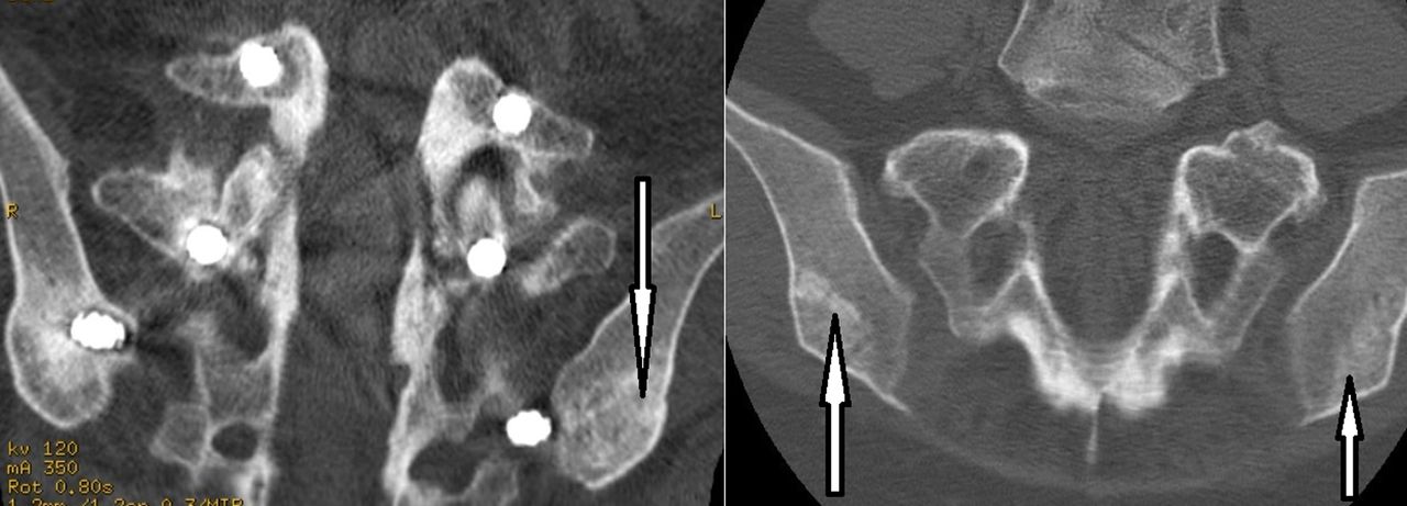
Representative computed tomography image of reconstructed iliac bone graft (RIBG) prior to secondary surgery and reharvest. The left image demonstrates cancellous with interspersed regions of cortical bone appearance of RIBG. This patient had 3 prior surgeries for scoliosis, adjacent segment deformity, and later for adjacent segment stenosis over a 15-year period. Iliac fixation is identifiable. The right image demonstrates both cancellous bone (far right) and cancellous with interspersed regions of cortical bone (left arrow). Arrows indicate RIBG.
- Figure 2
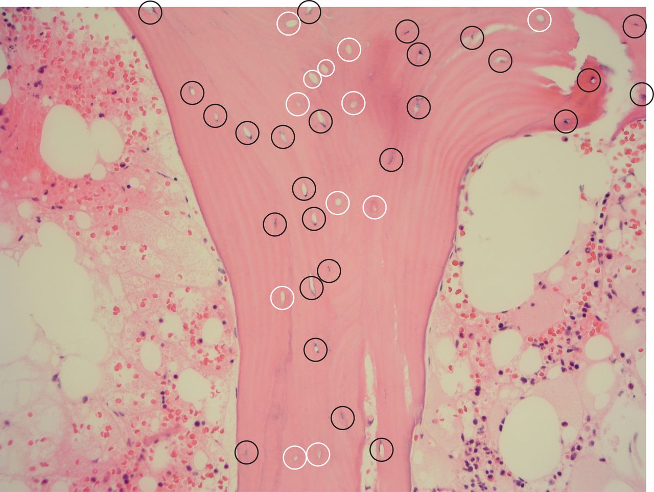
Representative reconstructed iliac bone graft histology (hematoxylin and eosin stain) of filled (dark circles) and unfilled (white circles) lacunae representing viable and necrotic bone, respectively. Lamellae are seen in the viable bone region. Original magnification ×200.
- Figure 3
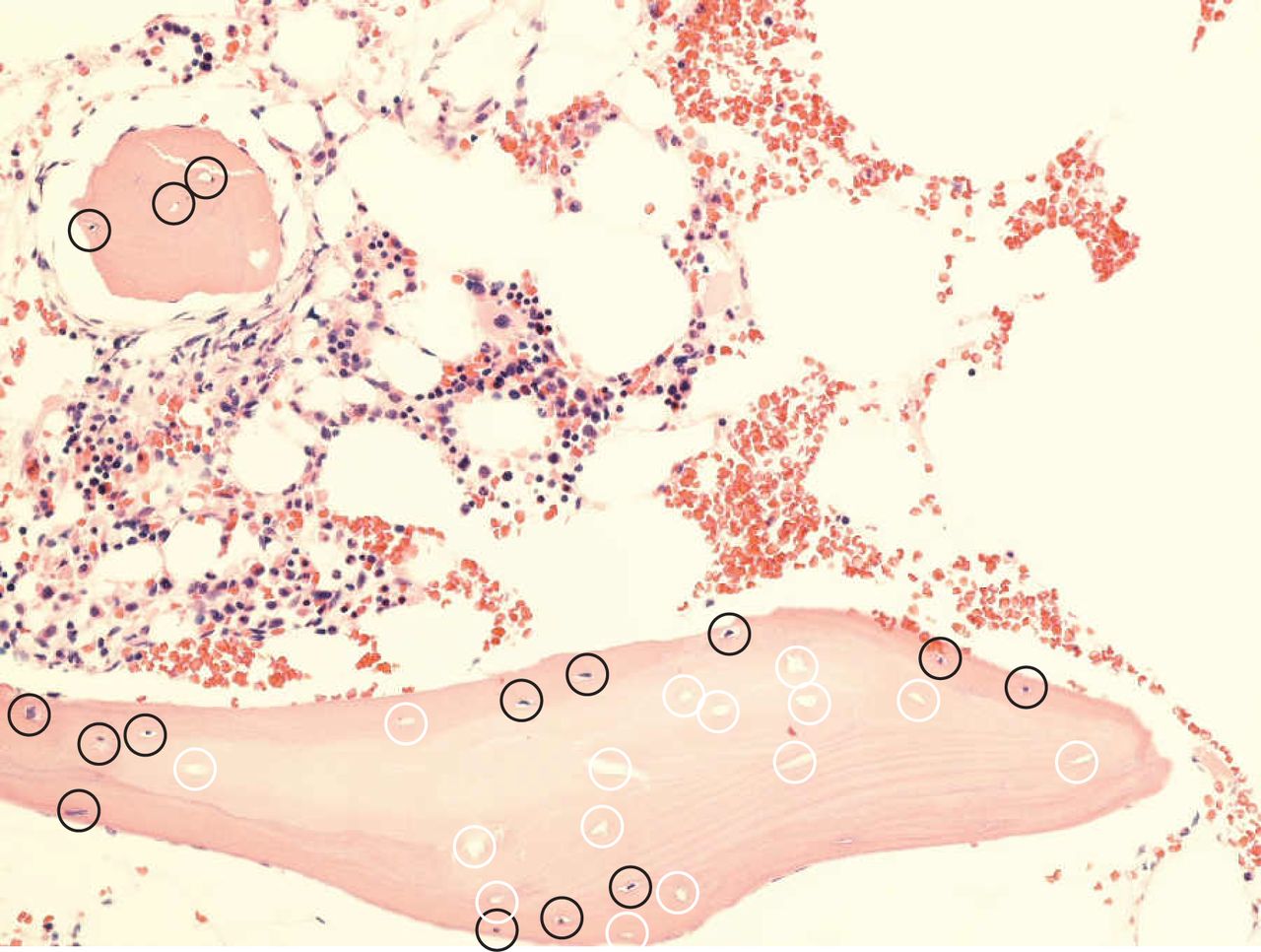
Histological section (hematoxylin and eosin stain) demonstrating region of viable reconstructed iliac bone graft bone with filled lacunae (dark circles) surrounding a region of necrotic bone with unfilled lacunae (white circles). Original magnification ×200.
- Figure 4
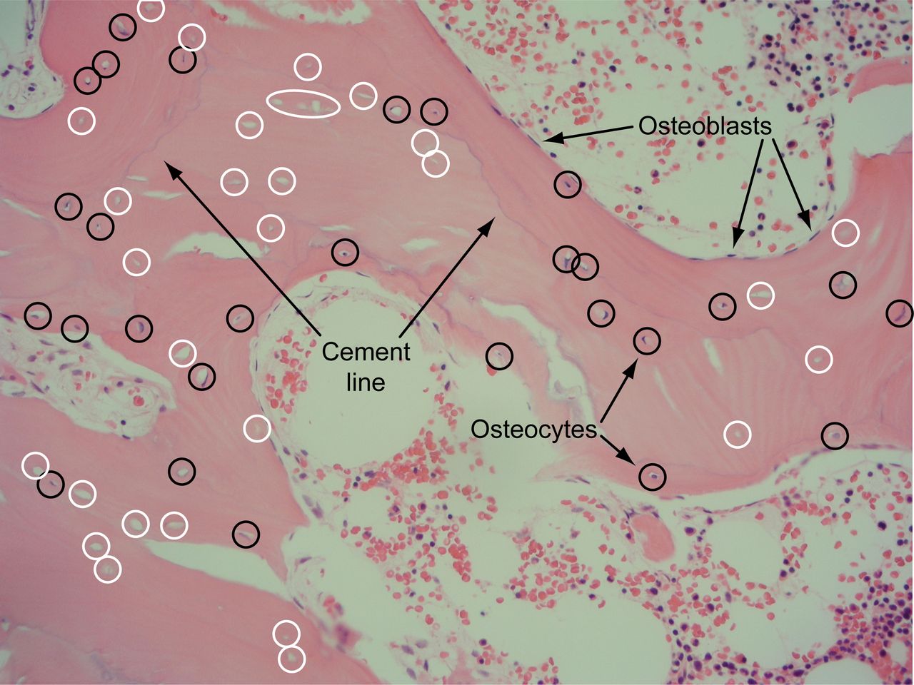
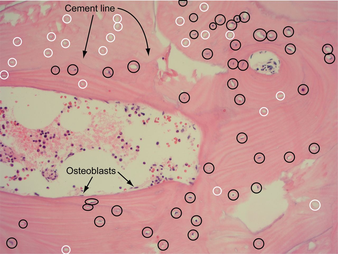
Histological reconstructed iliac bone graft section (hematoxylin and eosin stain) demonstrating “cement line” (arrows) between region of filled lacunae (dark circles) surrounding a region of predominately unfilled lacunae (white circles). Marrow is also represented. Original magnification ×200.
- Figure 5
Histological reconstructed iliac bone graft section (hematoxylin and eosin stain) demonstrating “cement line” (arrows) and region of filled lacunae (dark circles) predominately on lower side of cement line compared with a region of predominately unfilled lacunae (white circles) above the tidemark. This image also demonstrates a concentric arrangement of the lamellae in addition to longitudinally arranged lamellae. Original magnification ×200.
- Figure 6
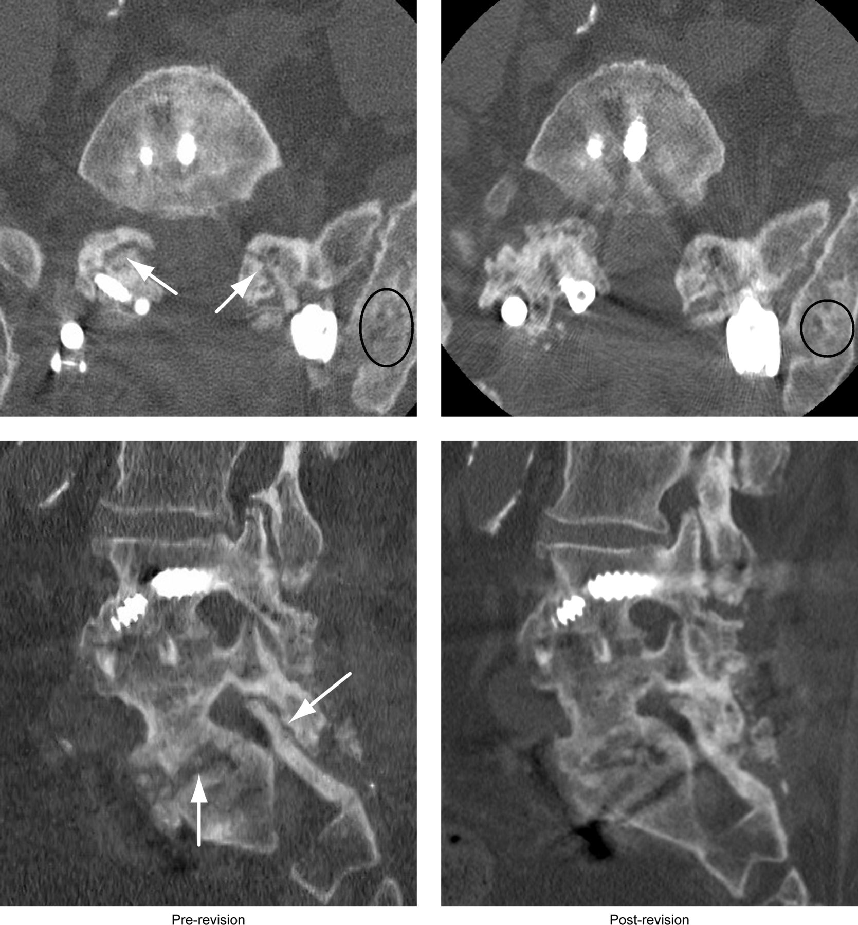
Example of a patient who underwent a second attempt at posterior pseudarthrosis repair of failed L4-S1 anterior-posterior fusion using posterior hybrid facet screws on right and pedicle screws on left. Computed tomography image axial and sagittal reconstruction prior to second revision of pseudarthrosis (left), arrows indicate bilateral facet joint and interbody nonunion. Axial and sagittal reconstruction (right) 1 year after successful pseudarthrosis repair using reconstructed iliac bone graft (RIBG) demonstrating solid arthrodesis of facet joint and interbody region. Ovals indicate revised RIBG.


Tables
Type of Secondary PSF Bonegraft Type n Age, y, Mean ± SD No. of Levels Fused, Median (Range) Smokers
No. (%)Supplement Treatment Solid PSF
No. (%), Mean mo to CTcBMP No. (%) Internal Bone Growth Stimulator No. (%) Pseudo repair RIBGa 7 53.7 ± 12.8 1 (1–6) 5 (71%) 6 (86%) 4 (57%) 7 (100%), 8.4 mo IBGb 17 49.7 ± 17.0 1 (1–4) 7 (41%) 0 (0%) 5 (29%) 12 (71%), 11.6 mo BMP 22 56.6 ± 17.1 1 (1–3) 10 (45%) 22 (100%) 10 (45%) 18 (82%), 11.3 mo IBG + BMP 8 55.4 ± 15.6 1 (1–3) 4 (50%) 8 (100%) 5 (63%) 7 (88%), 12.3 mo Local autograft 6 58.3 ± 15.9 1 (1) 3 (50%) 0 (0%) 2 (33%) 3 (50%), 8.6 mo Extension of PSF RIBGa 10 56.5 ± 3.6 2 (1–6) 6 (60%) 4 (40%) 2 (20%) 9 (90%), 15.2 mo IBGb 19 56.5 ± 9.1 1 (1–3) 8 (40%) 0 (0%) 5 (29%) 15 (79%), 24.2 mo BMP 56 60.5 ± 15.0 1 (1–7) 19 (34%) 56 (100%) 12 (21%) 44 (79%), 12.5 mo IBG + BMP 1 52.6 1 (1) 1 (100%) 1 (100%) 1 (100%) 1 (100%), 29.2 mo Local autograft 4 53.8 ± 12.2 1 (1) 1 (25%) 0 (0%) 1 (25%) 4 (100%), 8.9 mo Bone Viability Parameters Reconstructed Iliac Bone Graft Group (n = 17) Control Group (n = 17) Age, y, mean ± SD 55.3 ± 8.6 61.8 ± 21.9 Sex, % women 77 75 Lacunae with osteocytes, %, mean ± SD 82.8 ± 13.7 87.8 ± 7.5 Trabeculae with ≥1 viable osteocyte, n 90%–100% 9 17 80%–90% 6 0 60%–80% 2 0 Marrow cellularity, %, mean ± SD 31.1% ± 19.9% 45.3% ± 18.8% Marrow cellularity, %, range 5%–60% 20%–80% Hypercellular, n 2 2 Normocellular, n 5 15 Hypocellular, n 10 0 Clinical Outcome Measures RIBG IBG Control P Value RIBG vs IBG Pseudarthrosis Repair (n = 7) PSF Extension (n = 10) Total(n = 17) P Value Preoperative vs Postoperative Pseudarthrosis Repair (n = 17) PSF Extension (n = 19) Total(n = 36) P Value Preoperative vs Postoperative Back pain VAS Preoperative 7.3 ± 1.5 7.8 ± 1.5 7.6 ± 1.5 7.8 ± 1.2 7.6 ± 1.2 7.7 ± 1.2 1-y Postoperative 4.0 ± 3.2 3.7 ± 2.0 3.7 ± 2.6 <0.001 6.1 ± 2.3 3.8 ± 2.5 4.8 ± 2.6 <0.001 >0.1 2-y Postoperative 4.4 ± 1.8 3.5 ± 1.5 3.9 ± 1.7 <0.001 5.6 ± 2.8 4.5 ± 2.8 5.3 ± 2.8 <0.001 >0.1 Leg pain VAS Preoperative 6.6 ± 1.9 6.5 ± 3.0 6.5 ± 2.5 6.6 ± 2.5 6.2 ± 2.5 6.4 ± 2.5 1-y Postoperative 3.9 ± 3.4 2.7 ± 2.5 3.2 ± 2.9 <0.001 4.4 ± 3.2 3.3 ± 2.5 3.8 ± 2.8 <0.001 >0.2 2-y Postoperative 4.5 ± 2.2 2.5 ± 2.2 3.2 ± 2.4 <0.001 4.7 ± 3.5 3.7 ± 3.0 4.2 ± 3.2 <0.001 >0.2 Oswestry Disability Index Preoperative 62.3 ± 9.8 63.8 ± 18.2 63.1 ± 14.7 64.1 ± 15.3 57.5 ± 15.5 60.4 ± 15.5 1-y Postoperative 54.3 ± 15.2 43.2 ± 12.0 48.1 ± 14.5 0.004 49.6 ± 18.6 40.9 ± 19.1 44.7 ± 19.1 <0.004 >0.4 2-y Postoperative 50.3 ± 18.5 42.3 ± 13.1 46.0 ± 15.8 <0.001 51.3 ± 19.8 44.3 ± 17.2 47.5 ± 18.5 <0.004 >0.4 Abbreviations: IBG, iliac bone graft; PSF, posterior spinal fusion; RIBG, reconstructed iliac bone graft; VAS, visual analog scale.
Note: Data presented as mean ± SD. P values describe change in values.
In this issue


Jump to section
Related Articles
Cited By...
- No citing articles found.