


Article Figures & Data
Figures
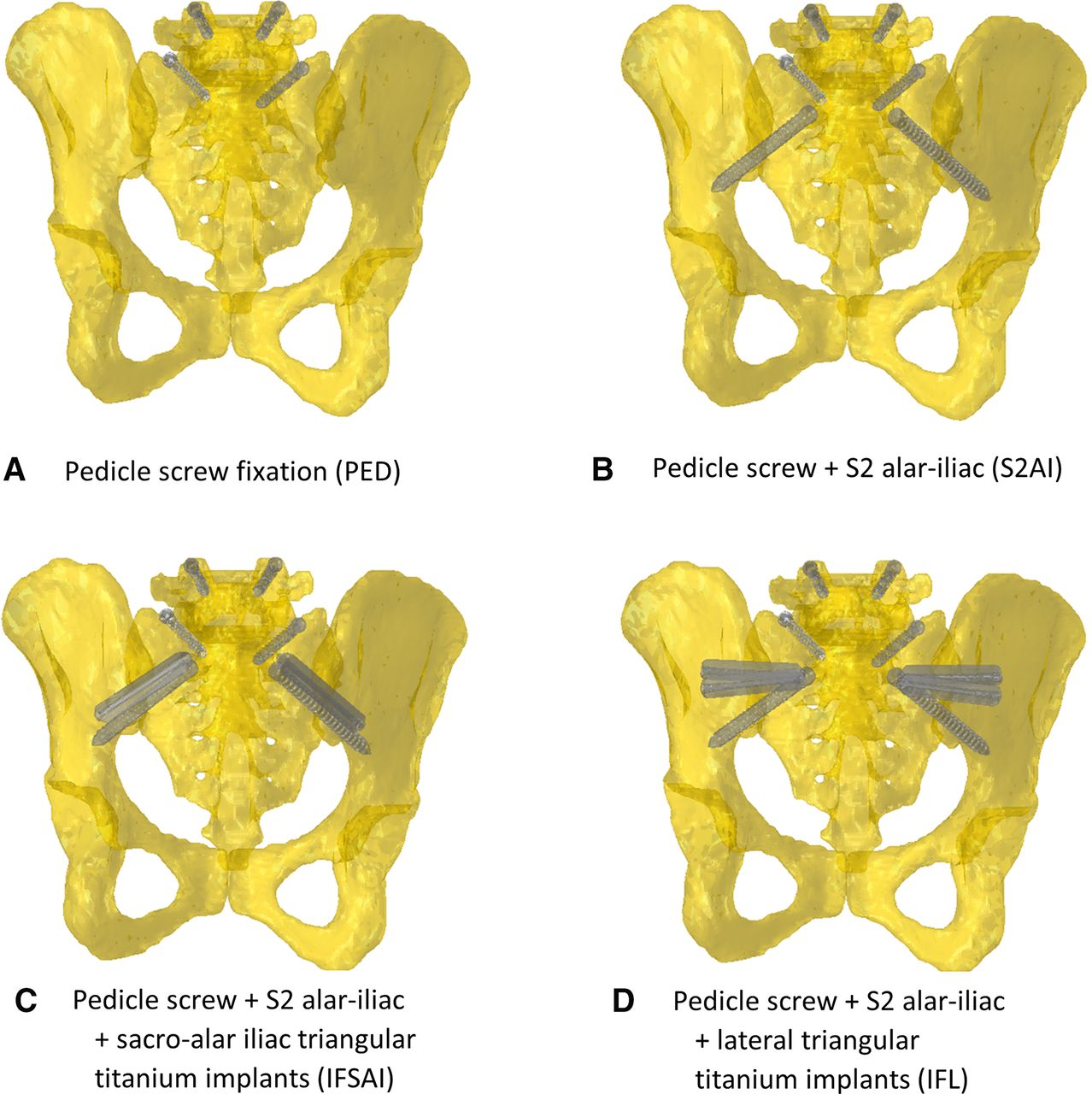
- Figure 1
The 4 configurations of the instrumentation in the sacropelvic region: (A) pedicle screw fixation (PED); (B) posterior fixation and S2 alar-iliac fixation (S2AI); (C) same as (B) bilaterally supplemented by a triangular titanium implant placed in a sacro-alar-iliac trajectory (IFSAI); (D) same as (B) supplemented by 2 bilateral laterally placed triangular titanium implants (IFL). Rods not shown.
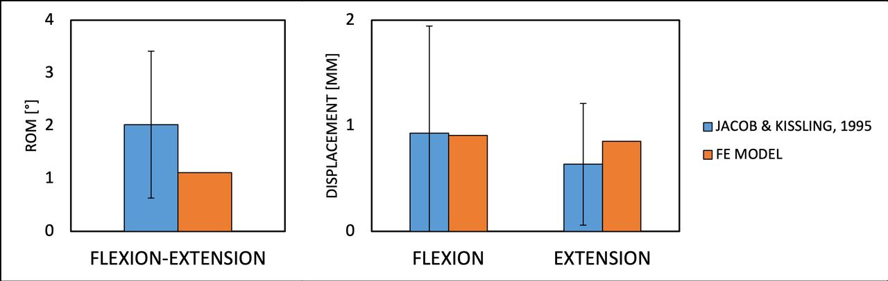
- Figure 2
Predicted ranges of motion (ROM) of the sacroiliac joint of the intact model in flexion-extension (left) and displacements of the sacrum in flexion and extension (right), as compared with data from in vitro experiments.7
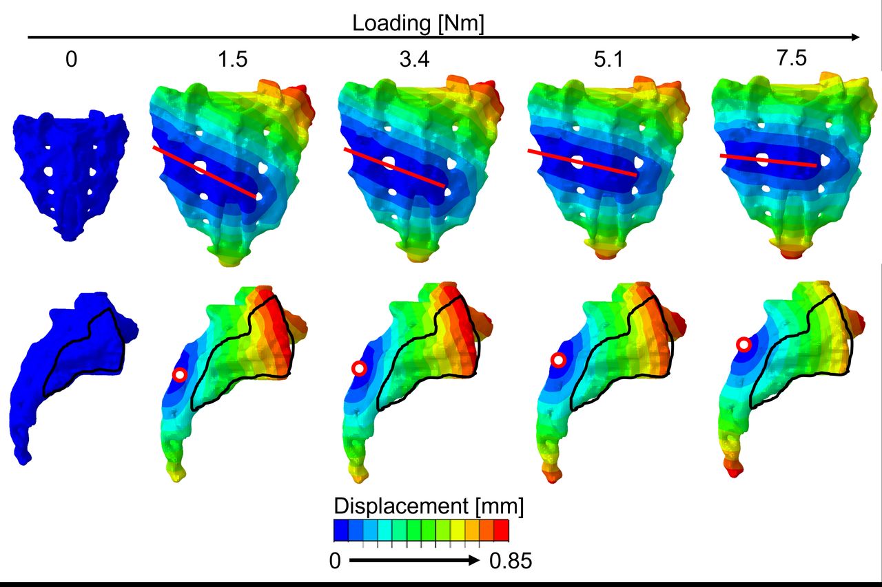
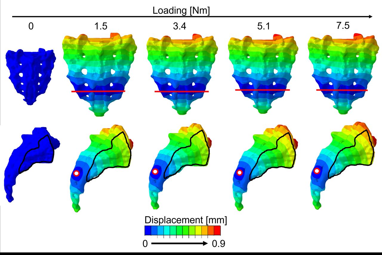
- Figure 3
Qualitative axis of rotation (line drawn) in the frontal plane of the sacrum and center of rotation (point) in the sagittal plane of the sacrum for 5 loading values (0, 1.5, 3.4, 5.1, 7.5 Nm) in extension for the intact model.
- Figure 4
Qualitative axis of rotation (line drawn) in the frontal plane of the sacrum and center of rotation (point) in the sagittal plane of the sacrum for 5 loading values (0, 1.5, 3.4, 5.1, 7.5 Nm) in flexion for the intact model.
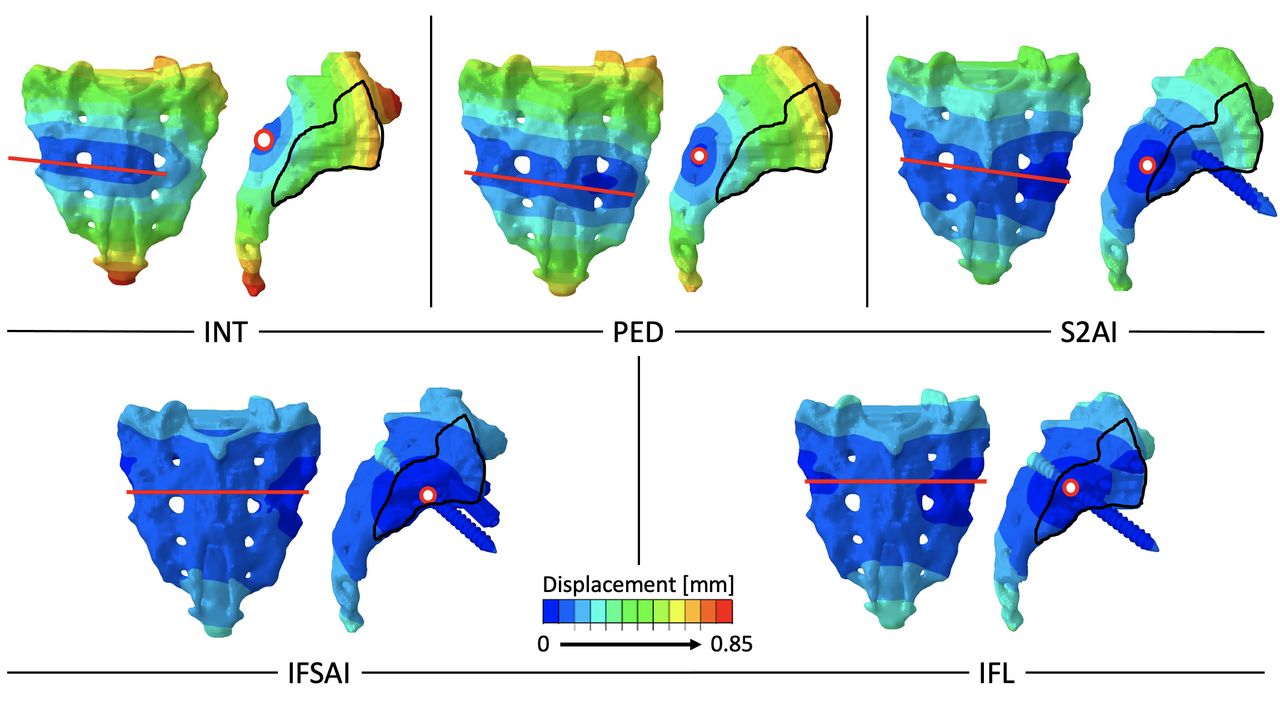
- Figure 5
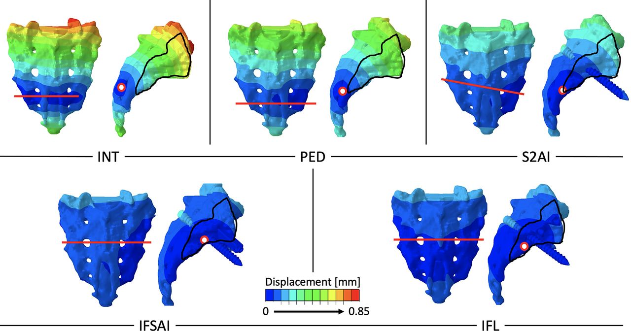
Qualitative axis of rotation (line drawn) in the frontal plane of the sacrum and center of rotation (point) in the sagittal plane of the sacrum for the 5 configurations: intact (INT), pedicle screws (PED), S2 alar-iliac screws (S2AI), bilateral S2AI and triangular implants inserted bilaterally in a sacral alar-iliac trajectory (IFSAI), bilateral S2AI and 2 bilateral triangular implants inserted in a lateral trajectory(IFL) in extension for the last step (7.5 Nm).
- Figure 6
Qualitative axis of rotation (line drawn) in the frontal plane of the sacrum and center of rotation (point) in the sagittal plane of the sacrum for the 5 configurations: intact (INT), pedicle screws (PED), S2 alar-iliac screws (S2AI),bilateral S2AI and triangular implants inserted bilaterally in a sacral alar-iliac trajectory (IFSAI), bilateral S2AI and 2 bilateral triangular implants inserted in a lateral trajectory (IFL) in flexion for the last step (7.5 Nm).


Tables
Ligament Stiffness, N/mm No. of Elements References Anterior longitudinal 700 3/Vertebral body 28,29 Anterior superior iliac 700 10 (×2) 28,29 Posterior short sacroiliac 400 10 (×2) 28,29 Posterior long sacroiliac 1000 4 (×2) 28,29 Pubic 500 10 30 Iliolumbar 1000 4 (×2) 30 Interosseous 2800 4 (×2) 28,29 - Table 2
Finite element modeling of the ligaments of the thoracolumbar spine (from calibration against Cook et al31).
Ligament Stiffness, N/mm No. of Elements Anterior longitudinal 3–30a 3 Posterior longitudinal 3–30a 3 Flaval 3–90a 3 Capsular 500–2000a 10 Supraspinous 1400a 4 Interspinous 3–21a 3 ↵a Depending on the local gray value in the computed tomography image.
- Table 3
Ranges of motion (in °) of the intact model, compared with data from experimental studies available in the literature and obtained under the same loading conditions. SDs of the literature data are reported in parentheses.
Level Flexion-Extension Lateral Bending Axial Rotation Model Literaturea Model Literaturea Model Literaturea L1-L2 7.3 8.5 (2.5) 7.1 8 (2.5) 6.5 3 (1.5) L2-L3 7.1 9.0 (2.0) 7.3 10.5 (3.0) 7.0 4.5 (2.5) L3-L4 8.7 10.0 (3.0) 9.6 11 (3.0) 5.1 5.5 (3.0) L4-L5 12.1 12.5 (3.5) 10.0 11 (2.5) 3.8 6 (3.0) L5-S1 13.8 14.0 (4.0) 5.8 8.5 (2.5) 3.2 4 (2.0) SIJ 1.2 1.3 (0.7) 0.7 0.9 (0.4) 0.8 1.2 (0.6) Abbreviation: SIJ, sacroiliac joint.
↵a Source: Cook et al31 for T11-S1 (91 specimens from female donors); Soriano-Baron et al32 for the SIJ (7 donors).
In this issue


Jump to section
Related Articles
Cited By...
- No citing articles found.