


Article Figures & Data
Figures
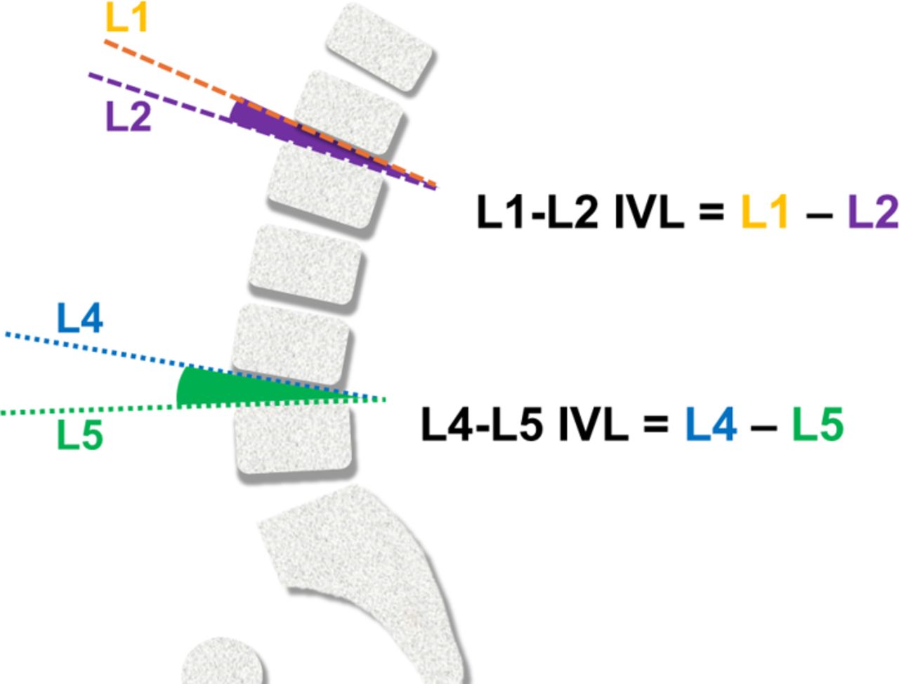
- Figure 1
Intervertebral lordosis (IVL) measurements.
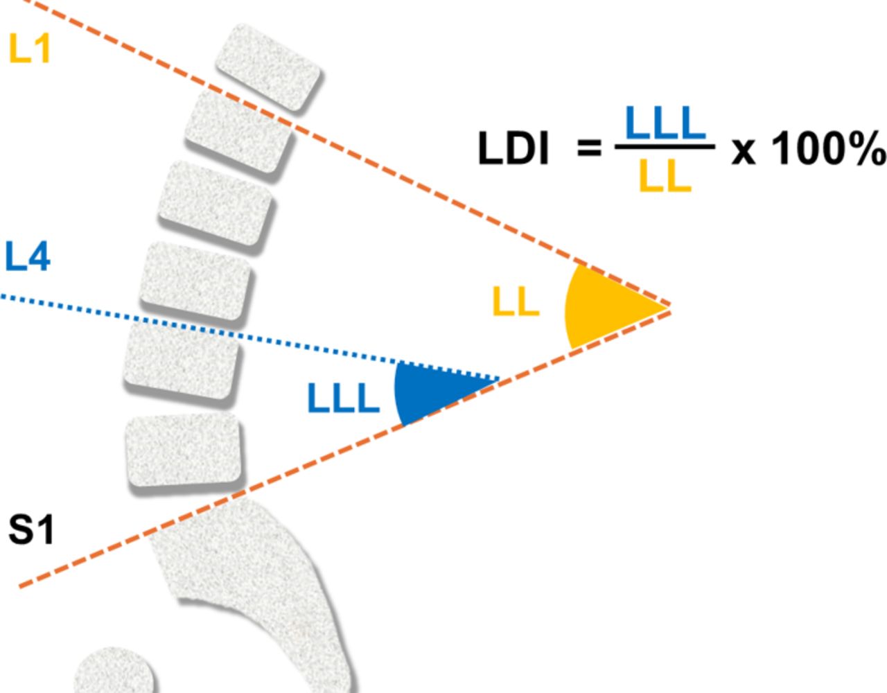
- Figure 2
Definition of lordosis distribution index (LDI). Abbreviations: LL, lumbar lordosis; LLL, lower lumbar lordosis
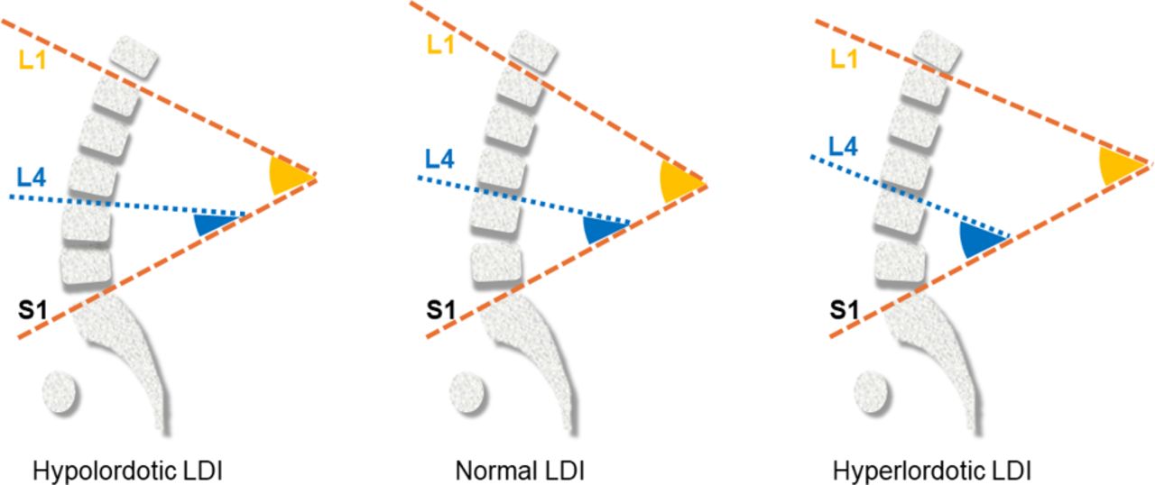
- Figure 3
Lordosis categories. Abbreviations: LDI, lordosis distribution index.
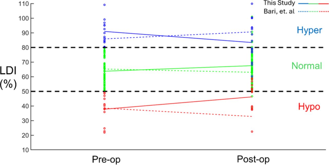
- Figure 4
Lordosis distribution index (LDI) pre- and postoperative comparisons for each patient color coded into different preoperative distribution groups.
- Figure 5
(A) Intervertebral lordosis (IVL) distribution in the lumbar region. (B) The ratio of IVL to lumbar lordosis (LL) distribution in the lumbar region.
- Figure 6
Lordosis distribution index (LDI) comparisons for different distribution groups. The pre- to postoperative changes in LDI for each group as reported by Bari et al14 are also shown.


Tables
Parameter Value Number of patients 111 Sex, women, n (%) 63 (56.8) Age, y, mean (SD) 62 ± 13.1 Follow-up time, d, median (range) 64 ± 38.5 PICs implanted, n 135 Approach, n (% of PICs) ALIF 85 (63) LLIF 7 (5) TLIF 43 (32) Number of levels fused, n (% of PICs) 1-level 72 (53) 2-levels 63 (47) Level of fusion, n (% of PICs) L4–L5 27 (20) L5–S1 45 (33) L4–S1 63 (47) Abbreviations: ALIF, anterior lumbar interbody fusion; LLIF, lateral lumbar interbody fusion; PICs, personalized interbody cages; TLIF, transforaminal lumbar interbody fusion.
Parameter Combined
(N = 111)Normal
(LDI 50–80%)
(n = 81)Hypolordotic
(LDI <50%)
(n = 14)Hyperlordotic
(LDI >80%)
(n = 16)PI (°) Preoperative 57.29 (12.56) 56.80 (12.25) 63.81 (12.48) 54.44 (13.20) Postoperative 58.45 (12.25) 58.24 (11.73) 64.71 (11.80) 54.07 (13.73) Change 0.89 1.26 0.18 −0.37 P 0.007 0.002 0.839 0.622 LL (°) Preoperative 51.46 (11.73) 53.49 (11.82) 50.29 (8.04) 42.23 (9.63) Postoperative 54.73 (12.69) 56.18 (13.20) 58.54 (5.24) 44.06 (9.00) Change 3.27 2.69 8.26 1.83 P <0.001 0.001 0.011 0.230 LLL (°) Preoperative 32.7 (9.37) 33.95 (8.10) 19.48 (6.68) 37.99 (7.29) Postoperative 35.96 (9.76) 37.41 (9.66) 27.11 (6.81) 36.37 (8.70) Change 3.26 3.47 7.63 −1.62 P <0.001 <0.001 0.009 0.404 Abbreviations: LDI, lordosis distribution index; LL, lumbar lordosis; LLL, lower lumbar lordosis; PI, pelvic incidence.
Note: Pre- and postoperative values are presented as mean (SD). Significant P values (<0.05) are shown in bold.
Parameter Combined
(N = 111)Normal
(LDI 50%–80%)
(n = 81)Hypolordotic
(LDI <50%)
(n = 14)Hyperlordotic
(LDI >80%)
(n = 16)PI−LL (°) Preoperative 5.98 (11.13) 3.58 (10.35) 13.04 (11.99) 12.21 (9.89) Postoperative 3.73 (10.92) 2.06 (9.71) 6.16 (13.12) 10.01 (12.64) Change −2.33 −1.49 −7.64 −2.20 P 0.003 0.071 0.031 0.241 LDI (%) Preoperative 64.37 (16.10) 63.72 (8.17) 37.77 (9.07) 90.91 (7.77) Postoperative 67.11 (16.29) 67.53 (13.34) 46.18 (10.43) 83.33 (14.62) Change 2.75 3.81 8.41 −7.58 P 0.045 0.010 0.030 0.092 Abbreviations: LDI, lordosis distribution index; PI−LL, difference between pelvic incidence and lumbar lordosis.
Note: Pre- and postoperative values are presented as mean (SD). Significant P values (<0.05) are shown in bold.
LDI Normal LDI Group Hypolordotic Group Hyperlordotic Group Preoperative, n (%) LDI >80% 0 0 16 (14%) LDI 50%–80% 81 (73%) 0 0 LDI <50% 0 14 (13%) 0 Postoperative, n (%) LDI >80% 8 (7%) 0 8 (7%) LDI 50%–80% 68 (61%) 6 (5%) 8 (7%) LDI <50% 5 (5%) 8 (7%) 0 Abbreviation: LDI, lordosis distribution index.
Parameter Combined
(N = 111)Normal
(LDI 50%–80%)
(n = 81)Hypolordotic
(LDI <50%)
(n = 14)Hyperlordotic
(LDI >80%)
(n = 16)L1−L2 IVL (°) Preoperative 6.75 (3.67) 7.02 (3.66) 7.55 (3.68) 4.34 (2.91) Postoperative 6.49 (3.38) 6.43 (3.34) 8.50 (3.60) 4.85 (2.51) Change −0.18 −0.49 1.28 0.19 P 0.603 0.246 0.106 0.817 L2−L3 IVL (°) Preoperative 8.62 (4.51) 8.44 (4.08) 11.81 (6.09) 6.46 (3.79) Postoperative 8.34 (3.94) 8.19 (3.86) 11.46 (3.63) 6.06 (2.83) Change −0.30 −0.20 −0.64 −0.53 P 0.452 0.574 0.784 0.457 L3−L4 IVL (°) Preoperative 9.98 (4.42) 10.01 (4.10) 12.32 (5.71) 7.42 (3.74) Postoperative 9.88 (4.42) 10.09 (4.16) 11.92 (4.68) 6.66 (4.16) Change −0.05 0.08 −0.27 −0.61 P 0.878 0.832 0.820 0.472 L4−L5 IVL (°) Preoperative 9.57 (4.75) 9.98 (4.92) 7.95 (3.88) 8.74 (4.33) Postoperative 10.54 (4.37) 11.16 (4.55) 8.75 (3.64) 8.82 (2.99) Change 0.87 0.93 0.89 0.50 P 0.107 0.145 0.616 0.673 L5−S1 IVL (°) Preoperative 10.54 (6.35) 11.07 (6.67) 6.77 (4.23) 11.22 (5.13) Postoperative 14.52 (4.82) 15.12 (4.44) 11.79 (5.43) 13.95 (5.57) Change 4.27 4.24 5.62 3.12 P <0.001 <0.001 0.021 0.098 Abbreviations: IVL, intervertebral lordosis; LDI, lordosis distribution index.
Note: Pre- and postoperative values are presented as mean (SD). Significant P values (<0.05) are shown in bold. Shaded areas represent levels treated with PIC, L4−L5, L5−S1, or L4−S1.
In this issue


Jump to section
Related Articles
Cited By...
- No citing articles found.