
Main menu

  • Home
  • Content
    • Current Issue
    • Advance Online Publication
    • Archive
  • About Us
    • About ISASS
    • About the Journal
    • Author Instructions
    • Editorial Board
    • Reviewer Guidelines & Publication Criteria
  • More
    • Advertise
    • Subscribe
    • Alerts
    • Feedback
  • Join Us
  • Reprints & Permissions
  • Sponsored Content
  • Other Publications
    • ijss

User menu

  • My alerts

Search

  • Advanced search
International Journal of Spine Surgery
  • My alerts
International Journal of Spine Surgery

Advanced Search

  • Home
  • Content
    • Current Issue
    • Advance Online Publication
    • Archive
  • About Us
    • About ISASS
    • About the Journal
    • Author Instructions
    • Editorial Board
    • Reviewer Guidelines & Publication Criteria
  • More
    • Advertise
    • Subscribe
    • Alerts
    • Feedback
  • Join Us
  • Reprints & Permissions
  • Sponsored Content
  • Follow ijss on Twitter
  • Visit ijss on Facebook
Research ArticleEndoscopic Minimally Invasive Surgery

Augmenting Endoscopic Transforaminal Spinal Decompression Surgery (Full Endoscopic Spine Surgery) Using Stimulated Electromyography Neuromonitoring Dilators

Dickson Hong Him Chau, Dhivakaran Gengatharan and Walter-Soon-Yaw Wong
International Journal of Spine Surgery December 2024, 8692; DOI: https://doi.org/10.14444/8692
Dickson Hong Him Chau
1 Department of Orthopedic Surgery, Sengkang General Hospital, Sengkang, Singapore
2 Department of Orthopaedics, SingHealth Duke-NUS Musculoskeletal Sciences, Academic Clinical Programme, Boulevard, Singapore
MBBS, MRCS, MMᴇᴅ (Oʀᴛʜᴏ), FRCSEᴅ (Oʀᴛʜ)
  • Find this author on Google Scholar
  • Find this author on PubMed
  • Search for this author on this site
Dhivakaran Gengatharan
1 Department of Orthopedic Surgery, Sengkang General Hospital, Sengkang, Singapore
MD
  • Find this author on Google Scholar
  • Find this author on PubMed
  • Search for this author on this site
Walter-Soon-Yaw Wong
1 Department of Orthopedic Surgery, Sengkang General Hospital, Sengkang, Singapore
MBCʜB, MRCS
  • Find this author on Google Scholar
  • Find this author on PubMed
  • Search for this author on this site
  • For correspondence: walterwongsy@gmail.com
  • Article
  • Figures & Data
  • Info & Metrics
  • PDF
Loading

Article Figures & Data

Figures

  • Tables
  • Figure 1
    • Download figure
    • Open in new tab
    • Download powerpoint
    Figure 1

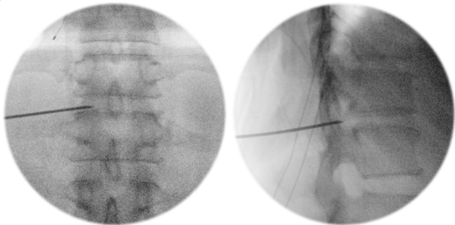
    Guide-needle insertion through the neural foramen onto the T11–T12 disc, confirmed with fluoroscopy.

  • Figure 2
    • Download figure
    • Open in new tab
    • Download powerpoint
    Figure 2

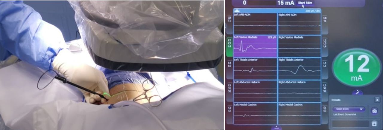
    A 6-mm neuromonitoring dilator, use adapted from the NuVasive NVM5 nerve monitoring system.

  • Figure 3
    • Download figure
    • Open in new tab
    • Download powerpoint
    Figure 3

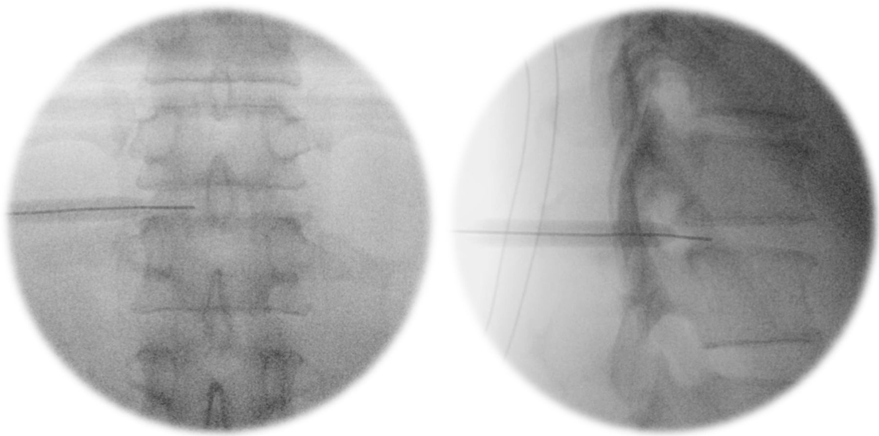
    Passing the neuromonitoring dilator over the guidewire into the foramen.

  • Figure 4
    • Download figure
    • Open in new tab
    • Download powerpoint
    Figure 4

    Rotating the electrode tip cranially and medially to obtain readings corresponding to neural tissue proximity.

  • Figure 5
    • Download figure
    • Open in new tab
    • Download powerpoint
    Figure 5

    Insertion of transforaminal endoscopic instruments via the “safe tract.”

  • Figure 6
    • Download figure
    • Open in new tab
    • Download powerpoint
    Figure 6

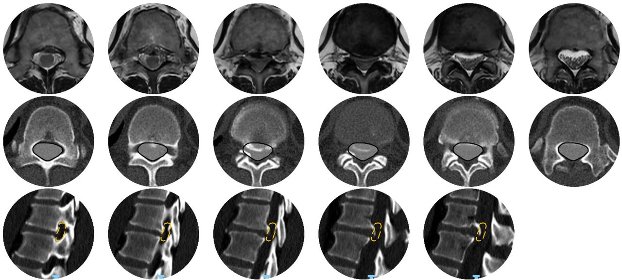
    Images showing a T11/T12 partially calcified disc causing spinal canal stenosis. Top row: Magnetic resonance imaging axial cuts. Middle row: Corresponding computed tomography (CT) axial cuts. Bottom row: CT sagittal cuts (left).
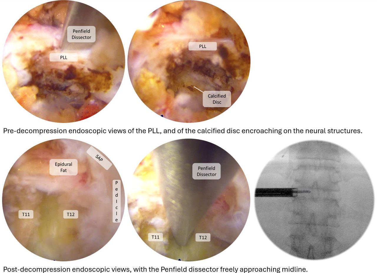
  • Figure 7
    • Download figure
    • Open in new tab
    • Download powerpoint
    Figure 7

    Endoscopic views pre- and postdecompression. Top: Predecompression endoscopic views of the posterior longitudinal ligament (PLL) and of the calcified disc encroaching on the neural structures. Bottom: Postdecompression endoscopic views, with the Penfield dissector freely approaching midline.

Tables

  • Figures
    • View popup
    Table

    Design features of the NVM5 6-mm dilator that render it suitable for FESS.

    Design FeatureIntraoperative Applicability
    Cannulated designEasily passes over the guide wire.
    6-mm diameterSnug fit with the endoscopic cannula, which is introduced without pinching tissue.
    Carbon material isolating the electrode tip (white triangle)“Real-time directionality”; receives signals only where electrode points. Carbon is brittle and needs to be swapped out for a metal dilator if malleting is required.
PreviousNext
Back to top

In this issue

International Journal of Spine Surgery: 19 (S2)
International Journal of Spine Surgery
Vol. 19, Issue S2
1 Apr 2025
  • Table of Contents
  • Index by author

Print
Download PDF
Article Alerts
Sign In to Email Alerts with your Email Address
Email Article

Thank you for your interest in spreading the word on International Journal of Spine Surgery.

NOTE: We only request your email address so that the person you are recommending the page to knows that you wanted them to see it, and that it is not junk mail. We do not capture any email address.

Enter multiple addresses on separate lines or separate them with commas.
Augmenting Endoscopic Transforaminal Spinal Decompression Surgery (Full Endoscopic Spine Surgery) Using Stimulated Electromyography Neuromonitoring Dilators
(Your Name) has sent you a message from International Journal of Spine Surgery
(Your Name) thought you would like to see the International Journal of Spine Surgery web site.
CAPTCHA
This question is for testing whether or not you are a human visitor and to prevent automated spam submissions.
Citation Tools
Augmenting Endoscopic Transforaminal Spinal Decompression Surgery (Full Endoscopic Spine Surgery) Using Stimulated Electromyography Neuromonitoring Dilators
Dickson Hong Him Chau, Dhivakaran Gengatharan, Walter-Soon-Yaw Wong
International Journal of Spine Surgery Dec 2024, 8692; DOI: 10.14444/8692

Citation Manager Formats

  • BibTeX
  • Bookends
  • EasyBib
  • EndNote (tagged)
  • EndNote 8 (xml)
  • Medlars
  • Mendeley
  • Papers
  • RefWorks Tagged
  • Ref Manager
  • RIS
  • Zotero
Share
Augmenting Endoscopic Transforaminal Spinal Decompression Surgery (Full Endoscopic Spine Surgery) Using Stimulated Electromyography Neuromonitoring Dilators
Dickson Hong Him Chau, Dhivakaran Gengatharan, Walter-Soon-Yaw Wong
International Journal of Spine Surgery Dec 2024, 8692; DOI: 10.14444/8692
Twitter logo Facebook logo Mendeley logo
  • Tweet Widget
  • Facebook Like
  • Google Plus One

Jump to section

  • Article
    • Abstract
    • Introduction
    • Surgical Technique
    • Case Presentation
    • Discussion
    • Conclusion
    • Footnotes
    • References
  • Figures & Data
  • Info & Metrics
  • PDF

Related Articles

  • No related articles found.
  • PubMed
  • Google Scholar

Cited By...

  • No citing articles found.
  • Google Scholar

More in this TOC Section

  • Effective Biportal Endoscopic Spine Surgery Technique With Better Facet Joint Preserving for Lumbar Lateral Recess Stenosis
  • Risk Analysis of Neurological Deterioration Associated With Fluid Insufflation in Uniportal Spine Endoscopy: A Case Series and Literature Review
  • Comparison of Pain and Functional Outcomes Among Geriatric and Nongeriatric Adults Following Full Endoscopic Spine Surgery for Degenerative Lumbar Pathology
Show more Endoscopic Minimally Invasive Surgery

Similar Articles

Keywords

  • endoscopic spine surgery
  • neuromonitoring
  • nerve irritation

Content

  • Current Issue
  • Latest Content
  • Archive

More Information

  • About IJSS
  • About ISASS
  • Privacy Policy

More

  • Subscribe
  • Alerts
  • Feedback

Other Services

  • Author Instructions
  • Join ISASS
  • Reprints & Permissions

© 2025 International Journal of Spine Surgery

International Journal of Spine Surgery Online ISSN: 2211-4599

Powered by HighWire