


Article Figures & Data
Figures
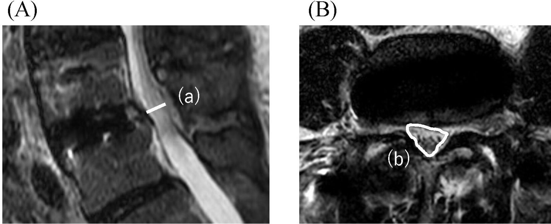
- Figure 1
Measurement of radiographic parameters. (A) Preoperative sagittal computed tomography (CT). (B and C) Postoperative sagittal CT. (a) Segmental lordosis. (b) Anterior disc height. (c) Posterior disc height. (d) Foraminal area was measured on CT images.
- Figure 2
Measurement of magnetic resonance imaging (MRI) findings. (A) Postoperative sagittal MRI. (B) Postoperative axial MRI. (a) Canal diameter and (b) central canal area were measured as the enclosed area of the spinal canal on sagittal and axial MRI.
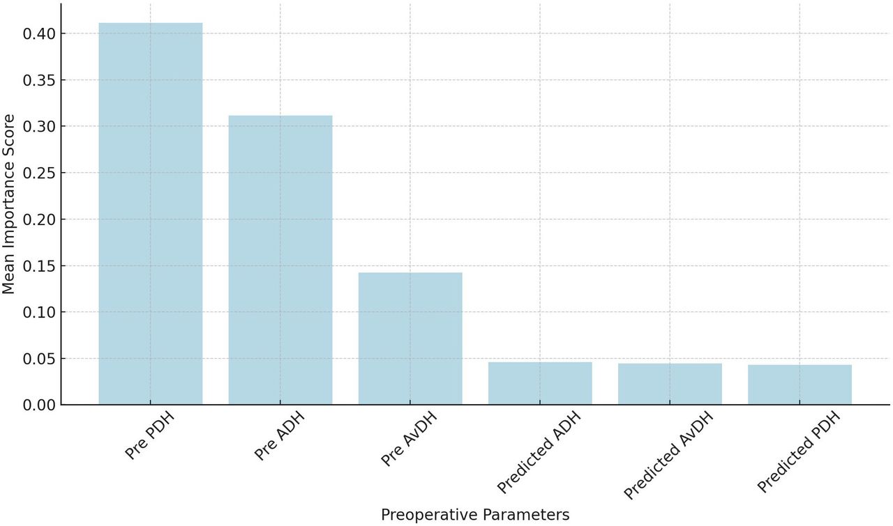
- Figure 3
Bar graph showing the feature importance scores derived from the random forest regression model used in the sensitivity analysis. The graph illustrates the relative impact of 6 key parameters. Pre-PDH demonstrated the highest importance, indicating its significant influence on postoperative SL outcomes. Abbreviations: ADH, anterior disc height; AvDH, average disc height; PDH, posterior disc height; SL, segmental lordosis;.
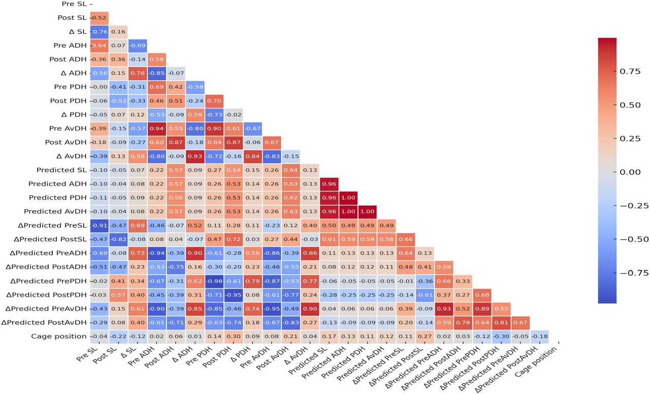
- Figure 4
Correlation matrix of parameters. The heatmap illustrates the correlation coefficients between key pre- and postoperative parameters. SL, ADH, PDH, AvDH, and cage position are evaluated for their linear relationships. Positive correlations are shown in shades of blue, while negative correlations are depicted in shades of red. The color’s intensity indicates the correlation’s strength, with darker shades representing stronger relationships. Abbreviations: ADH, anterior disc height; AvDH, average disc height; PDH, posterior disc height; SL, segmental lordosis;.
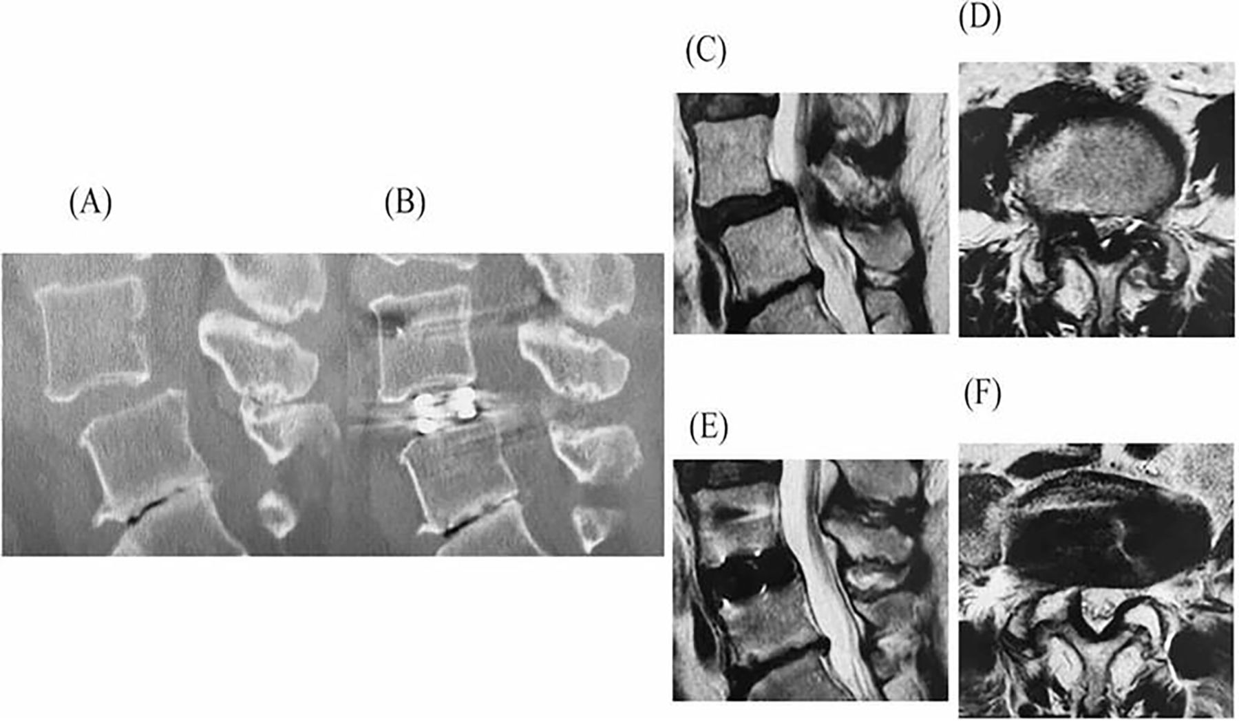
- Figure 5
A 55-year-old woman underwent LLIF with PPS for L4 spondylolisthesis. Sagittal CT images before (A) and after surgery (B) are shown. Sagittal MRIs before surgery are shown in (C), and after surgery in (E), while axial MRIs before surgery are shown in (D), and after surgery in (F). Preoperative ADH, PDH, AvDH, and SL were 8.7, 5.8, 7.3, and 6.9 mm, respectively. The predicted values for ADH, PDH, AvDH, and SL were 11.0, 7.9, 9.5, and 9.8 mm, respectively. Postoperative CT showed actual ADH, PDH, AvDH, and SL measurements of 12.8, 9.5, 11.2, and 7.2 mm, respectively. While the DH was greater than predicted, the SL was smaller than expected. MRI revealed significant improvement in dural sac compression. Abbreviations: ADH, anterior disc height; AvDH, average disc height; CT, computed tomography; LLIF, lateral lumbar interbody fusion; MRI, magnetic resonance imaging; PDH, posterior disc height; PPS, percutaneous pedicle screw; SL, segmental lordosis.


Tables
Outcome Measure No. of Driver Revolutions 0 1 2 3 4 5 6 7 8 9 10 11 12 13 14 ADH, mm 7 7.5 8 8.5 9 9.5 10 10.5 11 11.5 12 12.5 13 13.5 14 PDH, mm 6 6.25 6.5 6.75 6.9 7.15 7.4 7.65 7.9 8.1 8.3 8.55 8.8 9.05 9.3 SL (°) 3 3.85 4.7 5.55 6.4 7.25 8.1 8.95 9.8 10.65 11.5 12.35 13.2 14.1 15 Abbreviations: ADH, anterior disc height; PDH, posterior disc height; SL, segmental lordosis.
Note: This table shows the change in anterior-posterior disc height and segmental lordosis angle according to the number of drive screw turns.
Characteristic Value Age, y 70.6 (11.7) Sex, men/women, n 28/23 Height, cm, mean (SD) 159.8 (9.1) Body weight, kg, mean (SD) 63.9 (13.3) BMI, mean (SD) 24.8 (3.7) Tobacco use, n (%) 7 (16) Steroid use, n (%) 5 (12) Indications, n (%) LCS+ (LDS) 44 (86) Degenerative lumbar scoliosis 4 (8) Lumbar disc herniation 2 (4) Foraminal stenosis 1 (2) Levels treated, n (%) L1–L2 0 (0) L2–L3 8 (10) L3–L4 34 (43) L4–L5 37 (47) Overall 79 Number of levels fused, n (%) 1 level 26 (51) 2 levels 22 (43) 3 levels 3 (6) Mean (SD) 1.5 (0.6) Operation time, min, mean (SD) 128.1 (25.6) EBL, mL, mean (SD) 92.7 (58.8) LOS, d, mean (SD) 15.5 (4.1) Cage height, mm, mean (SD) 10.3 (1.2) Cage width, mm, mean (SD) 18 (0) Cage length, mm, mean (SD) 50.8 (4.2) Cage position, %, n (%) 51.4 (11.0) Cage placement, n (%) Anterior (<45%) 21 (27) Central (>45, <55%) 23 (29) Posterior (>55%) 35 (44) Cage subsidence, n (%) 4 (8) Early cage subsidence 0 (0) Delayed cage subsidence 4 (8) Transient motor weakness, n (%) 4 (8) Thigh pain and/or numbness, n (%) 6 (12) Revision surgery, n (%) 4 (8) Abbreviations: BMI, body mass index; EBL, estimated blood loss; LCS, lumbar canal stenosis; LDS, lumbar degenerative spondylolisthesis; LOS, length of stay.
- Table 3
Preoperative, postoperative, and change from pre- to postoperative sagittal measurements.
Outcome Measure Preoperative Postoperative ΔPost-Pre P a SL (°) 3.5 (4.2) 4.8 (2.8) 1.3 (3.6) 0.002b ADH, mm 5.9 (3.3) 10.7 (1.8) 4.9 (2.7) <0.001b PDH, mm 3.4 (2.6) 7.7 (1.8) 4.4 (1.8) <0.001b AvDH, mm 4.6 (2.7) 9.2 (1.5) 4.6 (2.0) <0.001b FA, mm2 98.4 (37.1) 131.2 (39.9) 32.7 (29.9) <0.001b Variable Pre-ADH Pre-PDH Pre-AvDH Pre-SL ΔCD r −0.091 −0.078 −0.108 0.082 P 0.432 0.501 0.349 0.478 ΔCCA r −0.118 −0.106 −0.134 0.033 P 0.306 0.357 0.244 0.777 Variable Post-ADH Post-PDH Post-AvDH Post-SL ΔCD r −0.076 0.080 −0.012 −0.117 P 0.512 0.487 0.915 0.311 ΔCCA r 0.017 0.126 0.066 0.005 P 0.884 0.275 0.569 0.966 Variable ΔADH ΔPDH ΔAvDH ΔSL ΔCD r 0.013 0.195 0.095 −0.122 P 0.909 0.089 0.411 0.291 ΔCCA r 0.120 0.272a 0.199 −0.010 P 0.298 0.017 0.083 0.928 Abbreviations: ADH, anterior disc height; AvDH, average disc height; PDH, posterior disc height; SL, segmental lordosis; ΔCCA, change of central canal area; ΔCD, change of canal diameter.
↵a Statistically significant.
In this issue


Jump to section
Related Articles
Cited By...
- No citing articles found.