
Main menu

  • Home
  • Content
    • Current Issue
    • Advance Online Publication
    • Archive
  • About Us
    • About ISASS
    • About the Journal
    • Author Instructions
    • Editorial Board
    • Reviewer Guidelines & Publication Criteria
  • More
    • Advertise
    • Subscribe
    • Alerts
    • Feedback
  • Join Us
  • Reprints & Permissions
  • Sponsored Content
  • Other Publications
    • ijss

User menu

  • My alerts

Search

  • Advanced search
International Journal of Spine Surgery
  • My alerts
International Journal of Spine Surgery

Advanced Search

  • Home
  • Content
    • Current Issue
    • Advance Online Publication
    • Archive
  • About Us
    • About ISASS
    • About the Journal
    • Author Instructions
    • Editorial Board
    • Reviewer Guidelines & Publication Criteria
  • More
    • Advertise
    • Subscribe
    • Alerts
    • Feedback
  • Join Us
  • Reprints & Permissions
  • Sponsored Content
  • Follow ijss on Twitter
  • Visit ijss on Facebook
Research ArticleFull Length Article
Open Access

A Comparison of the Degree of Lateral Recess and Foraminal Enlargement With Facet Preservation in the Treatment of Lumbar Stenosis With Standard Surgical Tools Versus a Novel Powered Filing Instrument: A Cadaver Study

Murat Cosar, Larry T. Khoo, Christopher A. Yeung and Anthony T. Yeung
International Journal of Spine Surgery January 2007, 1 (4) 135-142; DOI: https://doi.org/10.1016/SASJ-2007-0107-NT-R1
Murat Cosar
1The Department of Neurosurgery, Canakkale 18 Mart University of Medical School, Canakkale, Turkey
MD
  • Find this author on Google Scholar
  • Find this author on PubMed
  • Search for this author on this site
Larry T. Khoo
2The Division of Neurosurgery, UCLA, Los Angeles, California
MD
  • Find this author on Google Scholar
  • Find this author on PubMed
  • Search for this author on this site
Christopher A. Yeung
3The Arizona Institute for Minimally Invasive Spine Care, Phoenix, Arizona
MD
  • Find this author on Google Scholar
  • Find this author on PubMed
  • Search for this author on this site
Anthony T. Yeung
3The Arizona Institute for Minimally Invasive Spine Care, Phoenix, Arizona
MD
  • Find this author on Google Scholar
  • Find this author on PubMed
  • Search for this author on this site
  • Article
  • Figures & Data
  • Info & Metrics
  • PDF
Loading

Article Figures & Data

Figures

  • Tables
  • Figure 1
    • Download figure
    • Open in new tab
    • Download powerpoint
    Figure 1

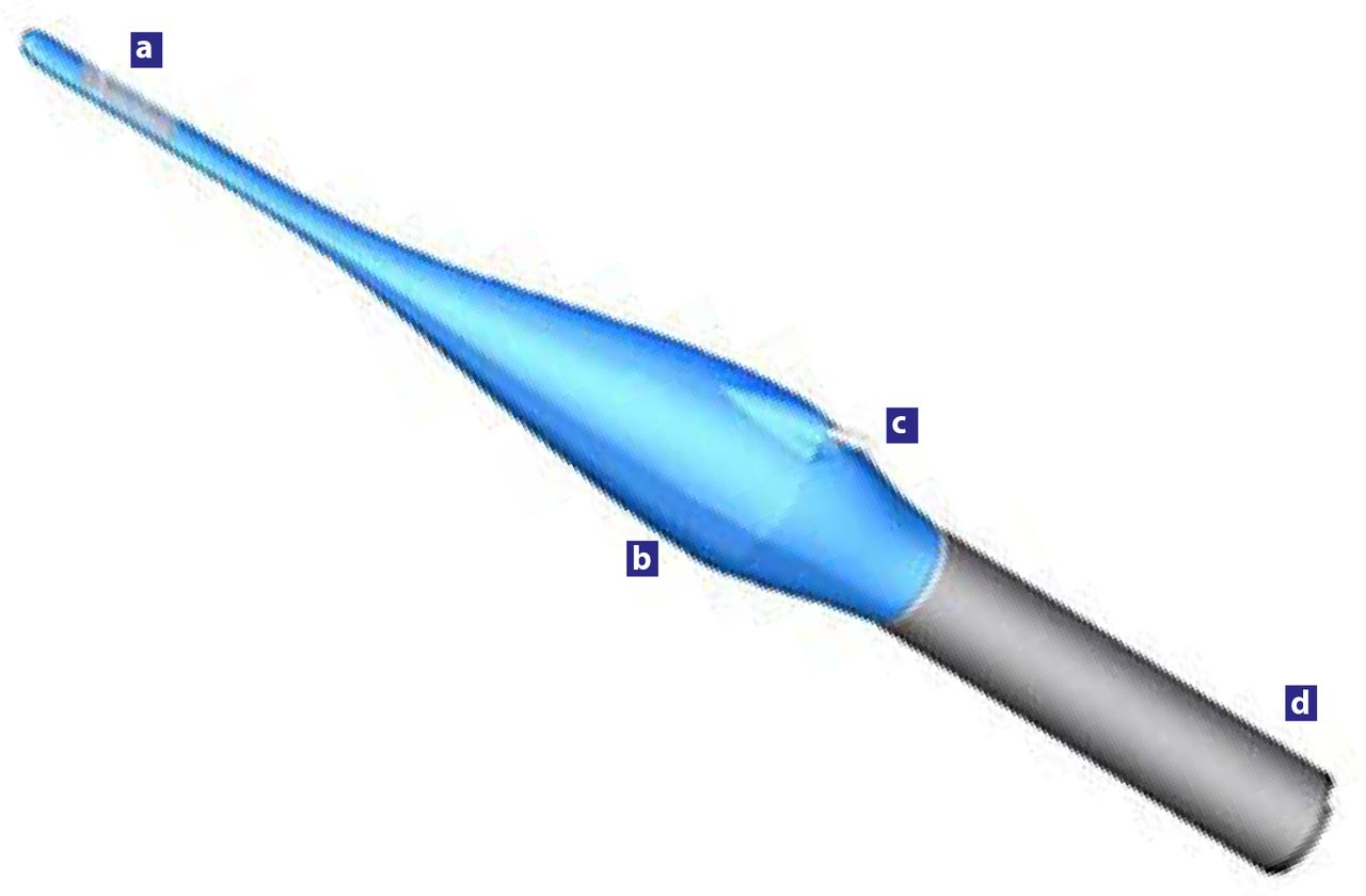
    The SurgiFile tool is demonstrated with the thin area of the exposed (a) cutting surface, (b) main toroidal drive body, (c) irrigation inlet, and (d) the surgical drill motor.

  • Figure 2
    • Download figure
    • Open in new tab
    • Download powerpoint
    Figure 2

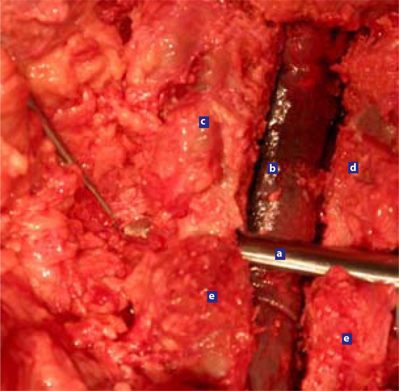
    (a) The SurgiFile tool is demonstrated here completing an inside-to-outside decompression along the entire length of the neural foraminal corridor. Because only the dorsal surface of the working blade is active, the SurgiFile tip can be used directly over the exposed (b) thecal sac and nerve root. (c) SurgiFile side. (d) Standard side. (e) Facet joint.

  • Figure 3
    • Download figure
    • Open in new tab
    • Download powerpoint
    Figure 3

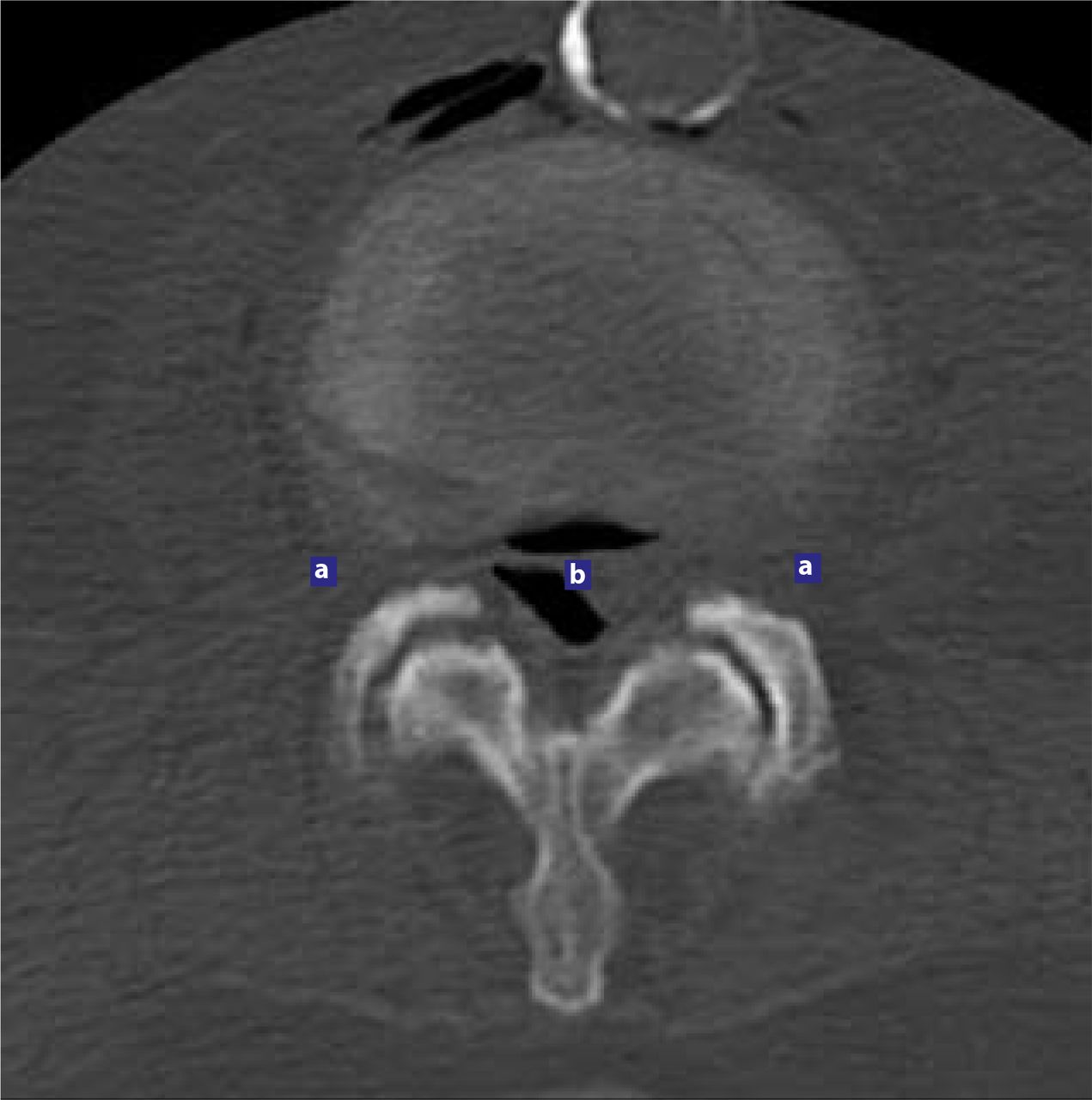
    The landmarks of the neural foramen are shown in the axial CT scan image. (a) Lateral foramen; (b) proximal recess.

  • Figure 4
    • Download figure
    • Open in new tab
    • Download powerpoint
    Figure 4

    (a) Axial A-P foraminal sizes; and (b) sagittal foraminal areas are shown.

  • Figure 5
    • Download figure
    • Open in new tab
    • Download powerpoint
    Figure 5

    Comparison of sagittal CT scans showing (a) medial proximal recess areas before (left) and after (right) use of SurgiFile tool; (b) lateral foraminal areas before (left) and after (right) use of SurgiFile tool; (c) medial foraminal areas before (left) and after (right) use of standard tools; and (d) lateral foraminal areas before (left) and after (right) use of standard tools.

  • Figure 6
    • Download figure
    • Open in new tab
    • Download powerpoint
    Figure 6

    (a) Postoperative change in Axial A-P foraminal distance; and (b) percentage of change in the sagittal foraminal area.

  • Figure 7
    • Download figure
    • Open in new tab
    • Download powerpoint
    Figure 7

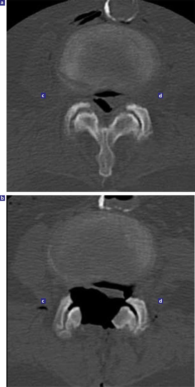
    (a) Preoperative macroscopic visual comparison of the SurgiFile versus the standard sides demonstrates the greater facet preservation overall in the SurgiFile side after neural decompression; (b) Postoperative comparisons of the; (c) Standard side on axial CT demonstrates greater qualitative preservation of the facet joints; and the (d) SurgiFile side.

Tables

  • Figures
    • View popup
    Table 1

    The Preoperative and Postoperative Foraminal Heights on Axial CT Scans (mm)

    Proximal RecessLateral Foramen
    SurgiFileStandardSurgiFileStandard
    PreopPostopPreopPostopPreopPostopPreopPostop
    Cadaver 1 L2—381119751108213295105
    Cadaver 1 L3—460132601007513075100
    Cadaver 1 L4—5931669813097160107130
    Cadaver 1 L5—S1951409512496162100120
    Cadaver 2 L2—362957075751008085
    Cadaver 2 L3—4701056585701007080
    Cadaver 2 L4—55010070957011075106
    Cadaver 2 L5—S18010580100100120100100
    Average73.875120.2576.625102.3883.125126.7587.75103.25
    • View popup
    Table 2

    The Preoperative and Postoperative Foraminal Area on Sagittal CT Scans (mm2)

    Proximal RecessLateral Foramen
    SurgiFileStandardSurgiFileStandard
    PreopPostopPreopPostopPreopPostopPreopPostop
    Cadaver 1 L2—318003700210030001900400024002900
    Cadaver 1 L3—417004300195032002200450028003200
    Cadaver 1 L4—514504100185031001700400027003000
    Cadaver 1 L5—S114003000160024001500350026002600
    Cadaver 2 L2—319003500175030002000290019001900
    Cadaver 2 L3—424003200155030003200380018002100
    Cadaver 2 L4—519002700135022002500290020002400
    Cadaver 2 L5—S119002600170024002300290018501900
    Average1,806.253,387.501,731.252,787.502,162.53,562.52,256.302,500.00
    • View popup
    Table 3

    Preop and Postop Foraminal Length Increase on Axial CT Scans (%)

    Proximal RecessLateral Foramen
    SurgiFileStandardSurgiFileStandard
    Cadaver 1 L2—31800370021003000
    Cadaver 1 L3—41700430019503200
    Cadaver 1 L4—51450410018503100
    Cadaver 1 L5—S11400300016002400
    Cadaver 2 L2—31900350017503000
    Cadaver 2 L3—42400320015503000
    Cadaver 2 L4—51900270013502200
    Cadaver 2 L5—S11900260017002400
    Average1,806.253,387.501,731.252,787.50
    • View popup
    Table 4

    Preop and Postop Foraminal Area Increase on Sagittal CT Scans (%)

    Proximal RecessLateral Foramen
    SurgiFileStandardSurgiFileStandard
    Cadaver 1 L2—31054211020
    Cadaver 1 L3—41504510415
    Cadaver 1 L4—51826513511
    Cadaver 1 L5—S1114501330
    Cadaver 2 L2—38471450
    Cadaver 2 L3—4341002016
    Cadaver 2 L4—542621620
    Cadaver 2 L5—S13741262
    Average93,559,573,62510,5
PreviousNext
Back to top

In this issue

International Journal of Spine Surgery
Vol. 1, Issue 4
1 Jan 2007
  • Table of Contents
  • Index by author

Print
Download PDF
Article Alerts
Sign In to Email Alerts with your Email Address
Email Article

Thank you for your interest in spreading the word on International Journal of Spine Surgery.

NOTE: We only request your email address so that the person you are recommending the page to knows that you wanted them to see it, and that it is not junk mail. We do not capture any email address.

Enter multiple addresses on separate lines or separate them with commas.
A Comparison of the Degree of Lateral Recess and Foraminal Enlargement With Facet Preservation in the Treatment of Lumbar Stenosis With Standard Surgical Tools Versus a Novel Powered Filing Instrument: A Cadaver Study
(Your Name) has sent you a message from International Journal of Spine Surgery
(Your Name) thought you would like to see the International Journal of Spine Surgery web site.
CAPTCHA
This question is for testing whether or not you are a human visitor and to prevent automated spam submissions.
Citation Tools
A Comparison of the Degree of Lateral Recess and Foraminal Enlargement With Facet Preservation in the Treatment of Lumbar Stenosis With Standard Surgical Tools Versus a Novel Powered Filing Instrument: A Cadaver Study
Murat Cosar, Larry T. Khoo, Christopher A. Yeung, Anthony T. Yeung
International Journal of Spine Surgery Jan 2007, 1 (4) 135-142; DOI: 10.1016/SASJ-2007-0107-NT-R1

Citation Manager Formats

  • BibTeX
  • Bookends
  • EasyBib
  • EndNote (tagged)
  • EndNote 8 (xml)
  • Medlars
  • Mendeley
  • Papers
  • RefWorks Tagged
  • Ref Manager
  • RIS
  • Zotero
Share
A Comparison of the Degree of Lateral Recess and Foraminal Enlargement With Facet Preservation in the Treatment of Lumbar Stenosis With Standard Surgical Tools Versus a Novel Powered Filing Instrument: A Cadaver Study
Murat Cosar, Larry T. Khoo, Christopher A. Yeung, Anthony T. Yeung
International Journal of Spine Surgery Jan 2007, 1 (4) 135-142; DOI: 10.1016/SASJ-2007-0107-NT-R1
Twitter logo Facebook logo Mendeley logo
  • Tweet Widget
  • Facebook Like
  • Google Plus One

Jump to section

  • Article
    • Abstract
    • INTRODUCTION
    • MATERIALS AND METHODS
    • RESULTS
    • DISCUSSION
    • Footnotes
    • REFERENCES
  • Figures & Data
  • Info & Metrics
  • PDF

Related Articles

  • No related articles found.
  • Google Scholar

Cited By...

  • No citing articles found.
  • Google Scholar

More in this TOC Section

  • Kyphoplasty versus vertebroplasty for painful osteoporotic vertebral compression fractures—which one is better? A systematic review and meta-analysis
  • Overtightening of halo pins resulting in intracranial penetration, pneumocephalus, and epileptic seizure
  • Retrospective cost analysis of cervical laminectomy and fusion versus cervical laminoplasty in the treatment of cervical spondylotic myelopathy
Show more Full Length Article

Similar Articles

Keywords

  • foraminal stenosis
  • lateral recess
  • spinal stenosis
  • spinal surgery

Content

  • Current Issue
  • Latest Content
  • Archive

More Information

  • About IJSS
  • About ISASS
  • Privacy Policy

More

  • Subscribe
  • Alerts
  • Feedback

Other Services

  • Author Instructions
  • Join ISASS
  • Reprints & Permissions

© 2025 International Journal of Spine Surgery

International Journal of Spine Surgery Online ISSN: 2211-4599

Powered by HighWire