
Main menu

  • Home
  • Content
    • Current Issue
    • Advance Online Publication
    • Archive
  • About Us
    • About ISASS
    • About the Journal
    • Author Instructions
    • Editorial Board
    • Reviewer Guidelines & Publication Criteria
  • More
    • Advertise
    • Subscribe
    • Alerts
    • Feedback
  • Join Us
  • Reprints & Permissions
  • Sponsored Content
  • Other Publications
    • ijss

User menu

  • My alerts

Search

  • Advanced search
International Journal of Spine Surgery
  • My alerts
International Journal of Spine Surgery

Advanced Search

  • Home
  • Content
    • Current Issue
    • Advance Online Publication
    • Archive
  • About Us
    • About ISASS
    • About the Journal
    • Author Instructions
    • Editorial Board
    • Reviewer Guidelines & Publication Criteria
  • More
    • Advertise
    • Subscribe
    • Alerts
    • Feedback
  • Join Us
  • Reprints & Permissions
  • Sponsored Content
  • Follow ijss on Twitter
  • Visit ijss on Facebook
Research ArticleArticles

Finite Element Analysis of Sacroiliac Joint Fixation under Compression Loads

Claire Bruna-Rosso, Pierre-Jean Arnoux, Rohan-Jean Bianco, Yves Godio-Raboutet, Léo Fradet and Carl-Éric Aubin
International Journal of Spine Surgery January 2016, 10 16; DOI: https://doi.org/10.14444/3016
Claire Bruna-Rosso
1Department of Mechanical Engineering, Polytechnique Montréal, Montreal, Canada
2iLab - Spine International Laboratory - Spine Imaging and Biomechanics
MASc
  • Find this author on Google Scholar
  • Find this author on PubMed
  • Search for this author on this site
Pierre-Jean Arnoux
2iLab - Spine International Laboratory - Spine Imaging and Biomechanics
3Laboratoire de Biomécanique Appliquée, Aix-Marseille Université, Marseille, France
PhD
  • Find this author on Google Scholar
  • Find this author on PubMed
  • Search for this author on this site
Rohan-Jean Bianco
1Department of Mechanical Engineering, Polytechnique Montréal, Montreal, Canada
2iLab - Spine International Laboratory - Spine Imaging and Biomechanics
3Laboratoire de Biomécanique Appliquée, Aix-Marseille Université, Marseille, France
4Sainte-Justine University Hospital Center, Montreal, Canada
PhD
  • Find this author on Google Scholar
  • Find this author on PubMed
  • Search for this author on this site
Yves Godio-Raboutet
2iLab - Spine International Laboratory - Spine Imaging and Biomechanics
3Laboratoire de Biomécanique Appliquée, Aix-Marseille Université, Marseille, France
MEng
  • Find this author on Google Scholar
  • Find this author on PubMed
  • Search for this author on this site
Léo Fradet
1Department of Mechanical Engineering, Polytechnique Montréal, Montreal, Canada
2iLab - Spine International Laboratory - Spine Imaging and Biomechanics
3Laboratoire de Biomécanique Appliquée, Aix-Marseille Université, Marseille, France
PhD
  • Find this author on Google Scholar
  • Find this author on PubMed
  • Search for this author on this site
Carl-Éric Aubin
1Department of Mechanical Engineering, Polytechnique Montréal, Montreal, Canada
2iLab - Spine International Laboratory - Spine Imaging and Biomechanics
4Sainte-Justine University Hospital Center, Montreal, Canada
PEng, PhD
  • Find this author on Google Scholar
  • Find this author on PubMed
  • Search for this author on this site
  • Article
  • Figures & Data
  • Info & Metrics
  • PDF
Loading

Article Figures & Data

Figures

  • Tables
  • Fig. 1
    • Download figure
    • Open in new tab
    • Download powerpoint
    Fig. 1

    Model details (ligaments and mesh).
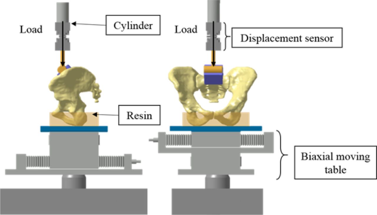
  • Fig. 2
    • Download figure
    • Open in new tab
    • Download powerpoint
    Fig. 2

    Experimental setup (clamps not represented for clarity).
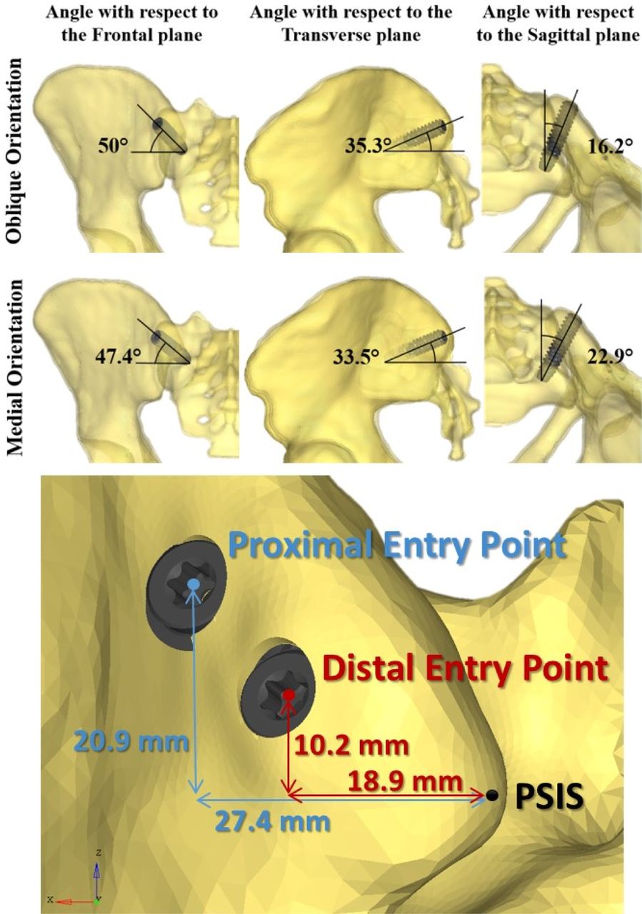
  • Fig. 3
    • Download figure
    • Open in new tab
    • Download powerpoint
    Fig. 3

    Screw trajectory parameters.
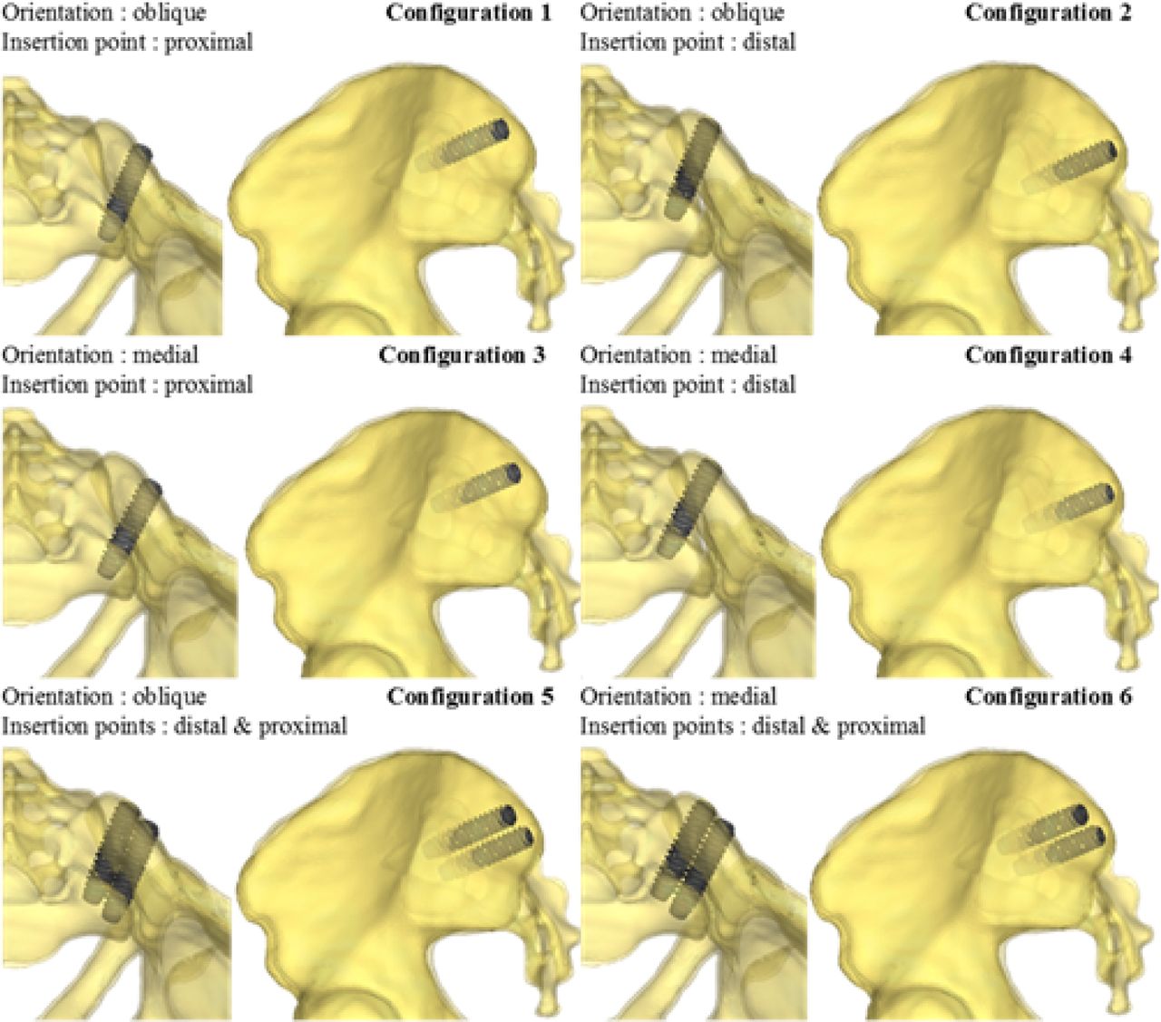
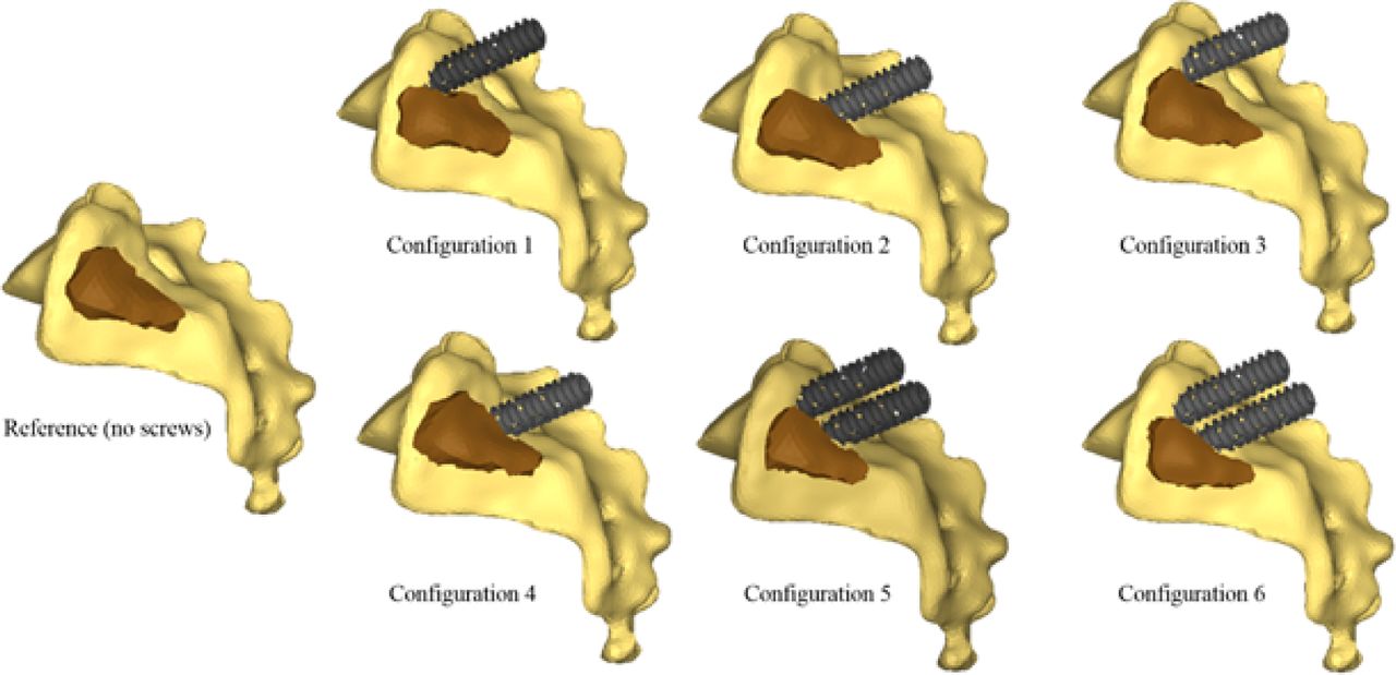
  • Fig. 4
    • Download figure
    • Open in new tab
    • Download powerpoint
    Fig. 4

    Six simulated configurations.
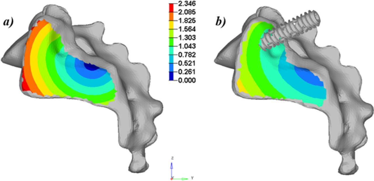
  • Fig. 5
    • Download figure
    • Open in new tab
    • Download powerpoint
    Fig. 5

    Sagittal view of the global displacements of the SIJ (translations in mm) for the simulations at 1000 N: a) uninstrumented (reference) and b) instrumented with one screw inserted obliquely at the proximal insertion point.
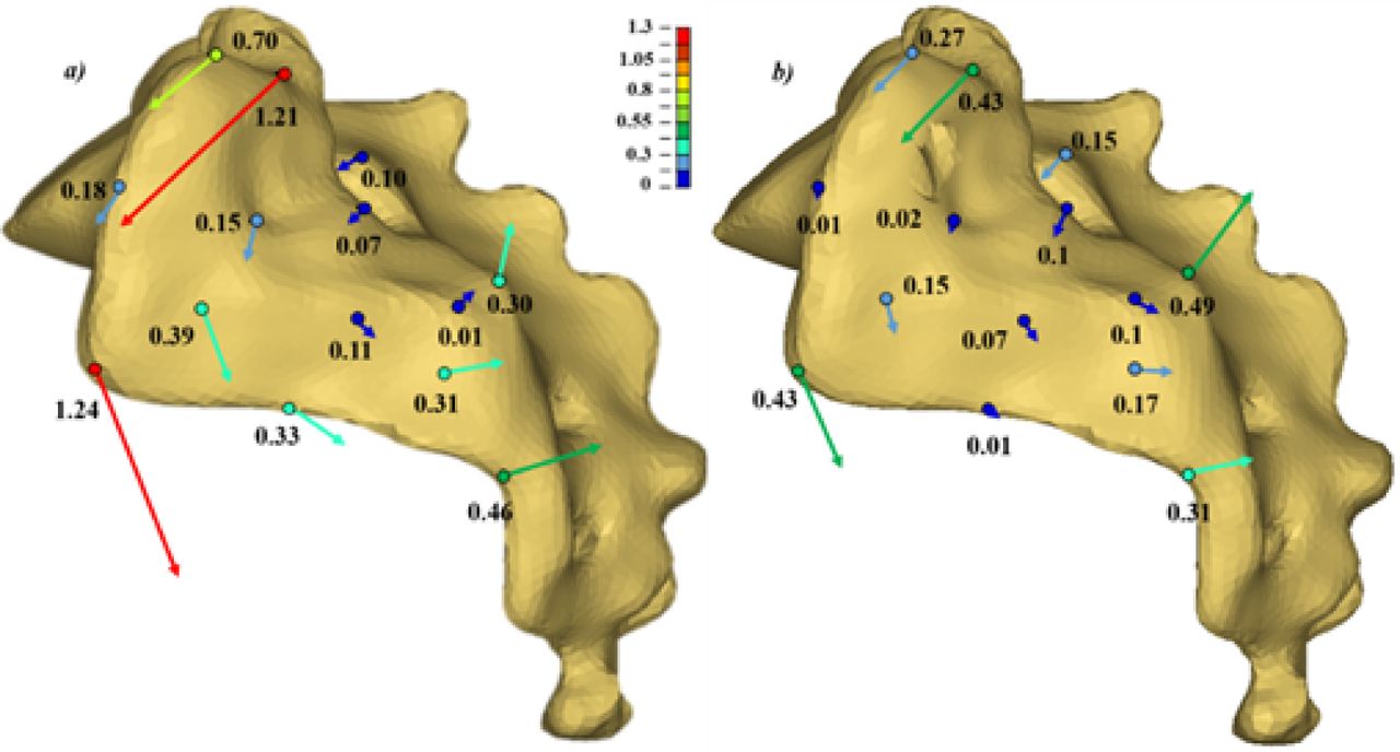
  • Fig. 6
    • Download figure
    • Open in new tab
    • Download powerpoint
    Fig. 6

    Local displacements (translations in mm) of 14 points of the SI facet of the sacrum with respect to the iliac bone after a vertical loading of the sacrum of 1000 N: a) unistrumented; b) instrumented (configuration 1). The displacement vectors are magnified for clarity.
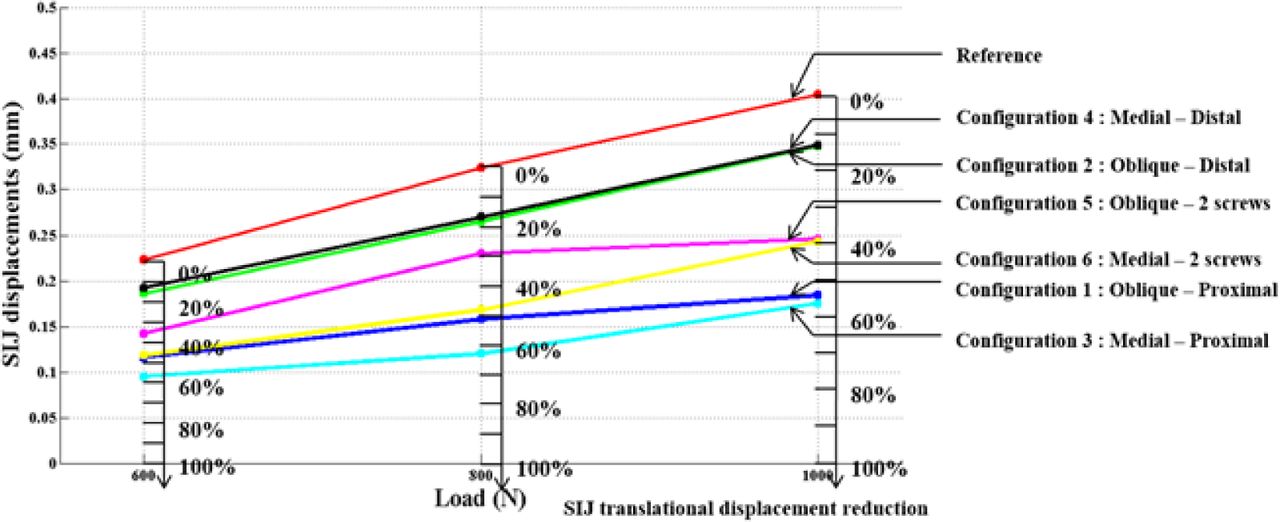
  • Fig. 7
    • Download figure
    • Open in new tab
    • Download powerpoint
    Fig. 7

    SIJ local displacements in the sagittal plane and % of reduction with respect to the uninstrumented reference.

  • Fig. 8
    • Download figure
    • Open in new tab
    • Download powerpoint
    Fig. 8

    Average SIJ local rotations and % of rotation reduction with respect to the uninstrumented reference.

  • Fig. 9
    • Download figure
    • Open in new tab
    • Download powerpoint
    Fig. 9

    Stresses (MPa) on the left ilium and sacrum trabecular bone (configuration 5, loaded at 800 N). The virtual axis of rotation is located below the figure.

  • Fig. 10
    • Download figure
    • Open in new tab
    • Download powerpoint
    Fig. 10

    Interosseous ligament modifications in the model to enable device insertion.

Tables

  • Figures
    • View popup
    Table 1

    Material properties used in the FEM.

    Cortical BoneTrabecular BoneLigamentsPubic SymphysisSIJ Articular Cartilage
    Density (kg.m-3)20.2221.05
    Young Modulus (MPa)262548.7540397150
    Poisson Ratio0.30.250.30.30.2
    Yield Stress (MPa)1051.95---
    Hardening modulus (MPa)87516.3---
    Hardening exponent11---
    Failure plastic strain0.040.04---
    Tangent Young Modulus (MPa)--10155-
    Tangent Poisson ratio--0.370.37-
    Viscoelastic constant--2828-
    Navier Constant--1.105 1.105 -
    • View popup
    Table 2

    Comparison of experimentally measured (mean) and simulated S1 endplate displacement reduction due to the screws.

    1 screw configuration2 screw configuration
    ExperimentalSimulationsDifferenceExperimentalSimulationsDifference
    600 N14.98%13.86%1.12%17.71%14.36%3.35%
    800 N12.09%14.75%2.66%14.27%15.46%1.19%
PreviousNext
Back to top

In this issue

International Journal of Spine Surgery
Vol. 10
1 Jan 2016
  • Table of Contents
  • Index by author

Print
Download PDF
Article Alerts
Sign In to Email Alerts with your Email Address
Email Article

Thank you for your interest in spreading the word on International Journal of Spine Surgery.

NOTE: We only request your email address so that the person you are recommending the page to knows that you wanted them to see it, and that it is not junk mail. We do not capture any email address.

Enter multiple addresses on separate lines or separate them with commas.
Finite Element Analysis of Sacroiliac Joint Fixation under Compression Loads
(Your Name) has sent you a message from International Journal of Spine Surgery
(Your Name) thought you would like to see the International Journal of Spine Surgery web site.
CAPTCHA
This question is for testing whether or not you are a human visitor and to prevent automated spam submissions.
Citation Tools
Finite Element Analysis of Sacroiliac Joint Fixation under Compression Loads
Claire Bruna-Rosso, Pierre-Jean Arnoux, Rohan-Jean Bianco, Yves Godio-Raboutet, Léo Fradet, Carl-Éric Aubin
International Journal of Spine Surgery Jan 2016, 10 16; DOI: 10.14444/3016

Citation Manager Formats

  • BibTeX
  • Bookends
  • EasyBib
  • EndNote (tagged)
  • EndNote 8 (xml)
  • Medlars
  • Mendeley
  • Papers
  • RefWorks Tagged
  • Ref Manager
  • RIS
  • Zotero
Share
Finite Element Analysis of Sacroiliac Joint Fixation under Compression Loads
Claire Bruna-Rosso, Pierre-Jean Arnoux, Rohan-Jean Bianco, Yves Godio-Raboutet, Léo Fradet, Carl-Éric Aubin
International Journal of Spine Surgery Jan 2016, 10 16; DOI: 10.14444/3016
Twitter logo Facebook logo Mendeley logo
  • Tweet Widget
  • Facebook Like
  • Google Plus One

Jump to section

  • Article
    • Abstract
    • Introduction
    • Methods
    • Results
    • Discussion
    • Conclusion
    • Disclosures & COI
    • References
  • Figures & Data
  • Info & Metrics
  • PDF

Related Articles

  • No related articles found.
  • Google Scholar

Cited By...

  • Minimally Invasive SI Joint Fusion Procedures for Chronic SI Joint Pain: Systematic Review and Meta-Analysis of Safety and Efficacy
  • Effect of Sacropelvic Hardware on Axis and Center of Rotation of the Sacroiliac Joint: A Finite Element Study
  • International Society for the Advancement of Spine Surgery Policy 2020 Update--Minimally Invasive Surgical Sacroiliac Joint Fusion (for Chronic Sacroiliac Joint Pain): Coverage Indications, Limitations, and Medical Necessity
  • Editor's Introduction: Update on Current Sacroiliac Joint Fusion Procedures: Implications for Appropriate Current Procedural Terminology Medical Coding
  • Biomechanics of the Sacroiliac Joint: Surgical Treatments
  • Google Scholar

More in this TOC Section

  • Validity and reliability of a novel patient reported outcome tool to evaluate post-operative dysphagia, odynophagia, and voice (DOV) disability after anterior cervical procedures
  • Surgical Revision after Sacroiliac Joint Fixation or Fusion
  • A Rare Case of T1-2 Thoracic Disc Herniation Mimicking Cervical Radiculopathy
Show more Articles

Similar Articles

Keywords

  • sacroiliac joint
  • finite element analysis
  • Biomechanics
  • arthrodesis

Content

  • Current Issue
  • Latest Content
  • Archive

More Information

  • About IJSS
  • About ISASS
  • Privacy Policy

More

  • Subscribe
  • Alerts
  • Feedback

Other Services

  • Author Instructions
  • Join ISASS
  • Reprints & Permissions

© 2025 International Journal of Spine Surgery

International Journal of Spine Surgery Online ISSN: 2211-4599

Powered by HighWire