


Article Figures & Data
Figures
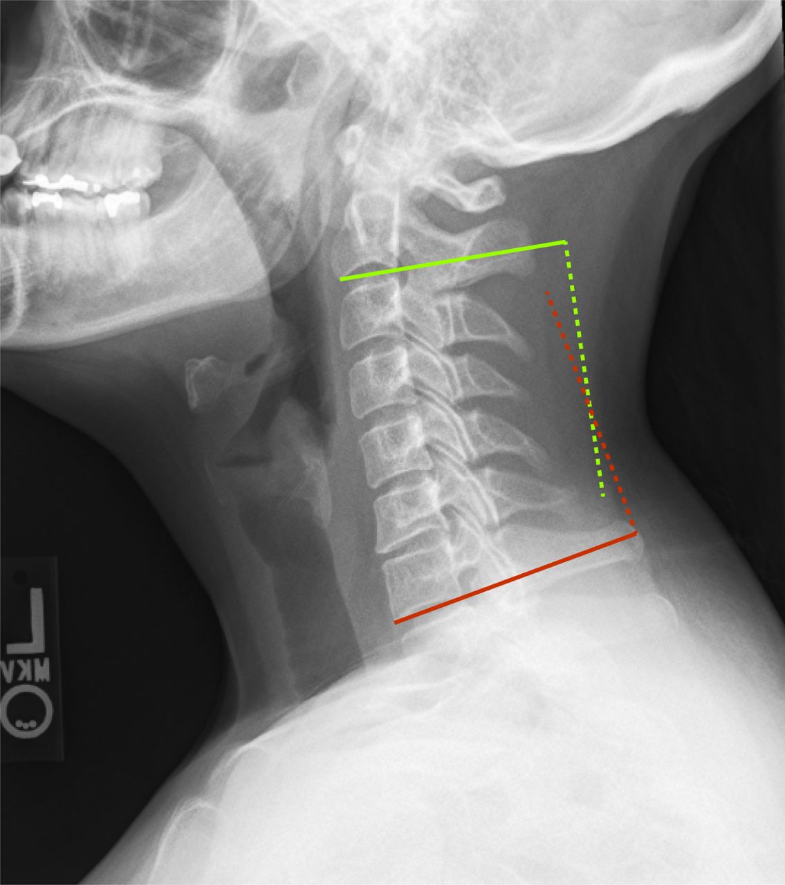
- Fig. 1
Curvature angle measured using the Cobb technique on plain radiograph. A line was placed along the inferior aspect of C2 (yellow solid line), and a perpendicular line (yellow dashed) was drawn. A line was placed along the inferior aspect of C7 (red line), and a perpendicular line (yellow dashed) was then drawn. The acute angle subtended between the two crossing lines is the curvature angle. In this example the measurement was 12.2 degrees of lordosis.
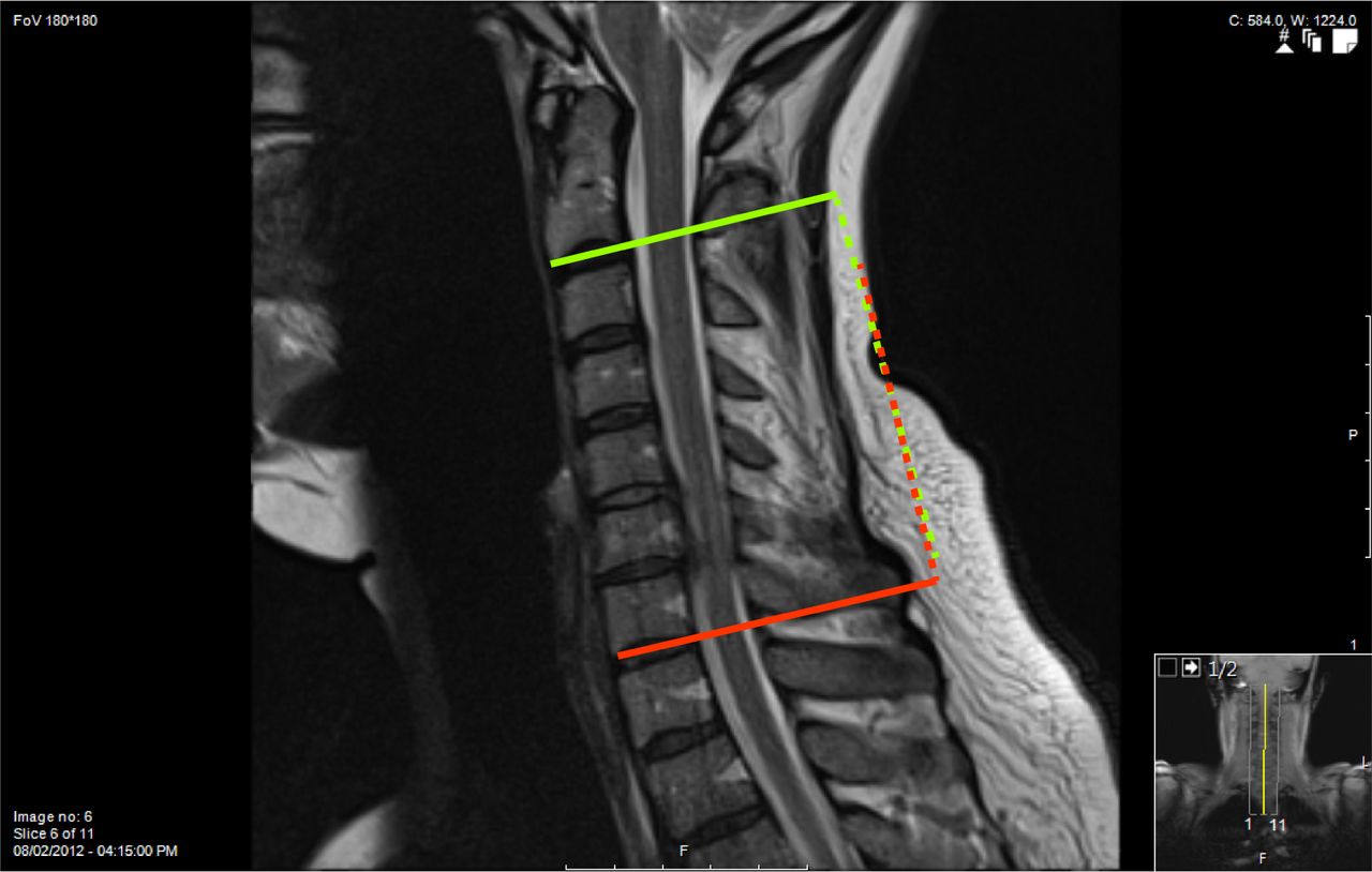
- Fig. 2
Curvature angle measured using the Cobb technique on MR, on the same individual seen in Figure 1. A line was placed along the inferior aspect of C2 (solid line), and a perpendicular line (yellow dashed) was drawn. A line was placed along the inferior aspect of C7 (red line), and a perpendicular line (yellow dashed) was then drawn. The acute angle subtended between the two crossing lines is the curvature angle. In this example the measurement was 1.6 degrees of kyphosis, notably different than the measurement performed on plain radiograph.
- Fig. 3
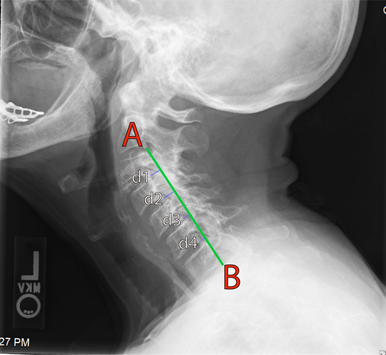
Curvature index (Ishihara) measured on plain radiograph. A line is connected between the most posterior-inferior portion of C2 (A) and the most posterior-inferior portion of C7 (B). The distance of line (AB) was recorded. The distances between the posterior-inferior aspects of C3, C4, C5, and C6 to the orthogonal intersection of the line from C2 to C7 was calculated (d1-4). The curvature index was calculated:

In this example the measurement was 31.1.
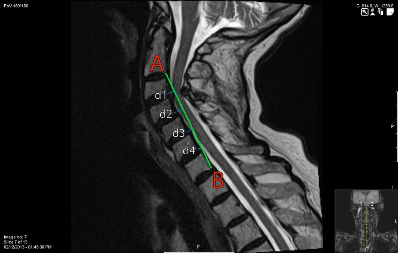
- Fig. 4
Curvature index (Ishihara) measured on MRI, on the same individual seen in Figure 3. A line is connected between the most posterior-inferior portion of C2 (A) and the most posterior-inferior portion of C7 (B). The distance of line (AB) was recorded. The distances between the posterior-inferior aspects of C3, C4, C5, and C6 to the orthogonal intersection of the line from C2 to C7 was calculated (d1-4). The curvature index was calculated:
In this example the measurement was 30.9.
- Fig. 5
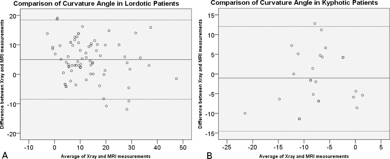
Bland-Altman plots of comparisons of curvature angle in lordotic (a) and kyphotic (b) patients. The average between pairs of measurements are plotted against their difference. The solid line indicates mean bias (average difference), and the dotted lines are limits of agreement (95% confidence limit lines).
- Fig. 6
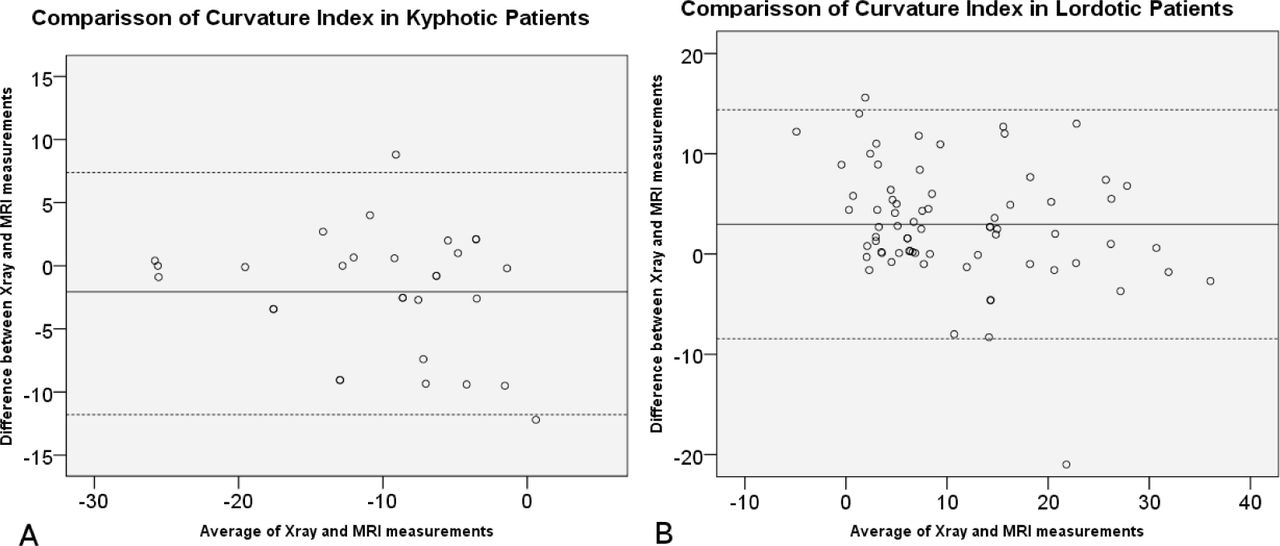
Bland-Altman plots of comparisons of curvature index in lordotic (a) and kyphotic (b) patients. The average between pairs of measurements are plotted against their difference. The solid line indicates mean bias (average difference), and the dotted lines are limits of agreement (95% confidence limit lines).


Tables
CA X-ray CA MRI CI X-ray CI MRI CA X-ray --- 0.853 0.839 0.790 CA MRI 0.853 --- 0.757 0.814 CI X-ray 0.839 0.757 --- 0.899 CI MRI 0.790 0.814 0.899 --- CA: Curvature Angle, CI: Curvature Index
All correlations were statistically significant (p< 0.05)
In this issue


Jump to section
Related Articles
Cited By...
- No citing articles found.