
Main menu

  • Home
  • Content
    • Current Issue
    • Advance Online Publication
    • Archive
  • About Us
    • About ISASS
    • About the Journal
    • Author Instructions
    • Editorial Board
    • Reviewer Guidelines & Publication Criteria
  • More
    • Advertise
    • Subscribe
    • Alerts
    • Feedback
  • Join Us
  • Reprints & Permissions
  • Sponsored Content
  • Other Publications
    • ijss

User menu

  • My alerts

Search

  • Advanced search
International Journal of Spine Surgery
  • My alerts
International Journal of Spine Surgery

Advanced Search

  • Home
  • Content
    • Current Issue
    • Advance Online Publication
    • Archive
  • About Us
    • About ISASS
    • About the Journal
    • Author Instructions
    • Editorial Board
    • Reviewer Guidelines & Publication Criteria
  • More
    • Advertise
    • Subscribe
    • Alerts
    • Feedback
  • Join Us
  • Reprints & Permissions
  • Sponsored Content
  • Follow ijss on Twitter
  • Visit ijss on Facebook
Research ArticleArticles

Clinical and Anatomical Features as well as Pathological Conditions of Surgically Treated Adult Patients with Occipitalization of the Atlas

Yasunori Tatara, Takachika Shimizu, Keisuke Fueki, Masatake Ino, Naofumi Toda, Tetsu Tanouchi and Nodoka Manabe
International Journal of Spine Surgery January 2016, 10 5; DOI: https://doi.org/10.14444/3005
Yasunori Tatara
1Yokohama Minami Kyosai Hospital, Yokohama, Kanagawa, Japan
MD
  • Find this author on Google Scholar
  • Find this author on PubMed
  • Search for this author on this site
Takachika Shimizu
2Gunma Spine Center, Harunaso Hospital, Takasaki, Japan
MD
  • Find this author on Google Scholar
  • Find this author on PubMed
  • Search for this author on this site
Keisuke Fueki
2Gunma Spine Center, Harunaso Hospital, Takasaki, Japan
MD
  • Find this author on Google Scholar
  • Find this author on PubMed
  • Search for this author on this site
Masatake Ino
2Gunma Spine Center, Harunaso Hospital, Takasaki, Japan
MD
  • Find this author on Google Scholar
  • Find this author on PubMed
  • Search for this author on this site
Naofumi Toda
2Gunma Spine Center, Harunaso Hospital, Takasaki, Japan
MD
  • Find this author on Google Scholar
  • Find this author on PubMed
  • Search for this author on this site
Tetsu Tanouchi
2Gunma Spine Center, Harunaso Hospital, Takasaki, Japan
MD
  • Find this author on Google Scholar
  • Find this author on PubMed
  • Search for this author on this site
Nodoka Manabe
2Gunma Spine Center, Harunaso Hospital, Takasaki, Japan
MD
  • Find this author on Google Scholar
  • Find this author on PubMed
  • Search for this author on this site
  • Article
  • Figures & Data
  • Info & Metrics
  • PDF
Loading

Article Figures & Data

Figures

  • Tables
  • Fig. 1
    • Download figure
    • Open in new tab
    • Download powerpoint
    Fig. 1

    Coronal CT reconstruction image of the occipital condyle demonstrating that the right occipital condyle (small arrow) is smaller in size than the left occipital condyle (large arrow) and this patient (case 10) manifests torticollis.
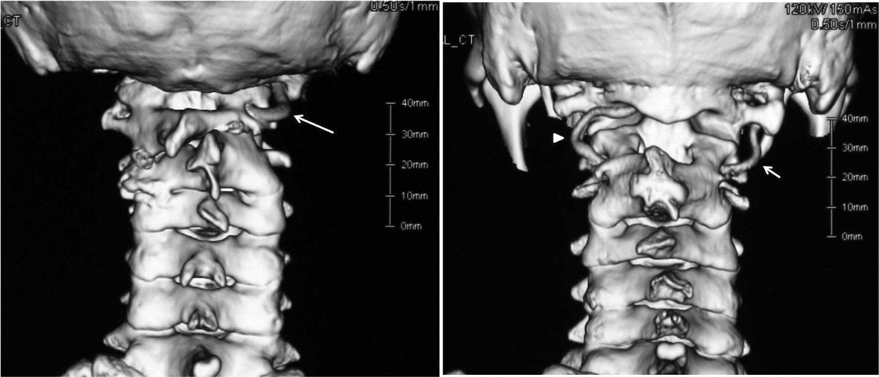
  • Fig. 2
    • Download figure
    • Open in new tab
    • Download powerpoint
    Fig. 2

    3D-CTA reconstruction images demonstrating typical cases of anomalous VAs. Left (case 5): The left VA is agenesis and the right VA enters the cranium via the extraspinal canal after exiting the transverse foramen of the axis. Right (case 8): The left VA (arrow head) enters the cranium via the intraspinal canal under the atlas and the right VA (arrow) enters the cranium via the extraspinal canal.

  • Fig. 3
    • Download figure
    • Open in new tab
    • Download powerpoint
    Fig. 3

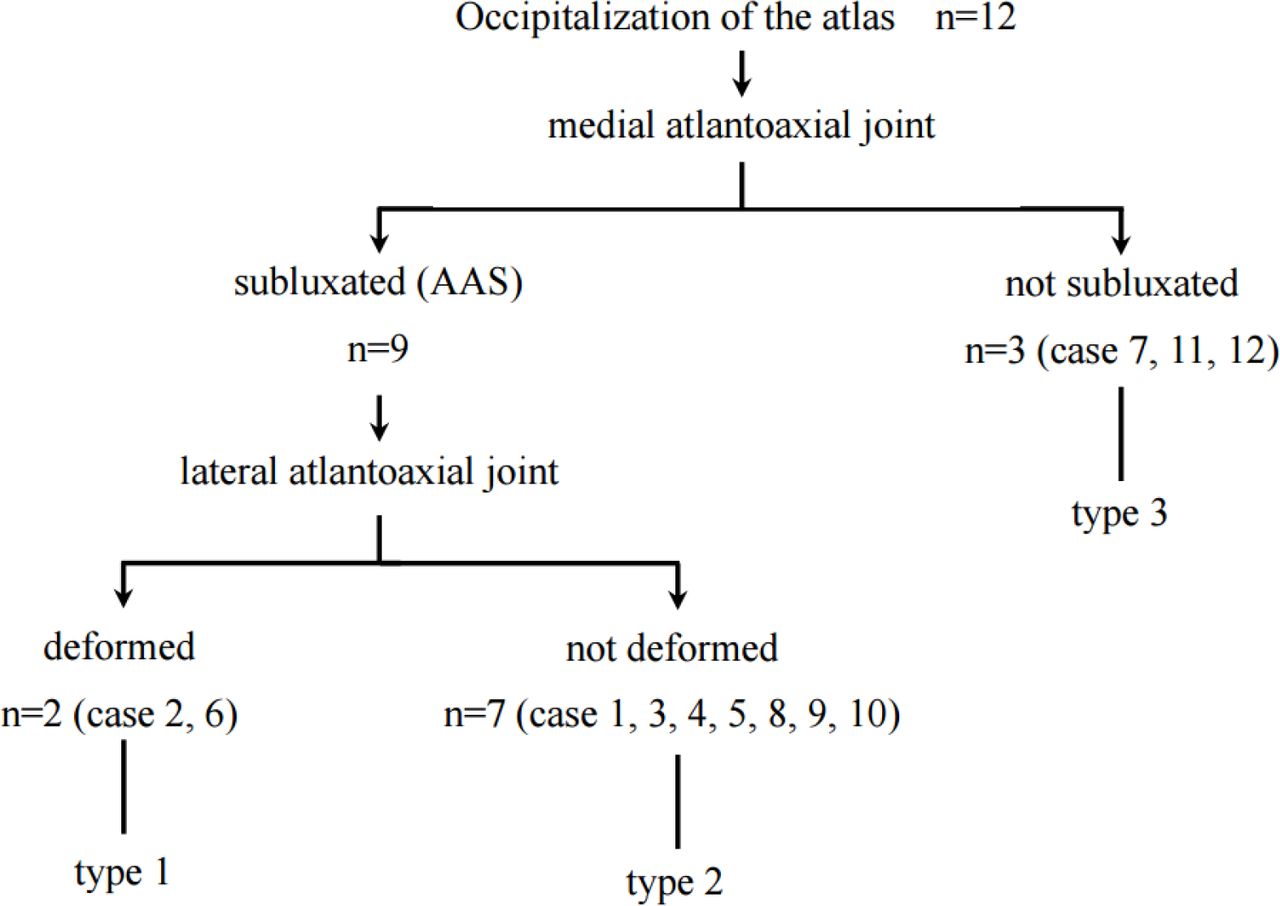
    Flow diagram of the classification for occipitalization of the atlas.
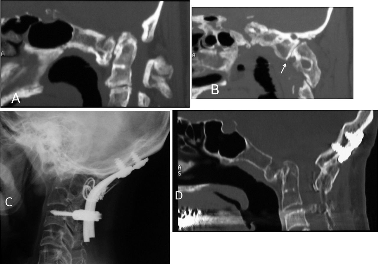
  • Fig. 4
    • Download figure
    • Open in new tab
    • Download powerpoint
    Fig. 4

    Type 1 (case 2). Preoperative midsagittal (A) and parasagittal (B) CT reconstruction images demonstrating that the atlas is completely fused with the occiput. The medial atlantoaxial joint is semi-dislocated. The lateral atlantoaxial joint (arrow) is severely deformed and the lateral mass is slipped anteroinferiorly against the superior facet of the axis. Postoperative x-ray film (C) and midsagittal CT reconstruction (D) images demonstrating that AAS remains to some degree but a stable bony arthrodesis is obtained.

  • Fig. 5
    • Download figure
    • Open in new tab
    • Download powerpoint
    Fig. 5

    Type 2. Preoperative midsagittal (A) and parasagittal (B) CT reconstruction images demonstrating that case 4 is a complete fusion type and the lateral atlantoaxial joint (arrow) is slightly loose but almost normal in shape. C2-3 fusion is also demonstrated. Postoperative x-ray film (C) and midsagittal CT reconstruction (D) images demonstrating that AAS is reduced and a stable bony arthrodesis is obtained.
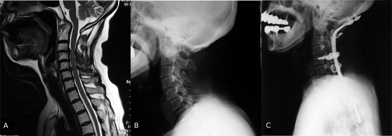
  • Fig. 6
    • Download figure
    • Open in new tab
    • Download powerpoint
    Fig. 6

    Type 3. Preoperative sagittal T2-weighted MR image (A) and x-ray film (B) of case 11 associated with rheumatoid arthritis demonstrating subaxial stenosis and subaxial instability but a wide space available for the cord at the CVJ. Postoperative x-ray film (C) demonstrating the cervical spine is successfully realigned and balanced.

Tables

  • Figures
    • View popup
    Table 1

    Summary of osseous anomalies and clinical presentation of 12 patients with occipitalization of the atlas.

    Case No.Age (yrs), SexComorbidityFusion TypeHypoplastic Occipital CondyleAtlantoaxial JointVertebral FusionClinical Presentation
    MedialLateral
    151, Fpartialsubluxated (mobile)hyperreflexia, paresthesia (extremities), pain in the nape of the neck
    247, Fcompletertsubluxated (irreducible)deformedC2-3hyperreflexia, paresthesia (occiput, extremities), motorweakness, gait disturbance
    363, Mpartialsubluxated (irreducible)C5-6hyperreflexia, paresthesia (extremities), pain in the nape of the neck
    456, Mcompletesubluxated (mobile)C2-3hyperreflexia, paresthesia (extremities), gait disturbance
    563, Fcompleteltsubluxated (irreducible)C2-3hyperreflexia, paresthesia (occiput, extremities), gait disturbance
    668, Fcompletesubluxated (irreducible)deformedC2-3hyperreflexia, paresthesia (extremities), gait disturbance
    774, MChiari malformationpartiallthyperreflexia, paresthesia (extremities), gait disturbance
    846, Mcompleteltsubluxated (irreducible)C2-3hyperreflexia, paresthesia (extremities), gait disturbance
    977, Fcompletesubluxated (irreducible)C2-3hyperreflexia, paresthesia (extremities), gait disturbance
    1066, Fpartialrtsubluxated (irreducible)hyperreflexia, paresthesia (extremities), gait disturbance
    1162, FRAcompletehyperreflexia, paresthesia (occiput, extremities), motorweakness, gait disturbance
    1245, FOPLLcompletehyperreflexia, paresthesia (extremities), motorweakness, gait disturbance
    • View popup
    Table 2

    Summary of surgical methods and outcomes in 12 patients with occipitalization of the atlas.

    Case No.Surgical MethodAASOutcome
    Preop.Postop.
    1Occ-C3 PSF and C1 laminectomysubluxated (mobile)reducedimprovement
    2Occ-C4 PSF, C1 laminectomy, and C4-6 laminoplastysubluxated (irreducible)not reducedimprovement
    3Occ-C3 PSF and C1 laminectomysubluxated (irreducible)reducedimprovement
    4Occ-C3 PSF, C1 laminectomy, and C4-6 laminoplastysubluxated (mobile)reducedimprovement
    5Occ-C4 PSF and C1 laminectomysubluxated (irreducible)not reducedimprovement
    6Occ-C7 PSF and C1 laminectomysubluxated (irreducible)not reducedimprovement
    7foramen magnum decompressionimprovement
    8Occ-C4 PSF and C1 laminectomysubluxated (irreducible)reducedimprovement
    9Occ-C5 PSF and C1 laminectomysubluxated (irreducible)reducedimprovement
    10Occ-C7 PSF and C1 laminectomysubluxated (irreducible)reducedimprovement
    11Occ-T2 PSFimprovement
    12Occ-T3 PSFimprovement
    • * Occ = Occiput, PSF = posterior spinal fusion

    • View popup
    Table 3

    Summary of VA anomalies in 12 patients with occipitalization of the atlas.

    Case No.Fusion TypeMorphology of the VAHypoplastic Side of the Transverse ForamenCourse of the V3 Segment of the VA into the Cranium
    1partialrt, hypoplasiaboth sides: extraspinal canal
    2completert, hypoplasiaboth sides: extraspinal canal
    3partialno lateralityboth sides: normal
    4completert, agenesisrtlt: intraspinal canal under the atlas
    5completelt, agenesisltrt: extraspinal canal
    6completert, hypoplasiaboth sides: extraspinal canal
    7partiallt, agenesisltrt: normal
    8completeno lateralityrt: extraspinal canal lt: intraspinal canal under the atlas
    9completert, hypoplasiaboth sides: extraspinal canal
    10partialrt, agenesisrtlt: normal
    11completert, hypoplasiaboth sides: extraspinal canal
    12completert, hypoplasiartboth sides: extraspinal canal
PreviousNext
Back to top

In this issue

International Journal of Spine Surgery
Vol. 10
1 Jan 2016
  • Table of Contents
  • Index by author

Print
Download PDF
Article Alerts
Sign In to Email Alerts with your Email Address
Email Article

Thank you for your interest in spreading the word on International Journal of Spine Surgery.

NOTE: We only request your email address so that the person you are recommending the page to knows that you wanted them to see it, and that it is not junk mail. We do not capture any email address.

Enter multiple addresses on separate lines or separate them with commas.
Clinical and Anatomical Features as well as Pathological Conditions of Surgically Treated Adult Patients with Occipitalization of the Atlas
(Your Name) has sent you a message from International Journal of Spine Surgery
(Your Name) thought you would like to see the International Journal of Spine Surgery web site.
CAPTCHA
This question is for testing whether or not you are a human visitor and to prevent automated spam submissions.
Citation Tools
Clinical and Anatomical Features as well as Pathological Conditions of Surgically Treated Adult Patients with Occipitalization of the Atlas
Yasunori Tatara, Takachika Shimizu, Keisuke Fueki, Masatake Ino, Naofumi Toda, Tetsu Tanouchi, Nodoka Manabe
International Journal of Spine Surgery Jan 2016, 10 5; DOI: 10.14444/3005

Citation Manager Formats

  • BibTeX
  • Bookends
  • EasyBib
  • EndNote (tagged)
  • EndNote 8 (xml)
  • Medlars
  • Mendeley
  • Papers
  • RefWorks Tagged
  • Ref Manager
  • RIS
  • Zotero
Share
Clinical and Anatomical Features as well as Pathological Conditions of Surgically Treated Adult Patients with Occipitalization of the Atlas
Yasunori Tatara, Takachika Shimizu, Keisuke Fueki, Masatake Ino, Naofumi Toda, Tetsu Tanouchi, Nodoka Manabe
International Journal of Spine Surgery Jan 2016, 10 5; DOI: 10.14444/3005
Twitter logo Facebook logo Mendeley logo
  • Tweet Widget
  • Facebook Like
  • Google Plus One

Jump to section

  • Article
    • Abstract
    • Introduction
    • Clinical Materials and Methods
    • Results
    • Discussion
    • Disclosures
    • References
  • Figures & Data
  • Info & Metrics
  • PDF

Related Articles

  • No related articles found.
  • Google Scholar

Cited By...

  • No citing articles found.
  • Google Scholar

More in this TOC Section

  • Validity and reliability of a novel patient reported outcome tool to evaluate post-operative dysphagia, odynophagia, and voice (DOV) disability after anterior cervical procedures
  • Surgical Revision after Sacroiliac Joint Fixation or Fusion
  • A Rare Case of T1-2 Thoracic Disc Herniation Mimicking Cervical Radiculopathy
Show more Articles

Similar Articles

Keywords

  • occipitalization of the atlas
  • atlantoaxial subluxation
  • craniovertebral junction
  • vertebral artery

Content

  • Current Issue
  • Latest Content
  • Archive

More Information

  • About IJSS
  • About ISASS
  • Privacy Policy

More

  • Subscribe
  • Alerts
  • Feedback

Other Services

  • Author Instructions
  • Join ISASS
  • Reprints & Permissions

© 2025 International Journal of Spine Surgery

International Journal of Spine Surgery Online ISSN: 2211-4599

Powered by HighWire