


Article Figures & Data
Figures
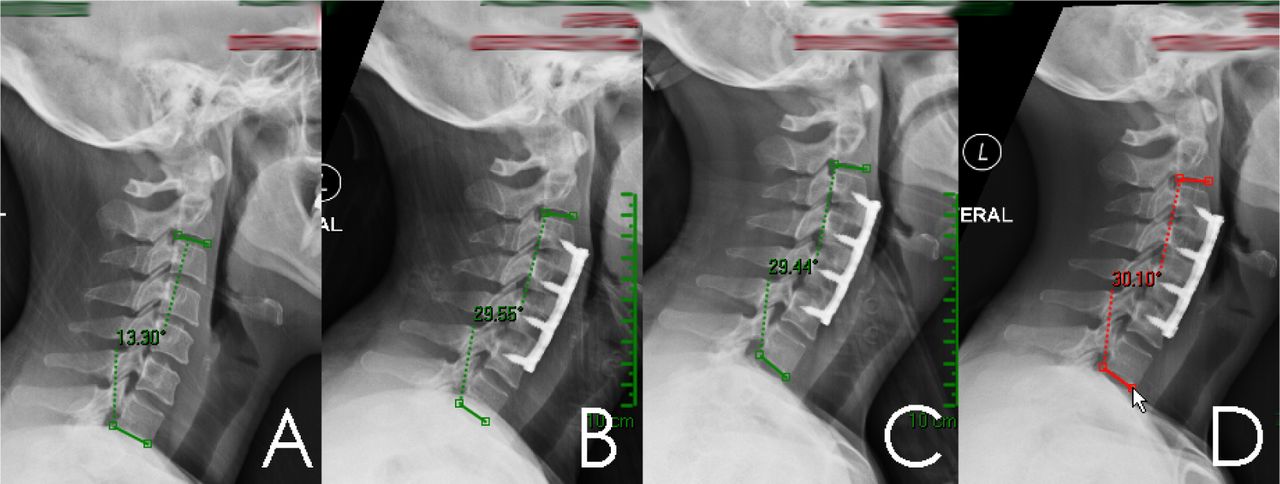
- Fig. 1
Case Example: Lateral Cervical x-rays obtained at Pre-op (A), 4 weeks (B), 10 weeks (C), and 6 months (D) showing increase in C2-C7 lordosis from 13°, to 29°, 29° and 30° respectively.
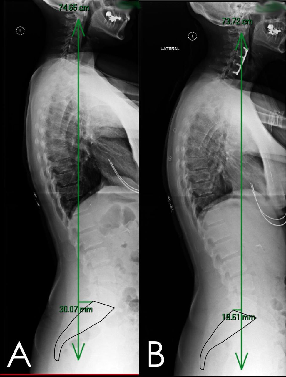
- Fig. 2
Case example continued: Lateral full 36 inch scoliosis films taken at pre-op (B) and 4 weeks post op (A) demonstrating increase in sagittal vertical axis from -30mm to 19mm.
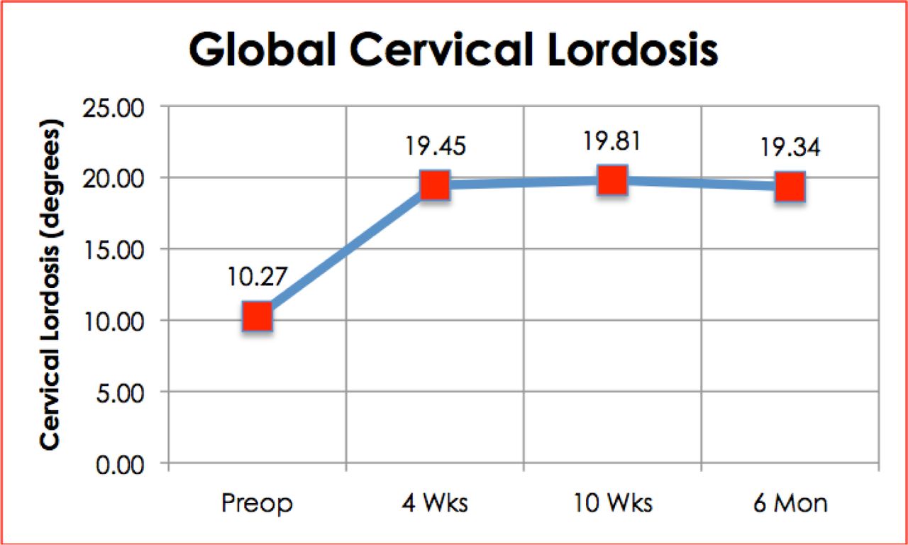
- Fig. 3
Line chart of the average degree of cervical lordosis measured at four time points (preoperative, 4 weeks, 10 weeks and 6 months postoperative).
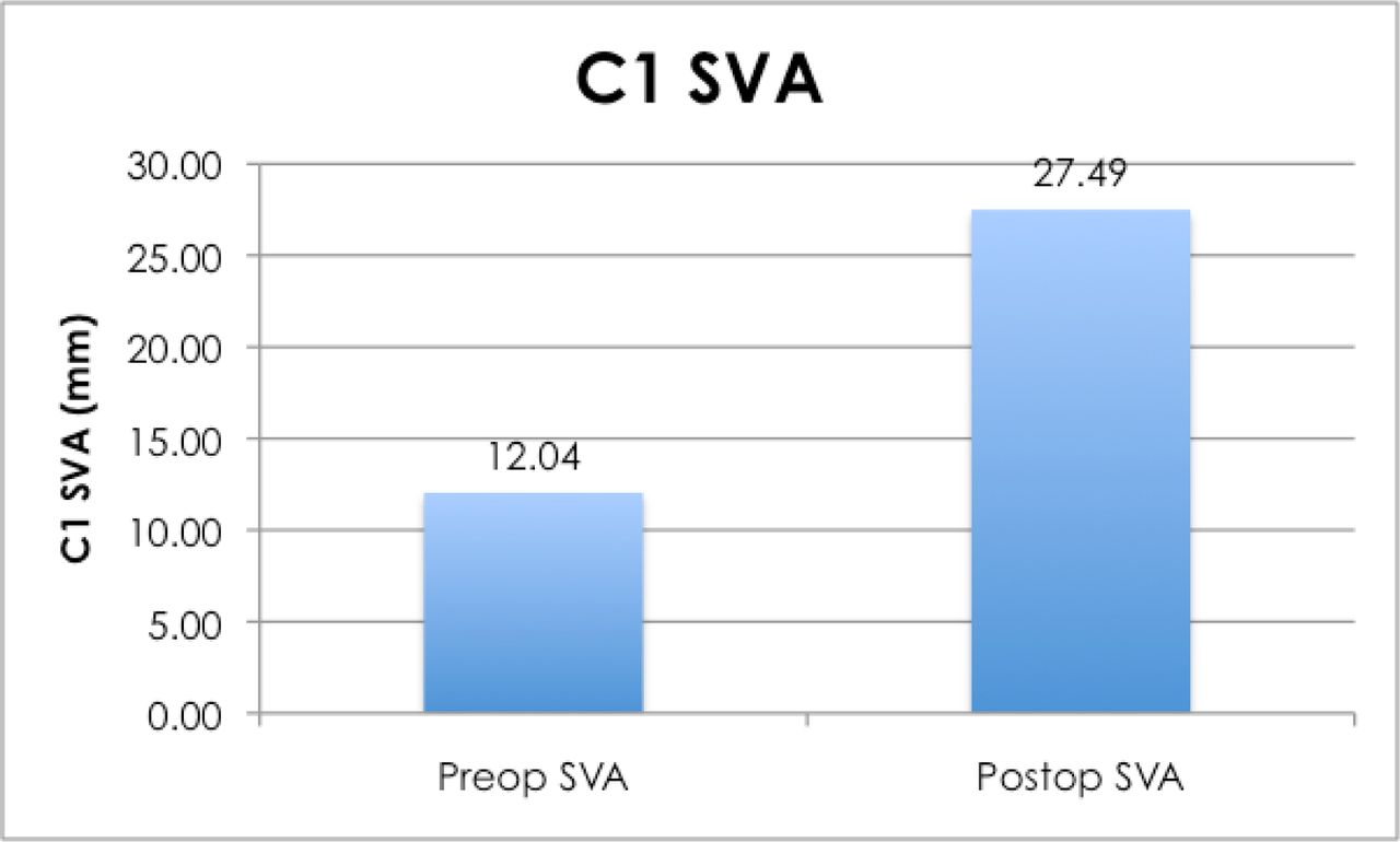
- Fig. 4
Bar graph showing the pre- and postoperative C1-SVA means.
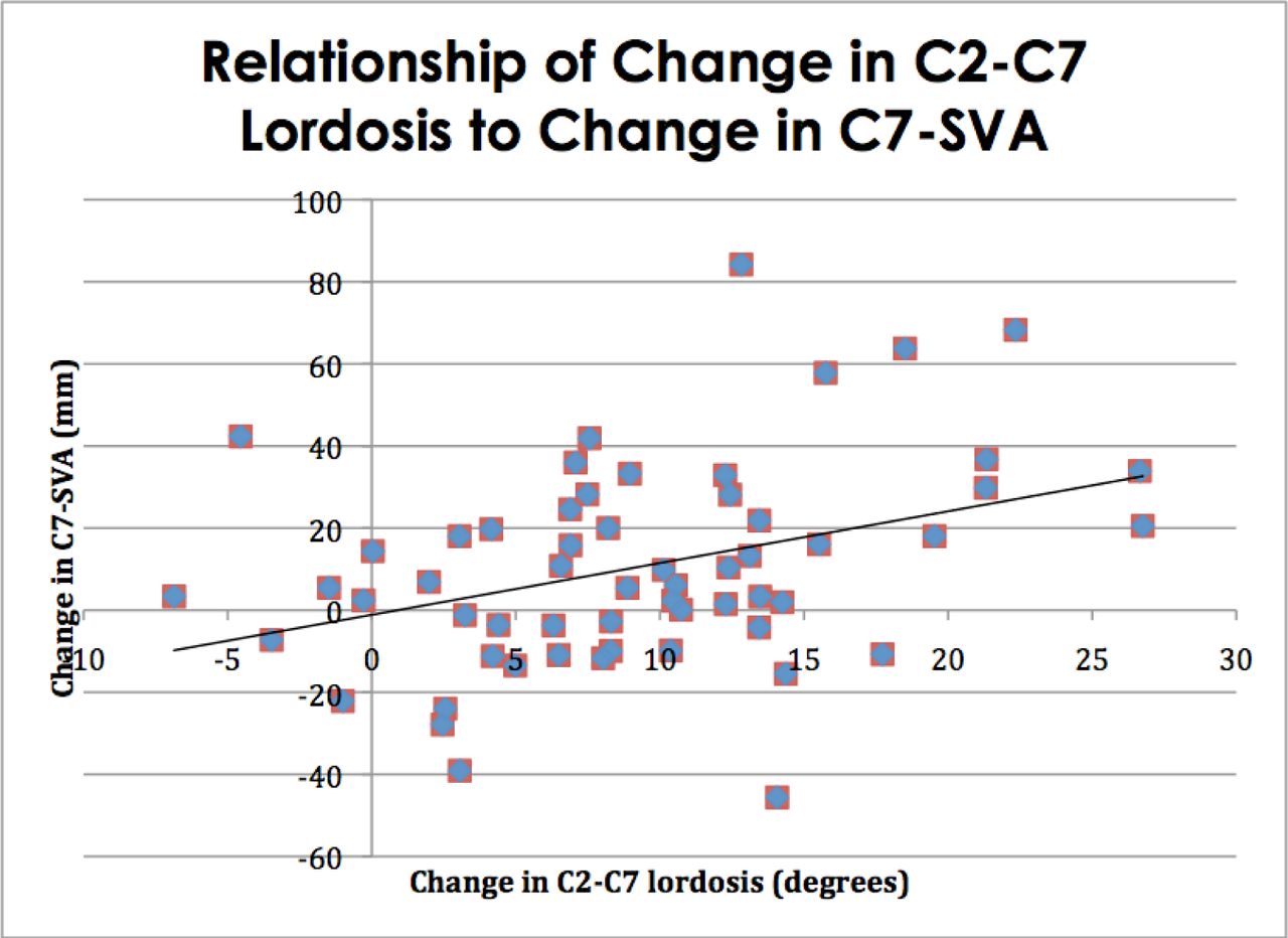
- Fig. 5
Scatter plot and regression analysis of change in C2-C7 Lordosis vs. change in C7-SVA in pre- to postoperative measurements.
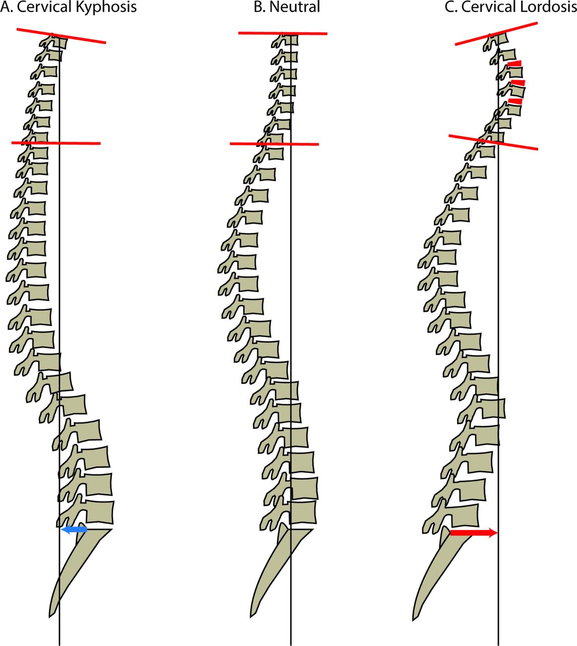
- Fig. 6
Diagram of Sagittal Vertical Axis (SVA, black line) shift with regards to increasing cervical lordosis (Cobb angle measured by red lines). As Cervical Lordosis increased from A to C, SVA also increases (blue and red arrow).


Tables
Demographic Mean Age 56 yrs Sex 47 female Smoking N= 32 Mean Weight 185.1 Lbs Mean Height 64.9 inches Levels Fused 4 levels, N=46
3 Levels, N=23Demographic Mean Age 56 yrs Sex 39 female, 19 Male Smoking N= 28 Mean Weight 182.1 Lbs Mean Height 65.1 inches Levels Fused 4 levels, N=39
3 Levels, N=19
In this issue


Jump to section
Related Articles
Cited By...
- No citing articles found.