
Main menu

  • Home
  • Content
    • Current Issue
    • Advance Online Publication
    • Archive
  • About Us
    • About ISASS
    • About the Journal
    • Author Instructions
    • Editorial Board
    • Reviewer Guidelines & Publication Criteria
  • More
    • Advertise
    • Subscribe
    • Alerts
    • Feedback
  • Join Us
  • Reprints & Permissions
  • Sponsored Content
  • Other Publications
    • ijss

User menu

  • My alerts

Search

  • Advanced search
International Journal of Spine Surgery
  • My alerts
International Journal of Spine Surgery

Advanced Search

  • Home
  • Content
    • Current Issue
    • Advance Online Publication
    • Archive
  • About Us
    • About ISASS
    • About the Journal
    • Author Instructions
    • Editorial Board
    • Reviewer Guidelines & Publication Criteria
  • More
    • Advertise
    • Subscribe
    • Alerts
    • Feedback
  • Join Us
  • Reprints & Permissions
  • Sponsored Content
  • Follow ijss on Twitter
  • Visit ijss on Facebook
Research ArticleCervical Spine

The Four Fixation Points of the Axis: Technique and Case Report

KRIS SIEMIONOW, PIOTR JANUSZ and STEVEN MARDJETKO
International Journal of Spine Surgery October 2018, 12 (5) 611-616; DOI: https://doi.org/10.14444/5073
KRIS SIEMIONOW
1Department of Orthopaedics, University of Illinois, Chicago, Illinois
MD
  • Find this author on Google Scholar
  • Find this author on PubMed
  • Search for this author on this site
PIOTR JANUSZ
1Department of Orthopaedics, University of Illinois, Chicago, Illinois
2Department of Spine Disorders and Pediatric Orthopedics, University of Medical Sciences, Poznan, Poland
MD
  • Find this author on Google Scholar
  • Find this author on PubMed
  • Search for this author on this site
STEVEN MARDJETKO
3Illinois Bone and Joint Institute, Chicago, Illinois
MD
  • Find this author on Google Scholar
  • Find this author on PubMed
  • Search for this author on this site
  • Article
  • Figures & Data
  • Info & Metrics
  • PDF
Loading

Article Figures & Data

Figures

  • Figure 1
    • Download figure
    • Open in new tab
    • Download powerpoint
    Figure 1

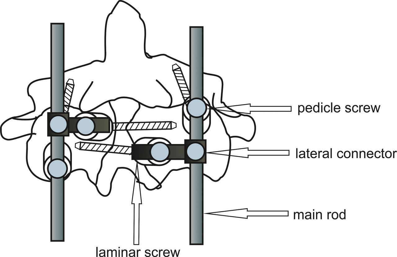
    Schematic diagram describing position of the screws with offset connectors.

  • Figure 2
    • Download figure
    • Open in new tab
    • Download powerpoint
    Figure 2

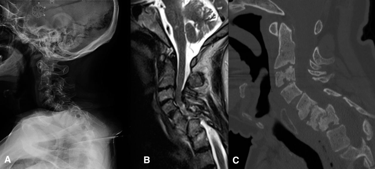
    (A) Lateral x-ray of the cervical spine of a 56-year-old male with Down syndrome and progressive myelopathy. (B) Sagittal magnetic resonance imaging demonstrates severe multilevel spinal cord compression with myelomalacia. (C) Sagittal computed tomography scan demonstrates C6-C7 spondylolishtesis with advanced degenerative changes and endplate erosions at C2-C3, C3-C4, C5-C6, and C6-C7.
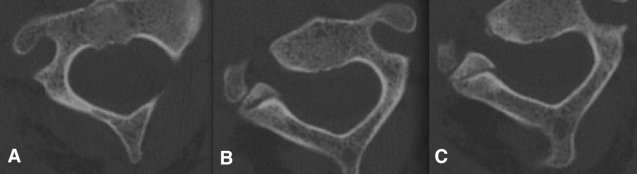
  • Figure 3
    • Download figure
    • Open in new tab
    • Download powerpoint
    Figure 3

    Axial computed tomography scan throughout the C2 vertebral body at the level of the pedicles demonstrating the right (A) pedicle, left (B) pedicle, and the lamina. Preoperative planning is critical to both implant and technique selection.

  • Figure 4
    • Download figure
    • Open in new tab
    • Download powerpoint
    Figure 4

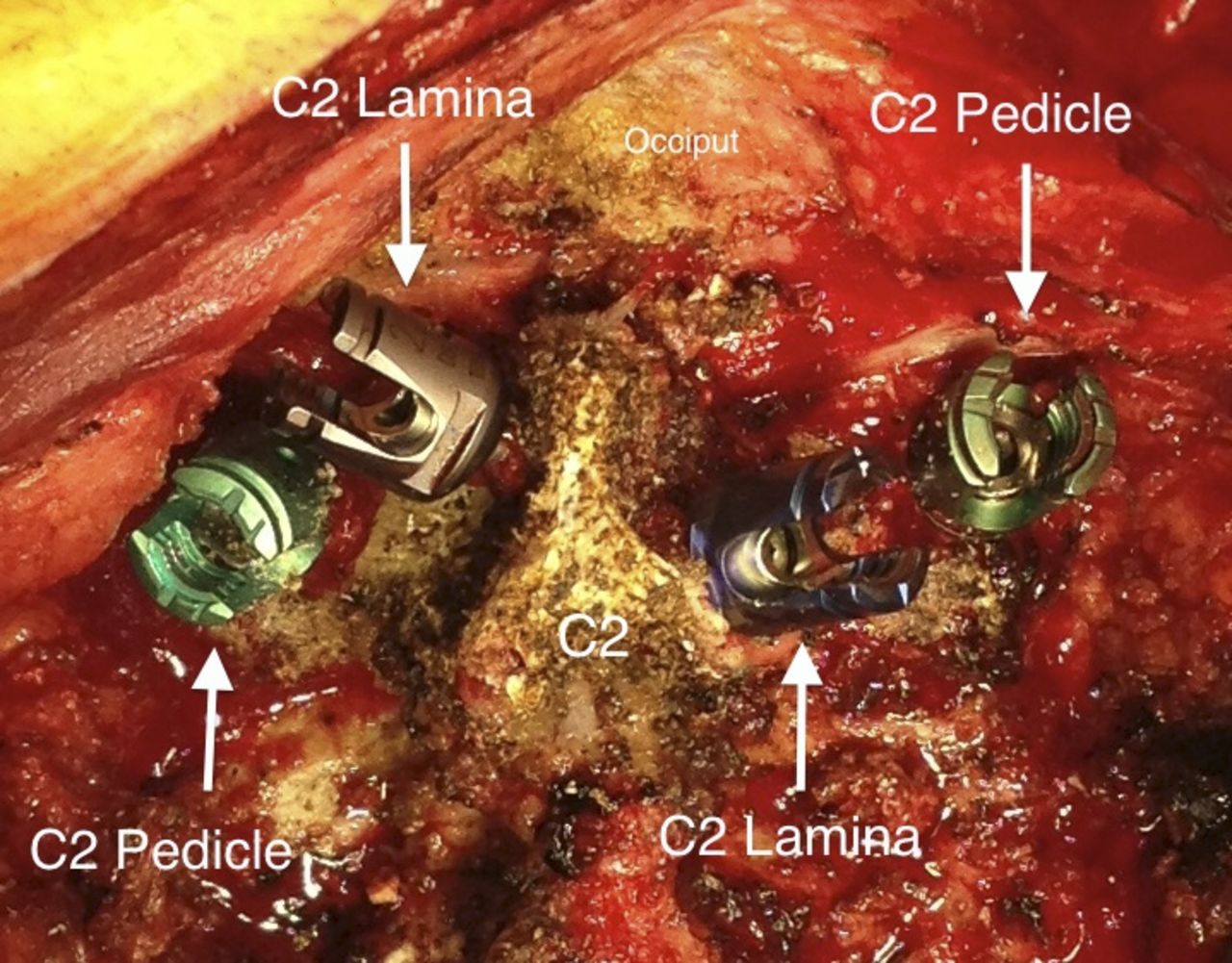
    Intraoperative photograph demonstrating placement of polyaxial pedicle screws into the pedicle and lamina of the axis. Preoperative planning is critical to ensure that all 4 screws can be safely placed into the pars/pedicle and lamina of the axis.
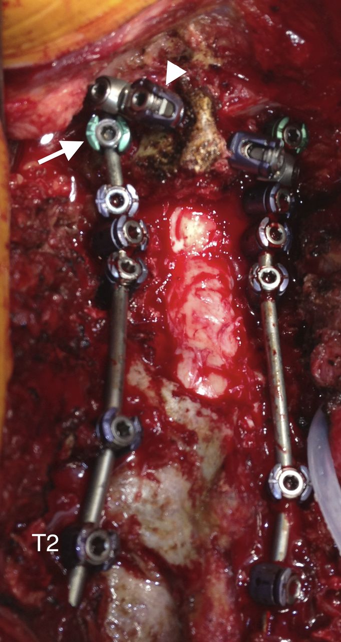
  • Figure 5
    • Download figure
    • Open in new tab
    • Download powerpoint
    Figure 5

    Intraoperative photograph demonstrating the use of lateral connectors to connect the rod to the translaminar screw. The left translaminar screw (arrowhead) is attached to the main rod via a cross connector that is anchored to the rod cephalad to the C2 pedicle screw (arrow). The pedicle screws are usually more in line with the rest of the construct and can be directly connected to the rod. Central laminectomy from C5 to C7 is visualized.

  • Figure 6
    • Download figure
    • Open in new tab
    • Download powerpoint
    Figure 6

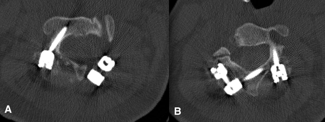
    Postoperative axial computed tomography scan of the C2 vertebrae demonstrating a right-sided C2 pedicle screw (A) and a left-sided laminar screw (B).
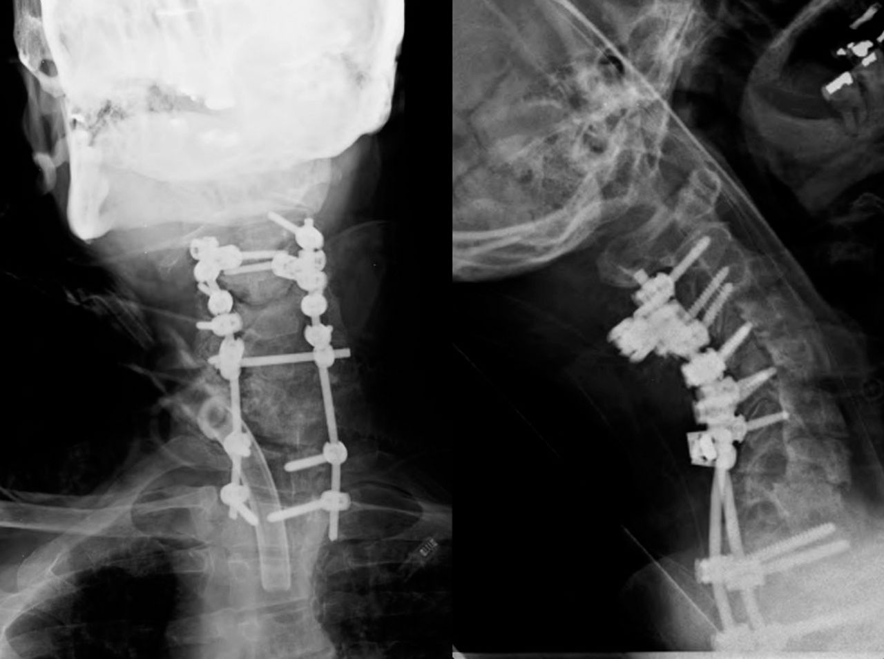
  • Figure 7
    • Download figure
    • Open in new tab
    • Download powerpoint
    Figure 7

    Postoperative antero-posterior and lateral x-rays taken 6 months postoperatively demonstrate maintenance of cervical lordosis.
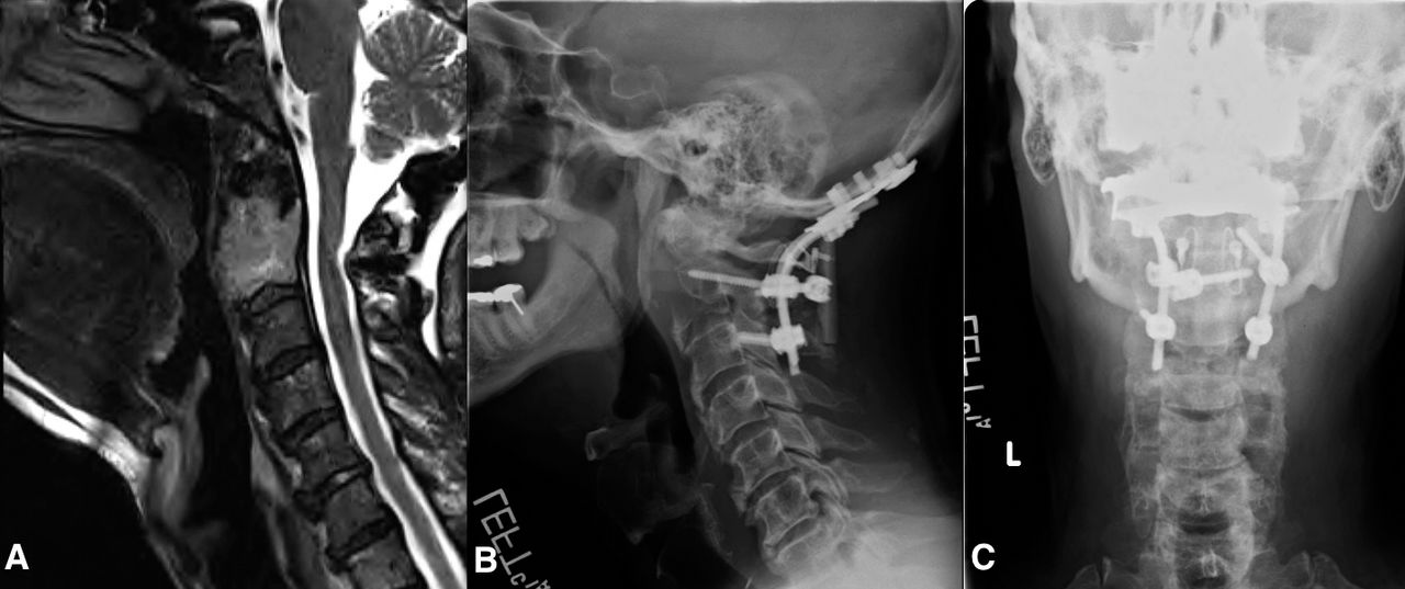
  • Figure 8
    • Download figure
    • Open in new tab
    • Download powerpoint
    Figure 8

    A 60-year-old male who was diagnosed with destructive meningioma of the C2 vertebral body and posterior spinal elements. The patient underwent an instrumented occiput to C3 posterior spinal fusion. There was destruction of the left C2 pedicle; therefore, no pedicle screw was placed on that side.

PreviousNext
Back to top

In this issue

International Journal of Spine Surgery
Vol. 12, Issue 5
1 Oct 2018
  • Table of Contents
  • Index by author

Print
Download PDF
Article Alerts
Sign In to Email Alerts with your Email Address
Email Article

Thank you for your interest in spreading the word on International Journal of Spine Surgery.

NOTE: We only request your email address so that the person you are recommending the page to knows that you wanted them to see it, and that it is not junk mail. We do not capture any email address.

Enter multiple addresses on separate lines or separate them with commas.
The Four Fixation Points of the Axis: Technique and Case Report
(Your Name) has sent you a message from International Journal of Spine Surgery
(Your Name) thought you would like to see the International Journal of Spine Surgery web site.
CAPTCHA
This question is for testing whether or not you are a human visitor and to prevent automated spam submissions.
Citation Tools
The Four Fixation Points of the Axis: Technique and Case Report
KRIS SIEMIONOW, PIOTR JANUSZ, STEVEN MARDJETKO
International Journal of Spine Surgery Oct 2018, 12 (5) 611-616; DOI: 10.14444/5073

Citation Manager Formats

  • BibTeX
  • Bookends
  • EasyBib
  • EndNote (tagged)
  • EndNote 8 (xml)
  • Medlars
  • Mendeley
  • Papers
  • RefWorks Tagged
  • Ref Manager
  • RIS
  • Zotero
Share
The Four Fixation Points of the Axis: Technique and Case Report
KRIS SIEMIONOW, PIOTR JANUSZ, STEVEN MARDJETKO
International Journal of Spine Surgery Oct 2018, 12 (5) 611-616; DOI: 10.14444/5073
Twitter logo Facebook logo Mendeley logo
  • Tweet Widget
  • Facebook Like
  • Google Plus One

Jump to section

  • Article
    • ABSTRACT
    • INTRODUCTION
    • MATERIALS AND METHODS
    • DISCUSSION
    • CONCLUSION
    • REFERENCES
  • Figures & Data
  • Info & Metrics
  • PDF

Related Articles

  • No related articles found.
  • PubMed
  • Google Scholar

Cited By...

  • No citing articles found.
  • Google Scholar

More in this TOC Section

  • Innovation and Adversity in Spine Surgery: A Retrospective
  • Cervical Myelopathy Secondary to Bilateral Atlantoaxial Pseudoarticulations in Rheumatoid Arthritis: A Case Report
  • Incidence of Traumatic Spinal Injury Following Public Policy Update on Moped Usage in South Carolina
Show more Cervical Spine

Similar Articles

Keywords

  • axis
  • pedicle screws
  • translaminar screws
  • posterior cervical fixation
  • C2 fixation
  • C2 screws

Content

  • Current Issue
  • Latest Content
  • Archive

More Information

  • About IJSS
  • About ISASS
  • Privacy Policy

More

  • Subscribe
  • Alerts
  • Feedback

Other Services

  • Author Instructions
  • Join ISASS
  • Reprints & Permissions

© 2025 International Journal of Spine Surgery

International Journal of Spine Surgery Online ISSN: 2211-4599

Powered by HighWire