


Article Figures & Data
Figures
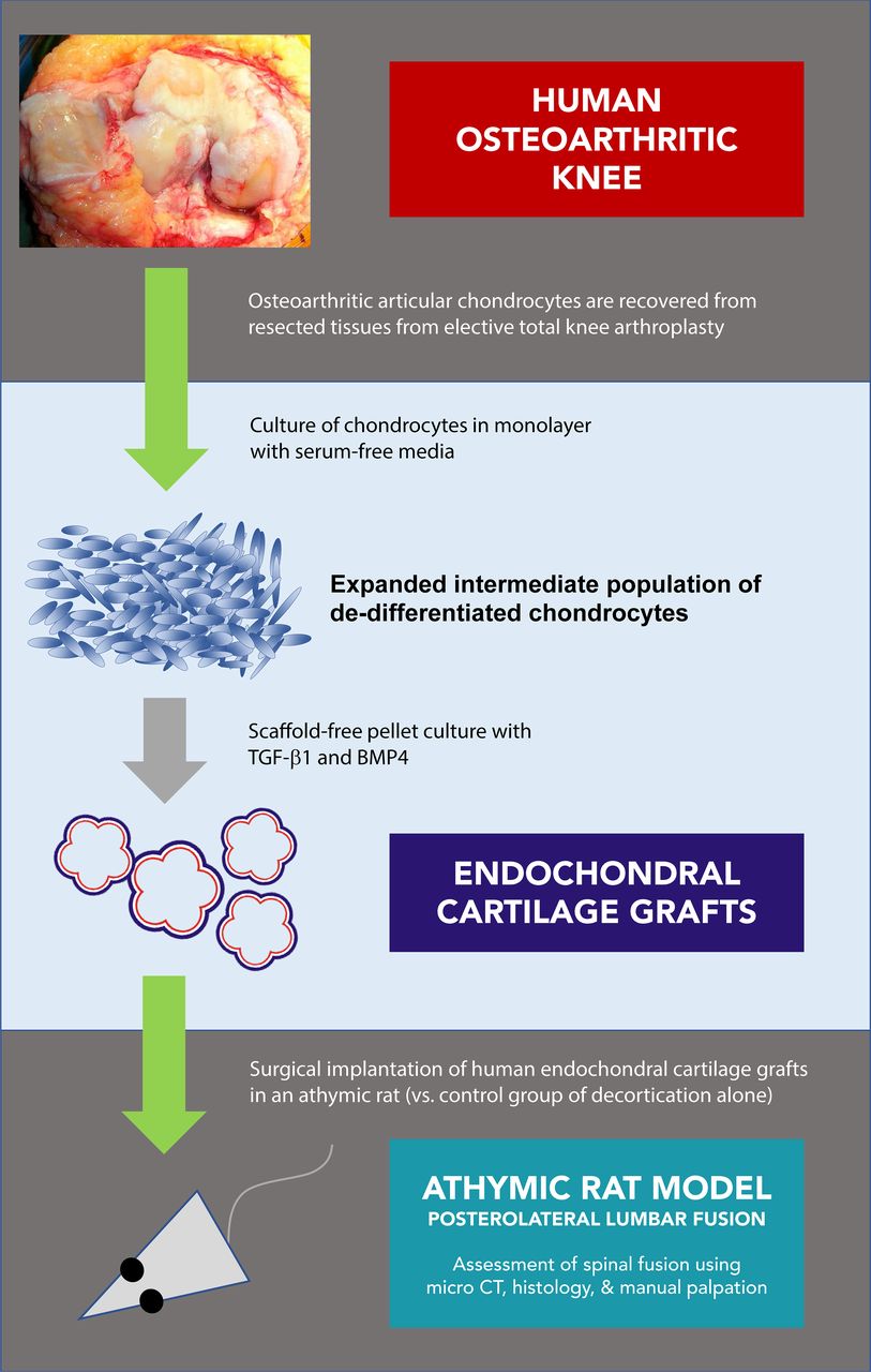
- Figure 1
Experimental outline.
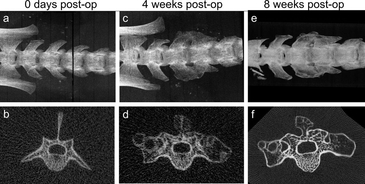
- Figure 2
Micro–computed tomography images showing time course of single-level posterolateral lumbar spinal fusion using hypertrophic chondrocyte pellet grafts in a single animal. (a, b) In vivo immediately after implantation. (c, d) In vivo 4 weeks postoperatively. (e, f) Ex vivo 8 weeks postoperatively.
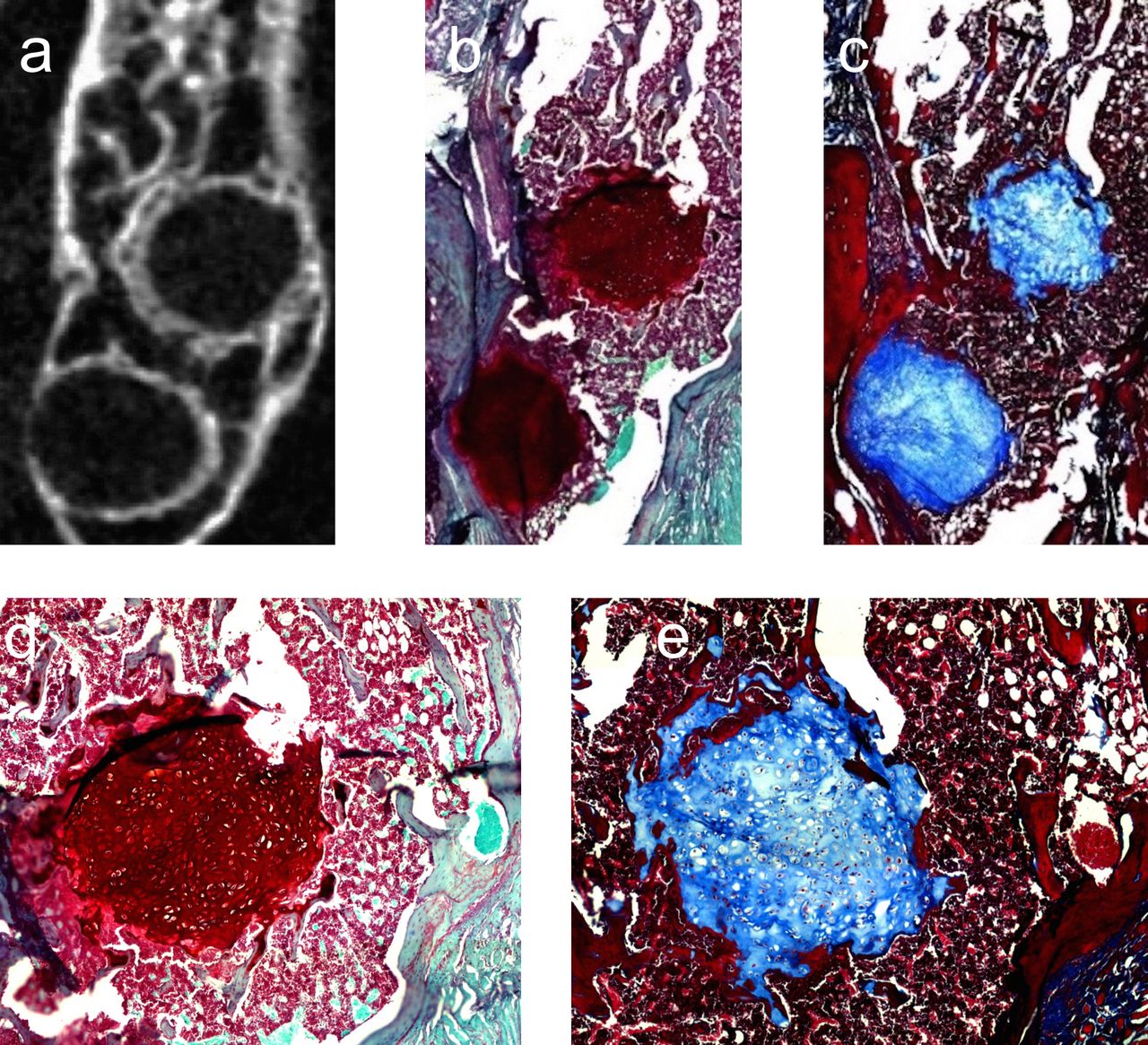
- Figure 3
Radiographic and histologic appearance of remnant cartilage grafts at 8 weeks. Micro–computed tomography (a), safranin-O staining (b, d), and Masson trichrome staining (c, e) of fusion masses show spherical areas of unmineralized cartilaginous tissue.
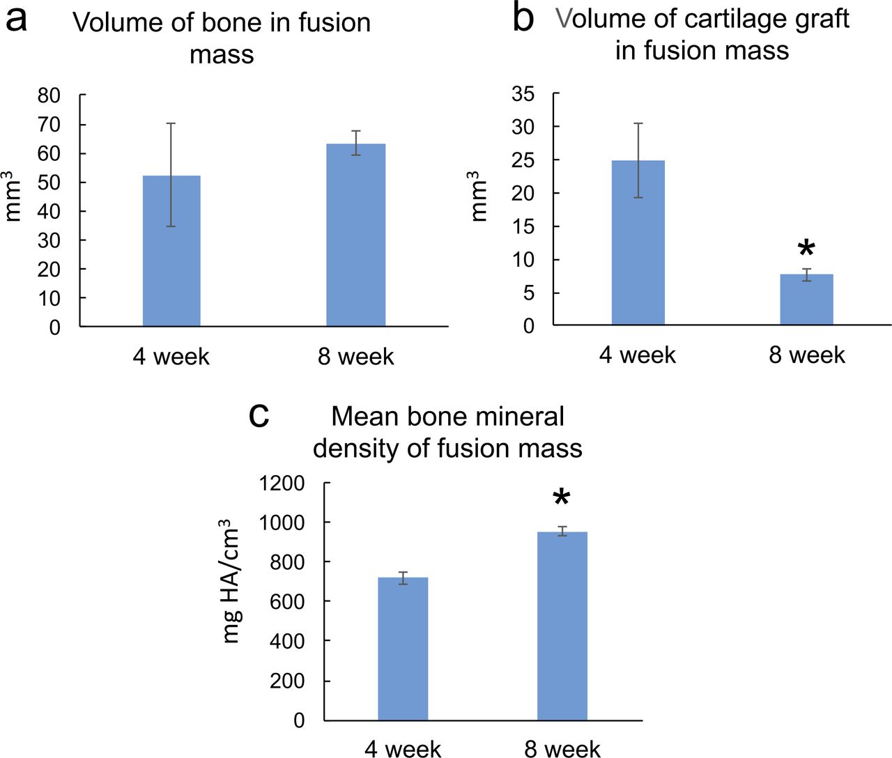
- Figure 4
Maturation of fusion masses. From 4 to 8 weeks, the amount of cartilage in the fusion masses decreased and mean bone mineral density increased. There was a trend toward increased bone volume with time; however, this did not reach statistical significance.


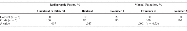
Tables
In this issue


