
Main menu

  • Home
  • Content
    • Current Issue
    • Advance Online Publication
    • Archive
  • About Us
    • About ISASS
    • About the Journal
    • Author Instructions
    • Editorial Board
    • Reviewer Guidelines & Publication Criteria
  • More
    • Advertise
    • Subscribe
    • Alerts
    • Feedback
  • Join Us
  • Reprints & Permissions
  • Sponsored Content
  • Other Publications
    • ijss

User menu

  • My alerts

Search

  • Advanced search
International Journal of Spine Surgery
  • My alerts
International Journal of Spine Surgery

Advanced Search

  • Home
  • Content
    • Current Issue
    • Advance Online Publication
    • Archive
  • About Us
    • About ISASS
    • About the Journal
    • Author Instructions
    • Editorial Board
    • Reviewer Guidelines & Publication Criteria
  • More
    • Advertise
    • Subscribe
    • Alerts
    • Feedback
  • Join Us
  • Reprints & Permissions
  • Sponsored Content
  • Follow ijss on Twitter
  • Visit ijss on Facebook
Research ArticleBiomechanics

Differences in Trabecular Bone, Cortical Shell, and Endplate Microstructure Across the Lumbar Spine

VIVEK PALEPU, SAI DEEPA RAYAPROLU and SRINIDHI NAGARAJA
International Journal of Spine Surgery August 2019, 13 (4) 361-370; DOI: https://doi.org/10.14444/6049
VIVEK PALEPU
US Food and Drug Administration, Center for Devices and Radiological Health, Office of Science and Engineering Laboratories, Division of Applied Mechanics, Silver Spring, Maryland
PHD
  • Find this author on Google Scholar
  • Find this author on PubMed
  • Search for this author on this site
SAI DEEPA RAYAPROLU
US Food and Drug Administration, Center for Devices and Radiological Health, Office of Science and Engineering Laboratories, Division of Applied Mechanics, Silver Spring, Maryland
MS
  • Find this author on Google Scholar
  • Find this author on PubMed
  • Search for this author on this site
SRINIDHI NAGARAJA
US Food and Drug Administration, Center for Devices and Radiological Health, Office of Science and Engineering Laboratories, Division of Applied Mechanics, Silver Spring, Maryland
PHD
  • Find this author on Google Scholar
  • Find this author on PubMed
  • Search for this author on this site
  • Article
  • Figures & Data
  • Info & Metrics
  • PDF
Loading

Article Figures & Data

Figures

  • Tables
  • Figure 1
    • Download figure
    • Open in new tab
    • Download powerpoint
    Figure 1

    Endplate thickness measurements at 5 different points on endplate from the mid-coronal slice of vertebra.

  • Figure 2
    • Download figure
    • Open in new tab
    • Download powerpoint
    Figure 2

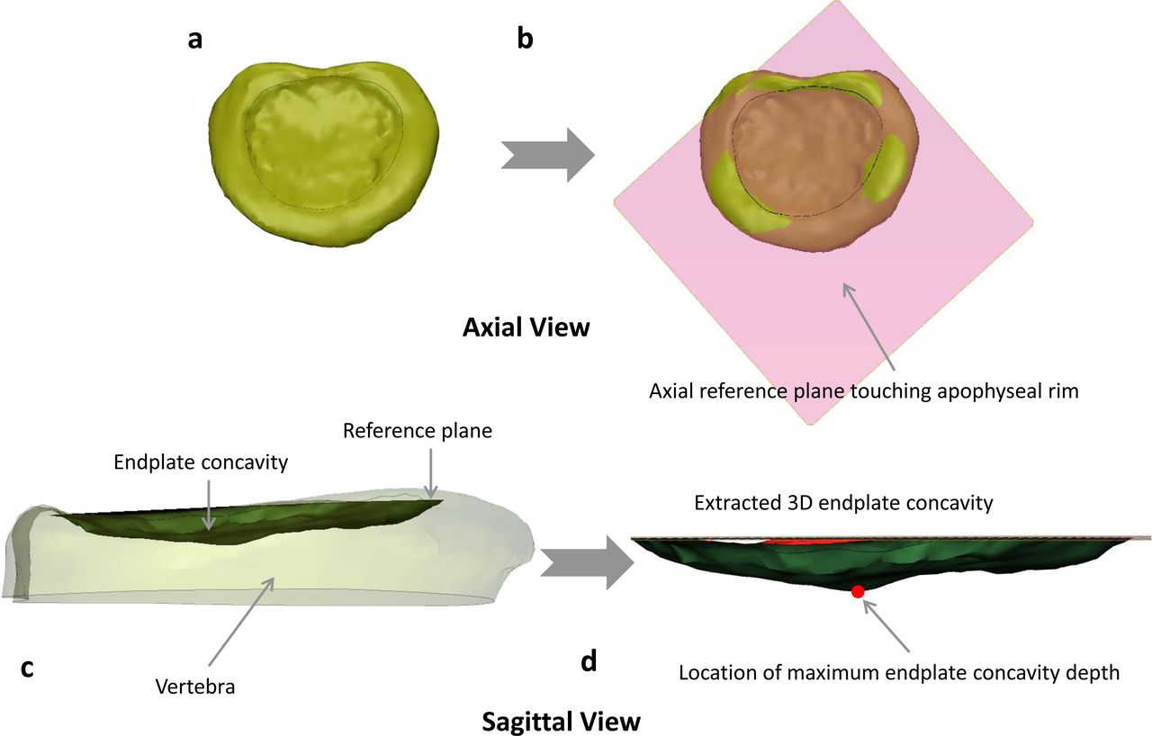
    Axial view of (a) 3-dimensional endplate structure, (b) axial reference plane created based on 3 points on apophyseal ring, and sagittal view of (c) axial reference plane on the endplate highlighting the endplate concavity space (shaded region) and (d) endplate concavity space with location of maximum concavity depth (highlighted with red dot).
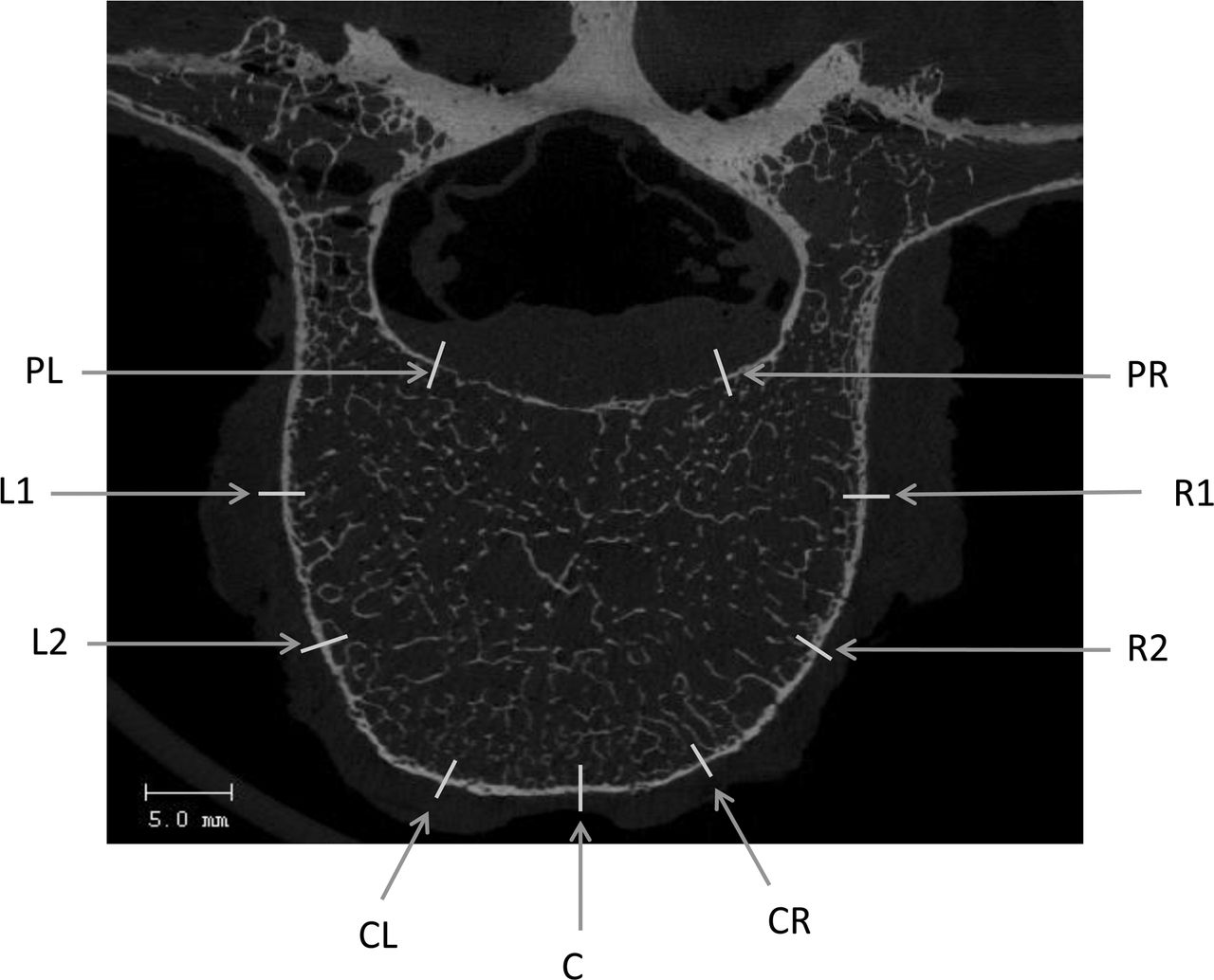
  • Figure 3
    • Download figure
    • Open in new tab
    • Download powerpoint
    Figure 3

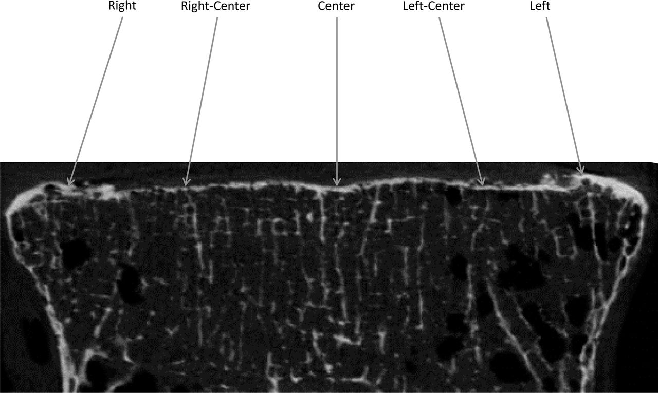
    Vertebral cortex thickness measurements at 9 different locations at the mid-axial slice of vertebra.
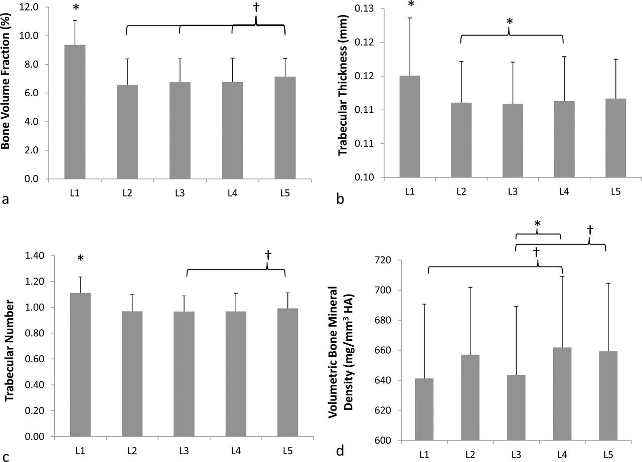
  • Figure 4
    • Download figure
    • Open in new tab
    • Download powerpoint
    Figure 4

    Mean and standard deviations of (a) trabecular bone volume fraction, (b) trabecular thickness, (c) trabecular number, and (d) volumetric bone mineral density for all lumbar vertebrae (*P ≤ .05; †P ≤ .1).
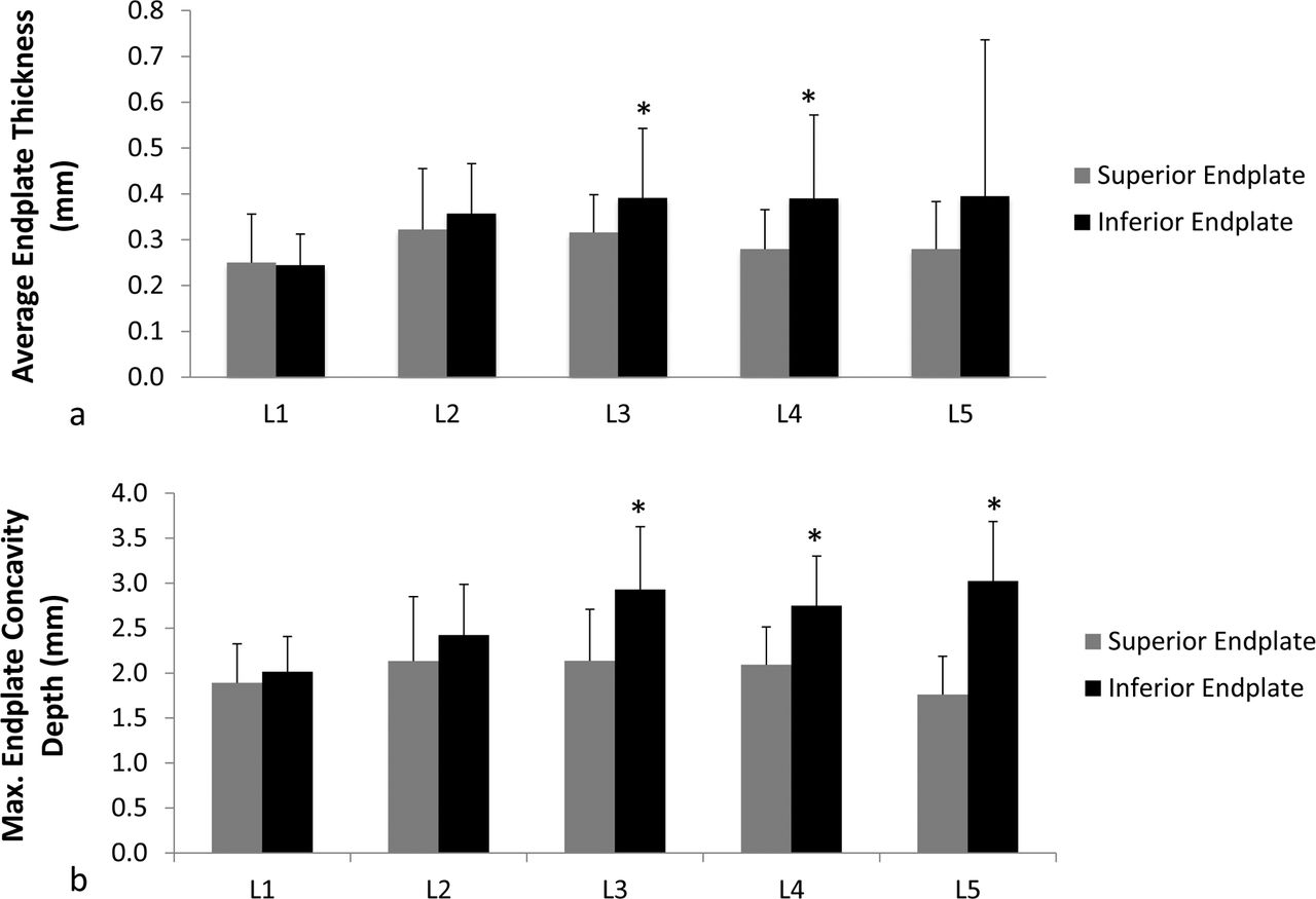
  • Figure 5
    • Download figure
    • Open in new tab
    • Download powerpoint
    Figure 5

    Mean and standard deviations of (a) average endplate thickness and (b) maximum endplate concavity depth in millimeters for superior and inferior endplates of all lumbar vertebrae. Student paired t test analyses were performed to obtain statistical significance between superior and inferior endplates (*P ≤ .05).
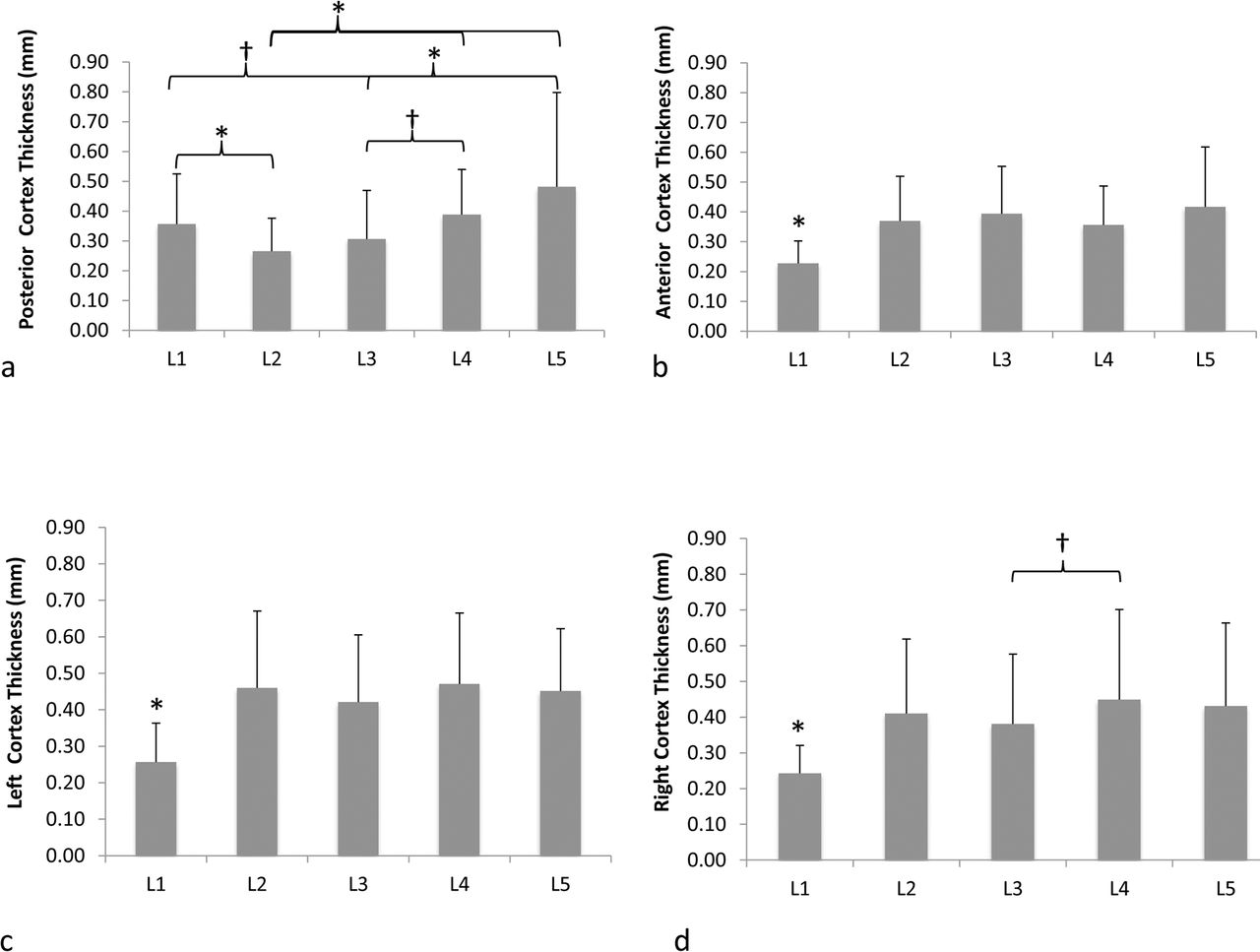
  • Figure 6
    • Download figure
    • Open in new tab
    • Download powerpoint
    Figure 6

    Mean and standard deviations of vertebral cortical thickness for (a) posterior, (b) anterior, (c) left, and (d) right cortex regions for all lumbar vertebrae (*P ≤ .05; †P < .1).
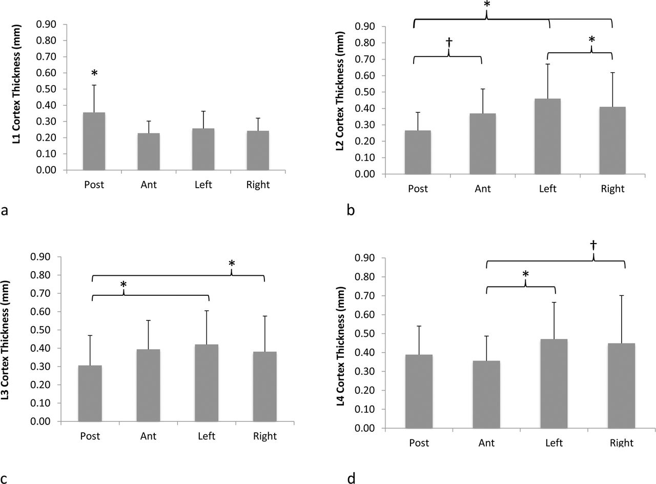
  • Figure 7
    • Download figure
    • Open in new tab
    • Download powerpoint
    Figure 7

    Mean and standard deviations of vertebral cortical thickness for (a) L1, (b) L2, (c) L3, (d) L4, and (e) L5 vertebrae depicting differences in thickness values between posterior, anterior, left, and right cortex regions of vertebral body (*P ≤ .05; †P < .1).

Tables

  • Figures
  • Table 1
PreviousNext
Back to top

In this issue

International Journal of Spine Surgery
Vol. 13, Issue 4
1 Aug 2019
  • Table of Contents
  • Index by author

Print
Download PDF
Article Alerts
Sign In to Email Alerts with your Email Address
Email Article

Thank you for your interest in spreading the word on International Journal of Spine Surgery.

NOTE: We only request your email address so that the person you are recommending the page to knows that you wanted them to see it, and that it is not junk mail. We do not capture any email address.

Enter multiple addresses on separate lines or separate them with commas.
Differences in Trabecular Bone, Cortical Shell, and Endplate Microstructure Across the Lumbar Spine
(Your Name) has sent you a message from International Journal of Spine Surgery
(Your Name) thought you would like to see the International Journal of Spine Surgery web site.
CAPTCHA
This question is for testing whether or not you are a human visitor and to prevent automated spam submissions.
Citation Tools
Differences in Trabecular Bone, Cortical Shell, and Endplate Microstructure Across the Lumbar Spine
VIVEK PALEPU, SAI DEEPA RAYAPROLU, SRINIDHI NAGARAJA
International Journal of Spine Surgery Aug 2019, 13 (4) 361-370; DOI: 10.14444/6049

Citation Manager Formats

  • BibTeX
  • Bookends
  • EasyBib
  • EndNote (tagged)
  • EndNote 8 (xml)
  • Medlars
  • Mendeley
  • Papers
  • RefWorks Tagged
  • Ref Manager
  • RIS
  • Zotero
Share
Differences in Trabecular Bone, Cortical Shell, and Endplate Microstructure Across the Lumbar Spine
VIVEK PALEPU, SAI DEEPA RAYAPROLU, SRINIDHI NAGARAJA
International Journal of Spine Surgery Aug 2019, 13 (4) 361-370; DOI: 10.14444/6049
Twitter logo Facebook logo Mendeley logo
  • Tweet Widget
  • Facebook Like
  • Google Plus One

Jump to section

  • Article
    • ABSTRACT
    • INTRODUCTION
    • METHODS
    • RESULTS
    • DISCUSSION
    • CONCLUSIONS
    • ACKNOWLEDGMENTS
    • Footnotes
    • REFERENCES
  • Figures & Data
  • Info & Metrics
  • PDF

Related Articles

  • No related articles found.
  • PubMed
  • Google Scholar

Cited By...

  • No citing articles found.
  • Google Scholar

More in this TOC Section

  • Sequential Anterior Longitudinal Ligament Release With Expandable Spacers for Lordosis Correction in Anterior-to-Psoas Lumbar Interbody Fusion: A Radiographic and Biomechanical Study
  • Comparative Biomechanical Analysis of Anterior Lumbar Interbody Fusion and Bilateral Expandable Transforaminal Lumbar Interbody Fusion Cages: A Finite Element Analysis Study
  • Impact of Different Operative Techniques for Patients With Adolescent Idiopathic Scoliosis on Frontal Curve Correction and Sagittal Balance
Show more Biomechanics

Similar Articles

Keywords

  • endplate concavity depth
  • trabecular bone microstructure
  • vertebral cortex thickness
  • endplate thickness
  • lumbar spine
  • anatomy

Content

  • Current Issue
  • Latest Content
  • Archive

More Information

  • About IJSS
  • About ISASS
  • Privacy Policy

More

  • Subscribe
  • Alerts
  • Feedback

Other Services

  • Author Instructions
  • Join ISASS
  • Reprints & Permissions

© 2025 International Journal of Spine Surgery

International Journal of Spine Surgery Online ISSN: 2211-4599

Powered by HighWire