


Article Figures & Data
Figures
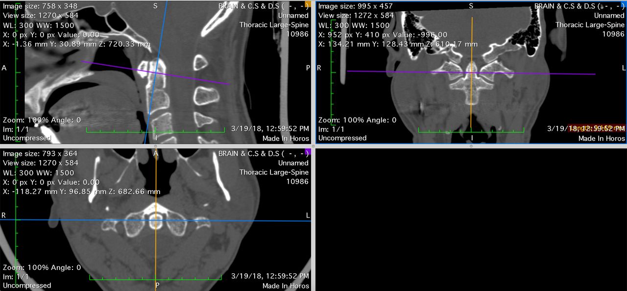
- Figure 1
This computerized tomography image shows the orientation of the sagittal (orange), axial (purple), and coronal (blue) sections of the odontoid in the Horos software.
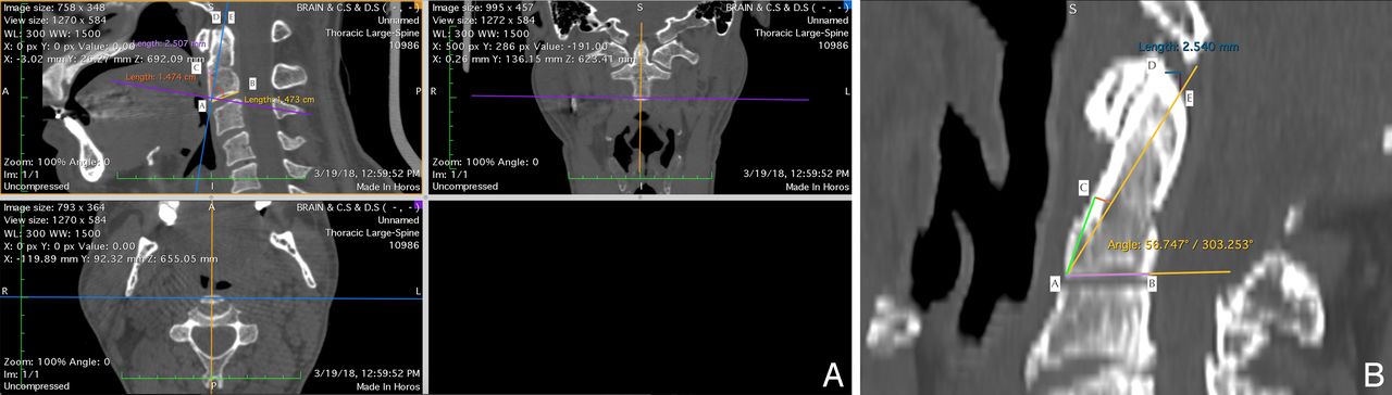
- Figure 2
(A) Computerized tomography image shows the points A, B, C, D, and E. Line AB (yellow), line AC (orange), line AD (blue), and angle DAC (screw insertion angle). (B) Computerized tomography image shows the axial cut is oriented perpendicular to the screw insertion axis.
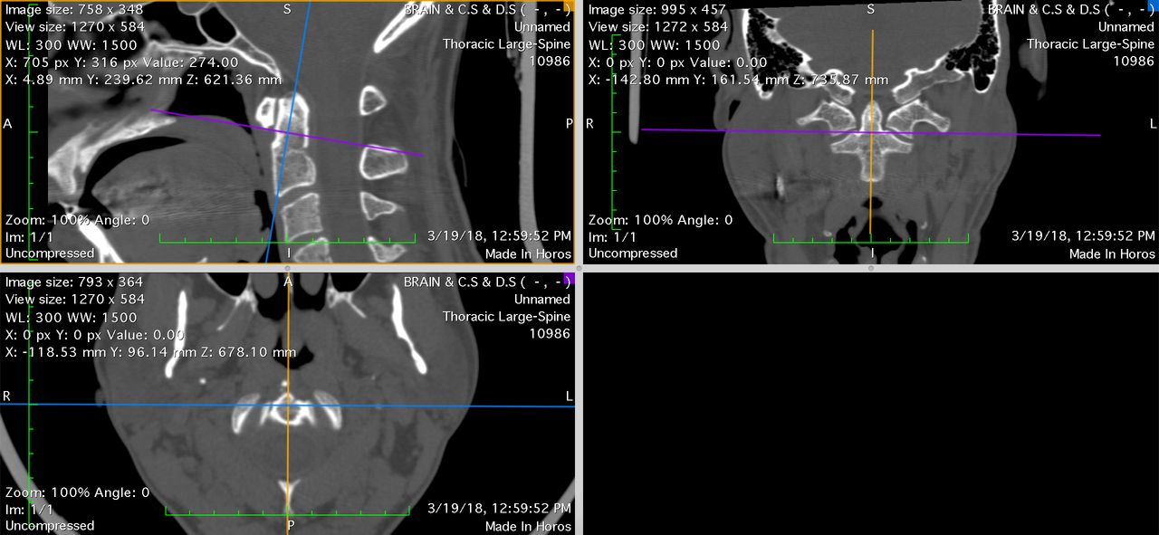
- Figure 3
Axial cut through the base of the odontoid and oriented perpendicular to the screw insertion axis.
- Figure 4
Measurement of the anteroposterior and transverse dimensions at the base of the odontoid in the axial cut.
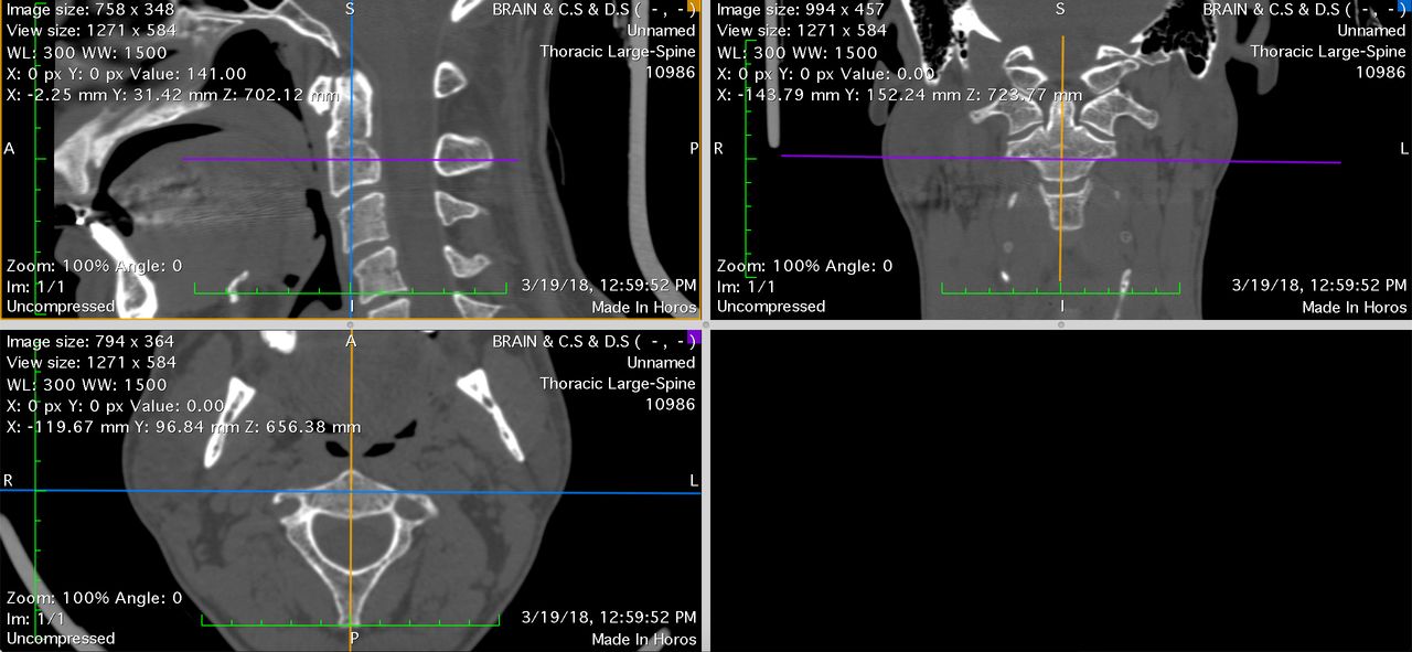
- Figure 5
The orientation of the images in the axial coronal and sagittal plane. The axial cutis through the waist of the odontoid and perpendicular to the screw insertion axis.
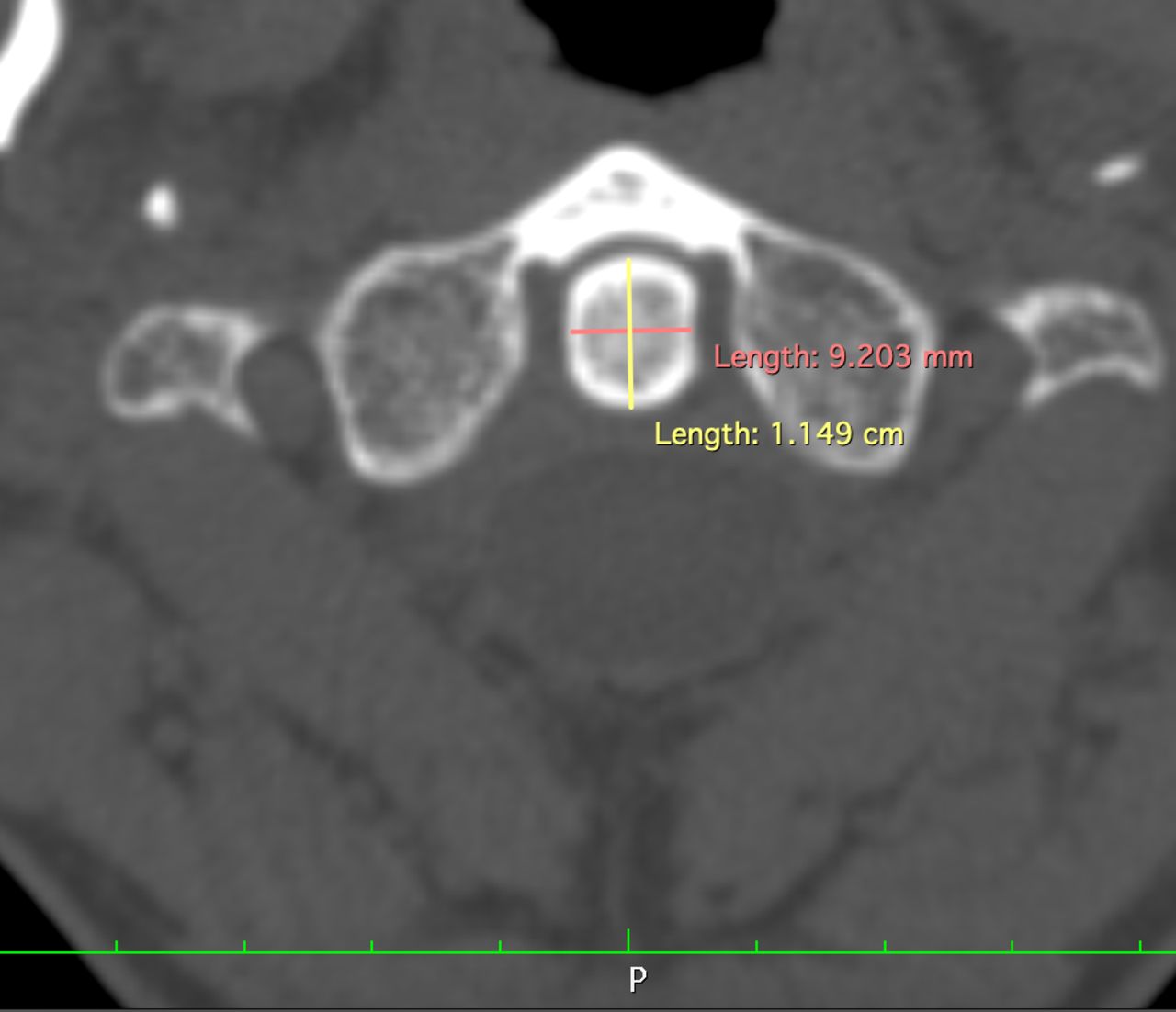
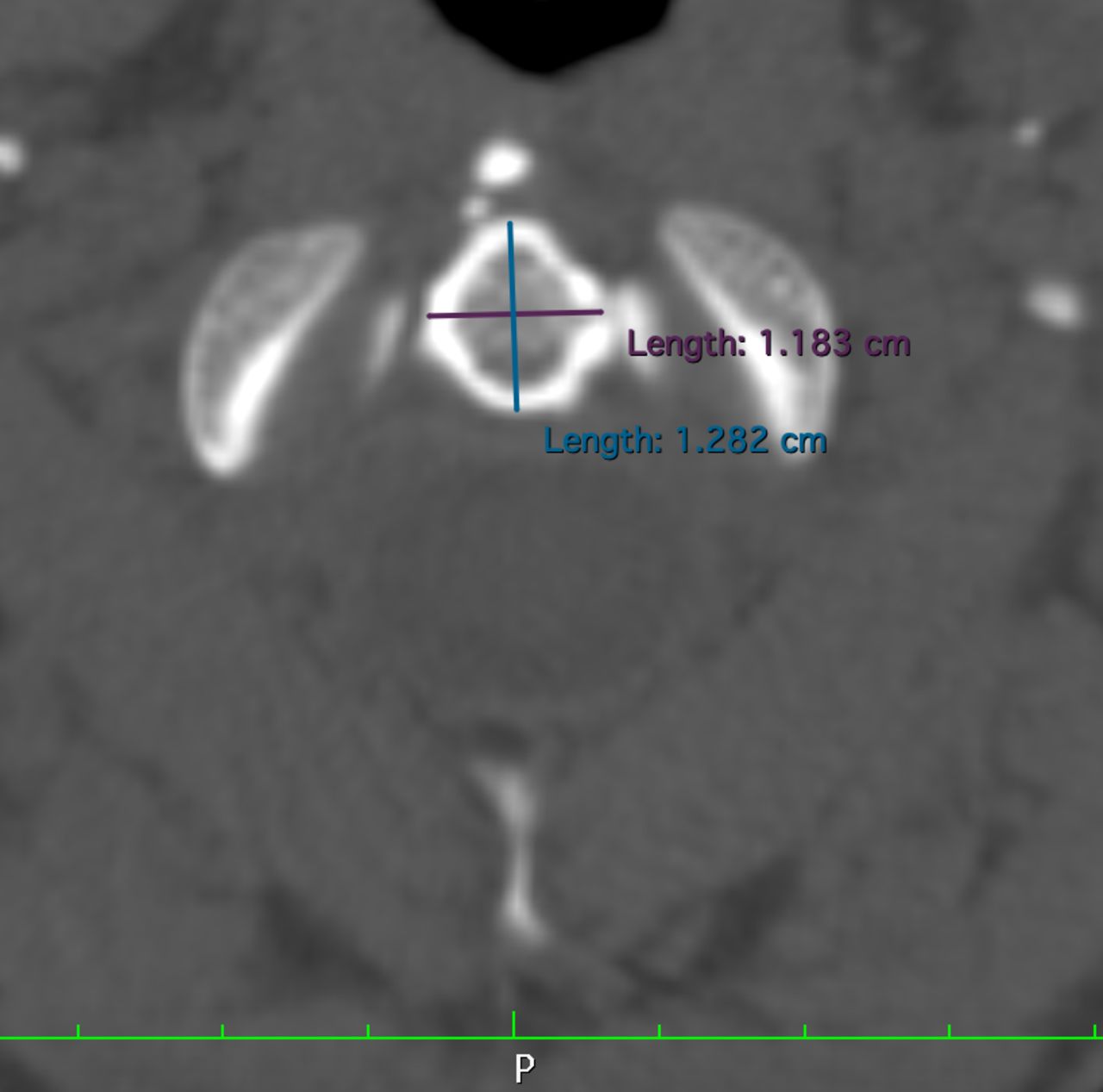
- Figure 6
Measurement of the anteroposterior and transverse dimensions at the waist of the odontoid in the axial cut.
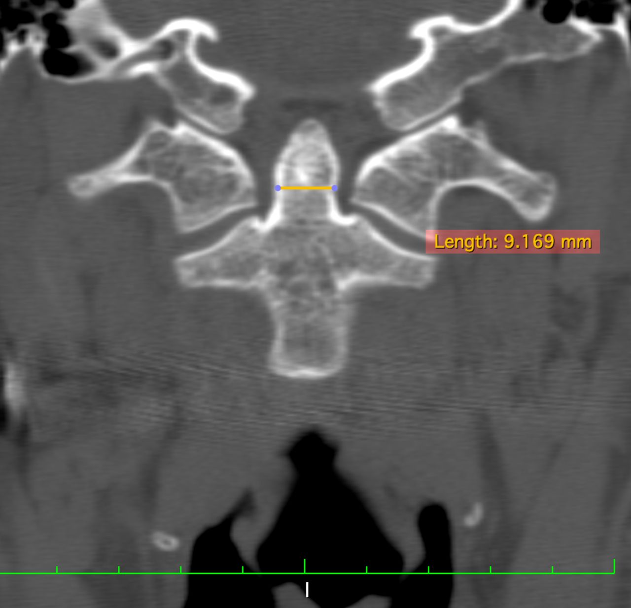
- Figure 7
Measurement of the width at the waist of the odontoid in the coronal cut.
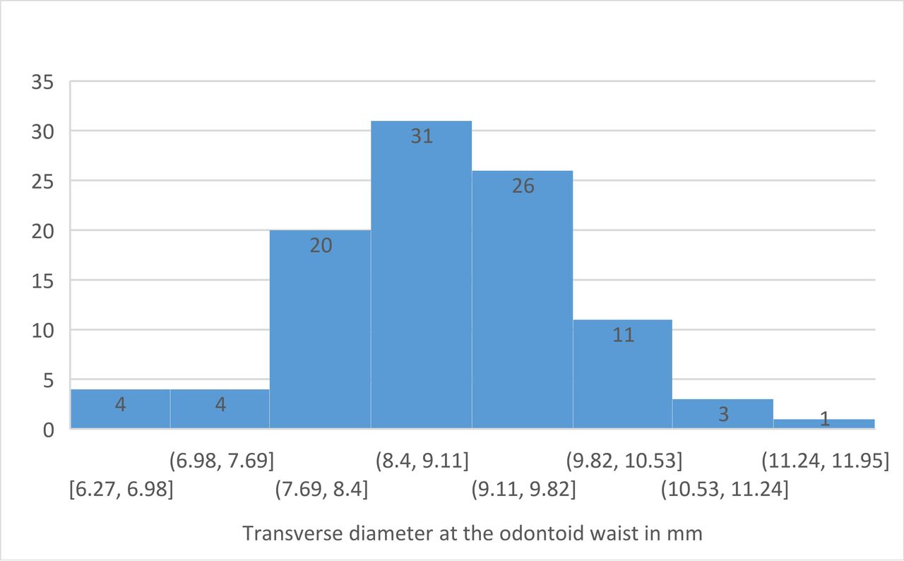
- Figure 8
Histogram showing the distribution of the measurement value of the transverse diameter of the odontoid at the waist taken in the axial cut.
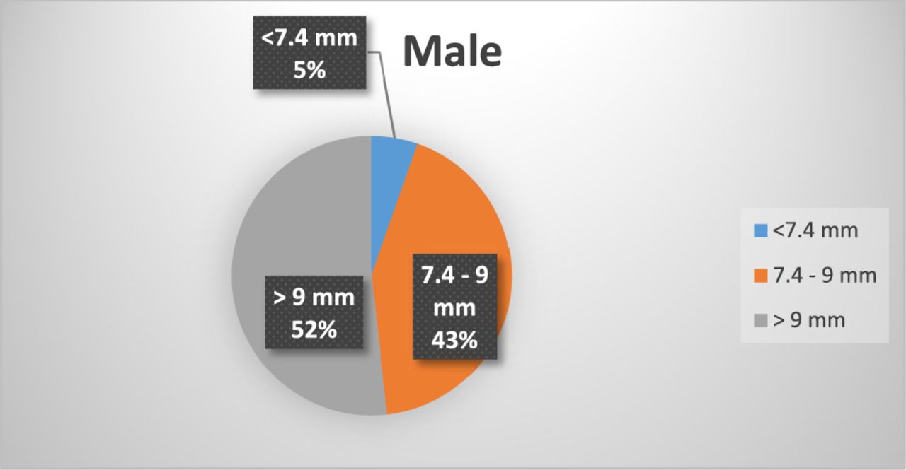
- Figure 9
A pie graph showing the distribution of the values for the transverse diameter of the odontoid measured at its waist in an axial cut perpendicular to the screw insertion axis.


Tables
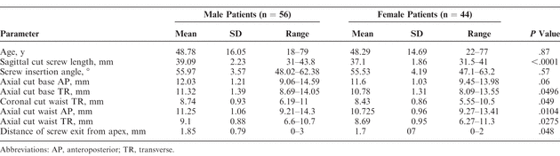
In this issue


Jump to section
Related Articles
Cited By...
- No citing articles found.