


Article Figures & Data
Figures
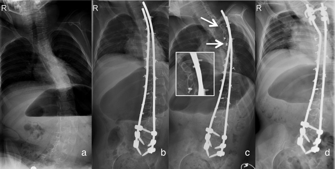
- Figure 1
Coronal radiographs. (a) Preoperative scoliosis. (b) Postoperative instrumented spine. (c) Observation of the thoracic broken sublaminar cables (arrows) at 5-month follow-up. (d) 3 years after revision surgery.
- Figure 2
(a) Severe metallosis surrounding the broken titanium (Ti) cables. (arrows). (b) Electron microscopy analysis of a broken Ti cable. The oblique surface of fracture demonstrates signs of metal fatigue failure.
- Figure 3
Coronal radiographs. (a) Preoperative curvature. (b) Postoperative instrumentation with correction of the curve. (c) Preserved stability of the instrumentation at 2-year follow-up showing the sliding effect at the most proximal rod/wire juncture (circles). Growth was determined as the difference in length between the proximal rod–cross-link distance 1-day postoperatively and at 2-year follow-up radiography.
- Figure 4
Coronal radiographs. (a) Preoperative curvature of the spine. (b) Postoperative radiograph. (c) Radiograph revealing the 2 broken cables at levels T4 and T5 (arrows). (d) Postoperative radiograph following revision surgery.
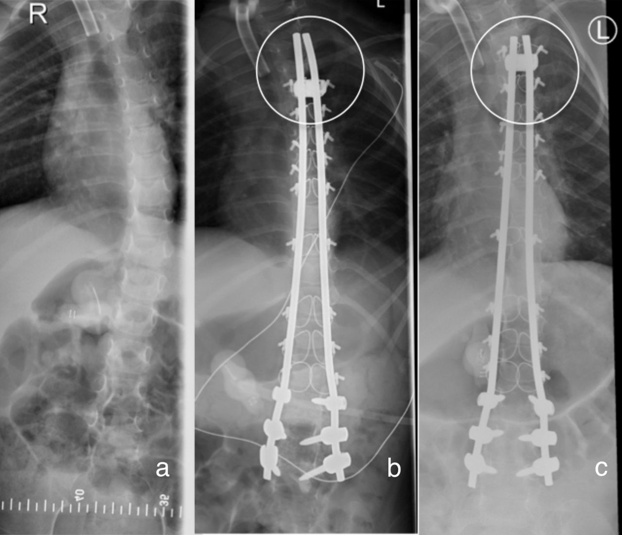
- Figure 5
Coronal and sagittal radiographs. (a) Preoperative curvature with a mild frontal Cobb angle and increased upper thoracic kyphosis. (b) Postoperative instrumentation. (c) At 6-year follow-up, the sliding effect facilitated substantial growth. The distal rods have grown out of the T4 sublaminar wire (circle). Growth was measured as the difference in length between the distance of the T4 sublaminar wires and rod ends 1-day postoperatively and at 6-year follow-up radiography.


In this issue


Jump to section
Related Articles
Cited By...
- No citing articles found.