


Article Figures & Data
Figures
- Figure 1
Intraoperative image showing the side and end-on view of placement of the right angled retractor.
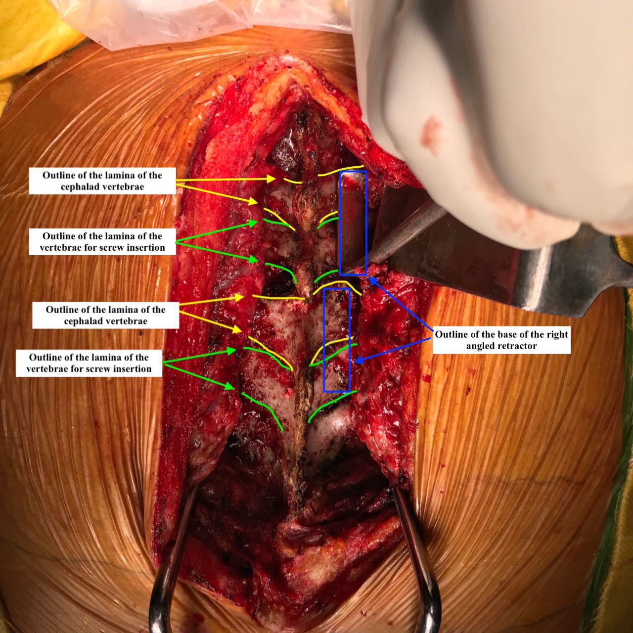
- Figure 2
Intraoperative image showing the outline of the lamina of the cephalad vertebrae and the vertebrae for screw insertion. A rectangular box is the outline for the base of the right-angled retractor. This must be placed on the surface of adjacent lamina on the contralateral side. The long limb of the retractor then guides the sagittal angulation of the screw.
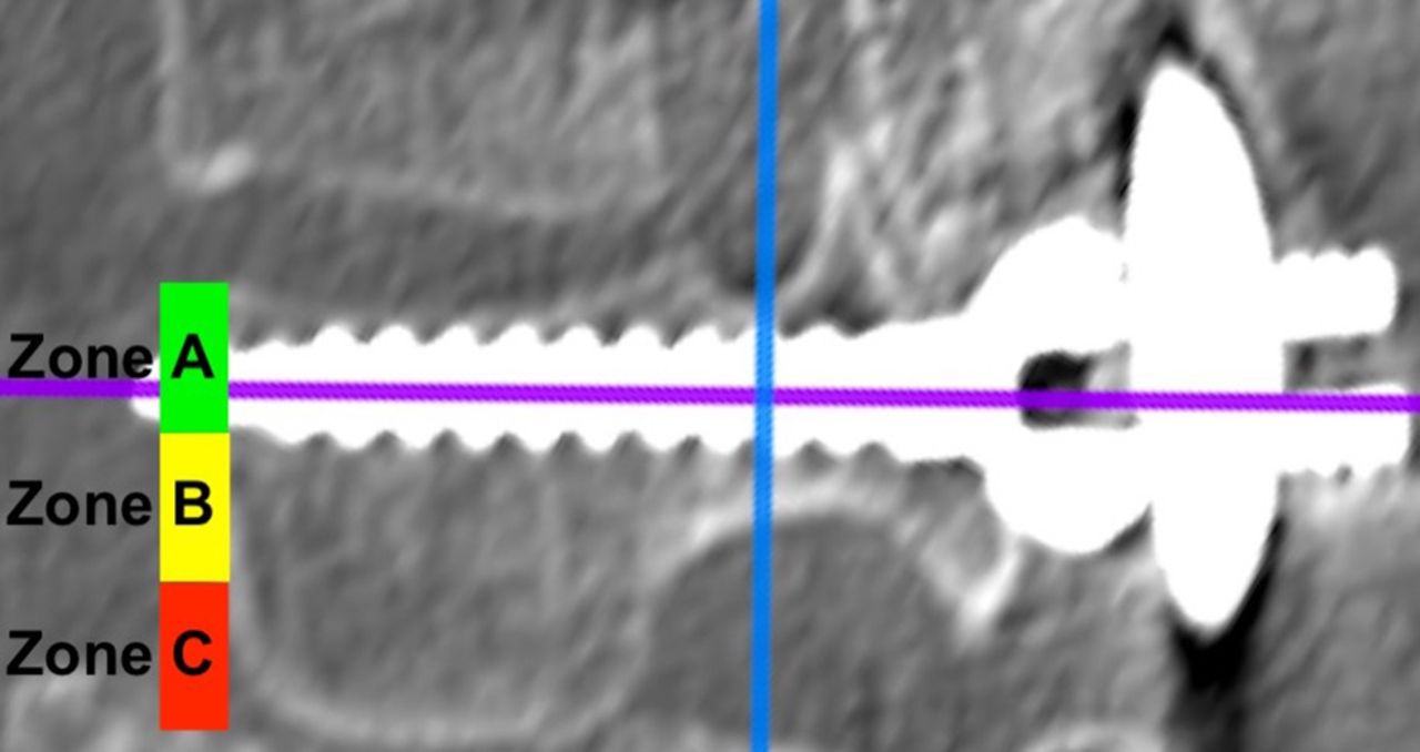
- Figure 3
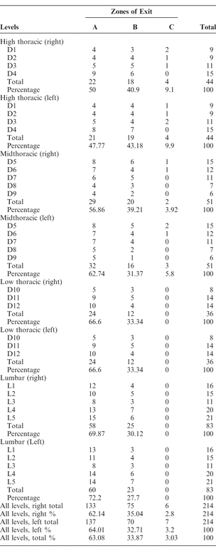
The 3 zones of the exit of the screws.
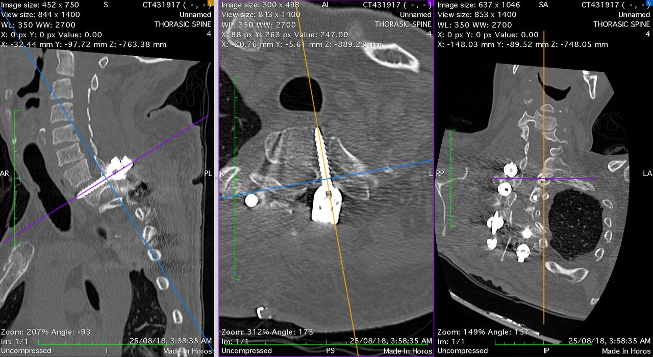
- Figure 4
Case of neglected old D3 vertebral body fracture treated with posterior spinal fusion and instrumentation from D1 to D5. Image shows the 3D MPR mode of Horos with cuts.
- Figure 5
L4-L5 transforaminal lumbar interbody fusion performed for a case of degenerative disc disease. The computed tomography scan shows the reconstructed postoperative images with the screw in zone A. The postoperative radiograph of the same patient is represented in Figure 7.
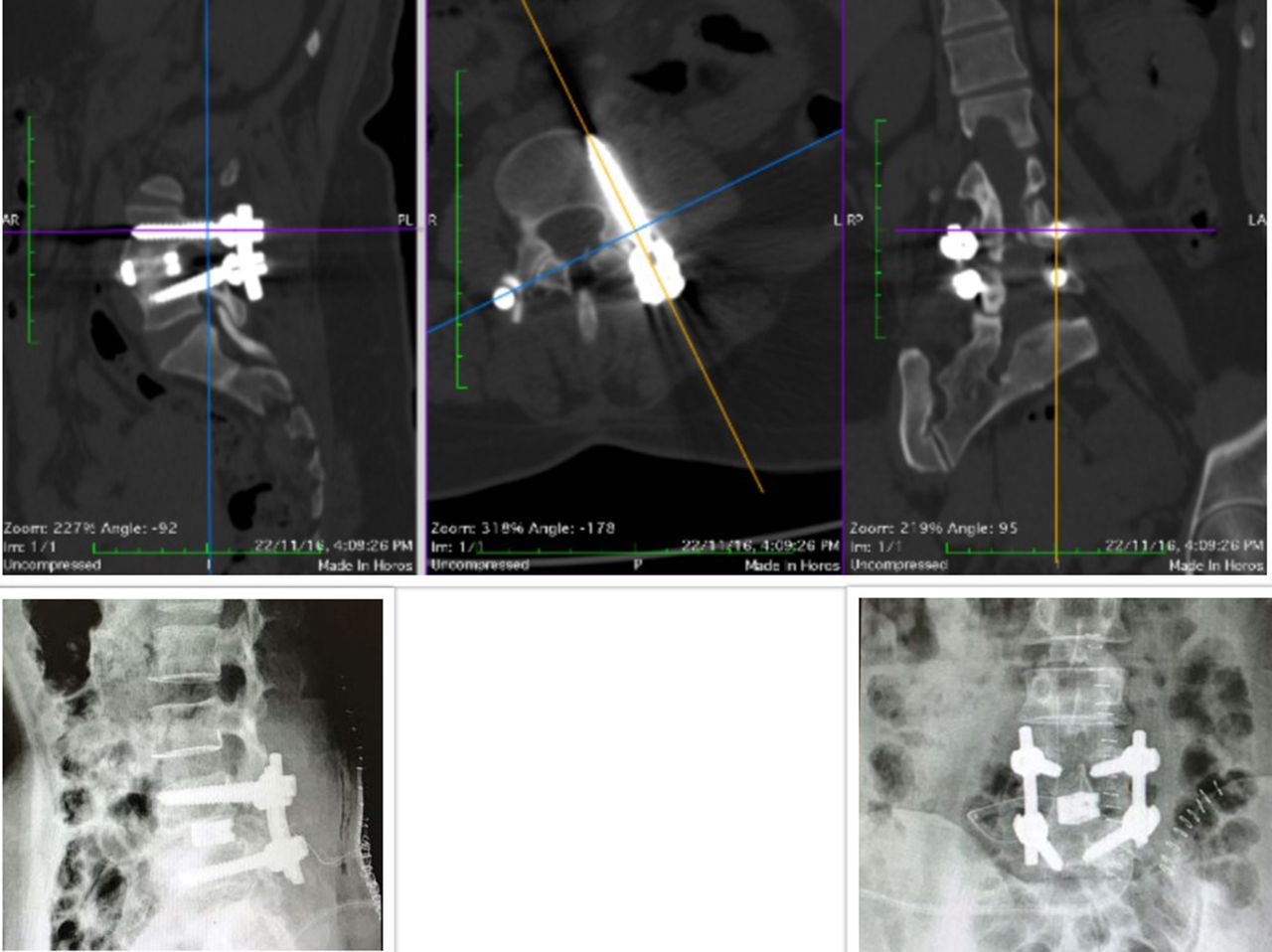
- Figure 6
A sagittal cut along the axis of the screw showing a grade 1 breach of the superior pedicle wall.
- Figure 7
(A) L2-L3 spondylodiscitis. (B) Interlaminar line and a line drawn perpendicular to the interlaminar line passing through the base of the superior articular facet simulating the base of the right angle). (C) Final postoperative x-ray.


Tables
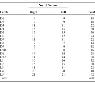
In this issue


Jump to section
Related Articles
Cited By...
- No citing articles found.