


Article Figures & Data
Figures
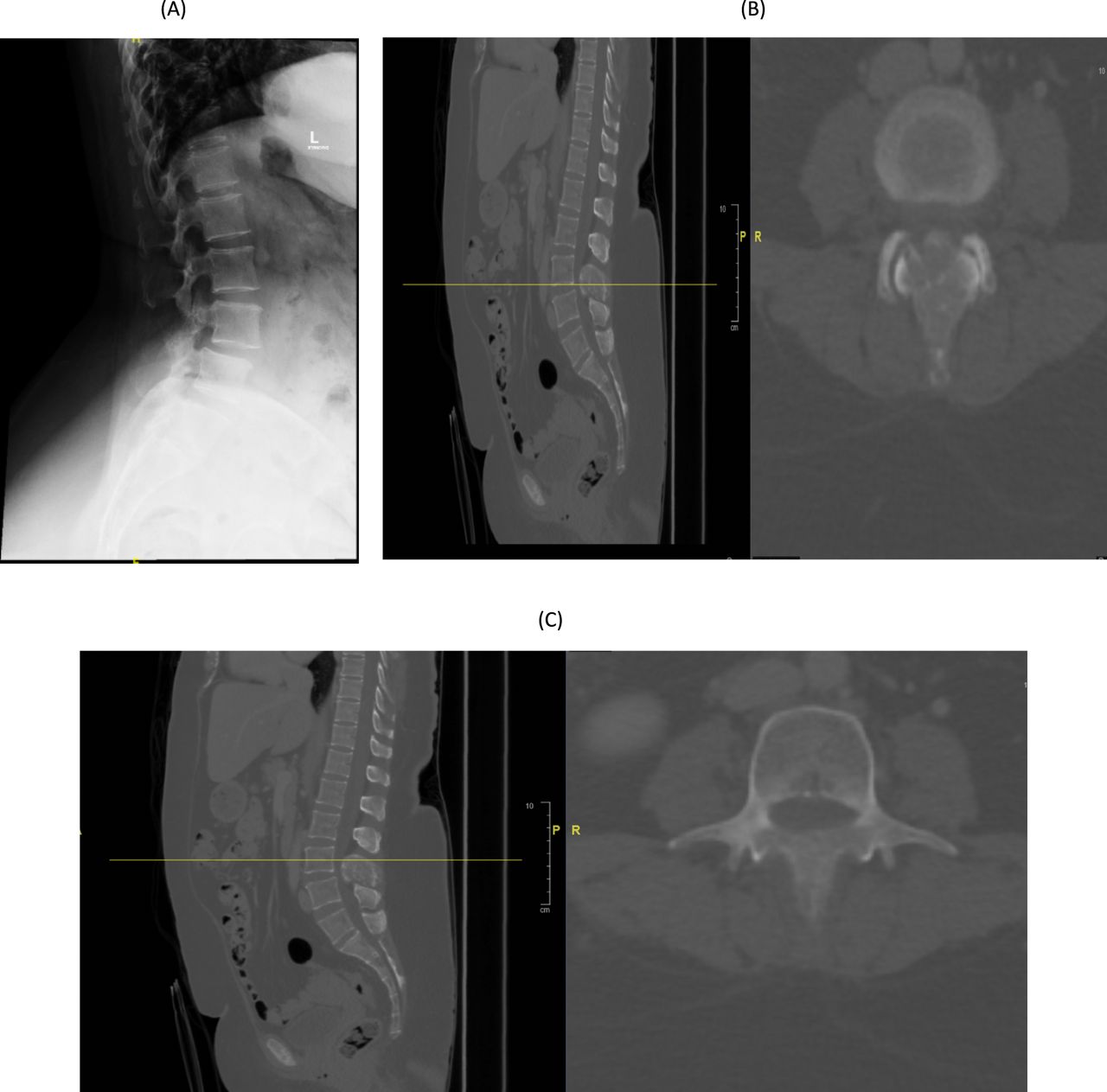
- Figure 1
(A) Sagittal lumbar spine x-ray demonstrates loss of normal outline of the spinous process. (B, C) Sagittal and axial spine computed tomography (CT), respectively, demonstrating a lesion involving the lamina and spinous process of L3 vertebra.
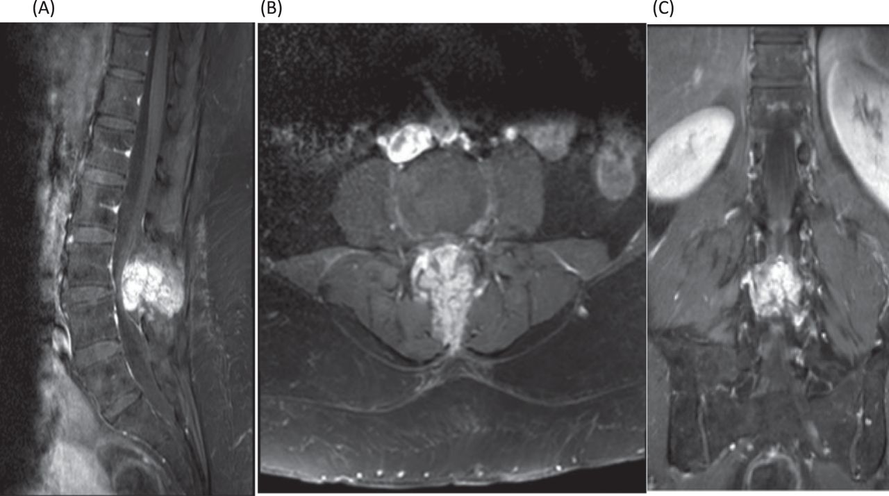
- Figure 2
(A) T1-weighted sagittal lumbar spine MRI with contrast showing expansile enhancing lesion involving the spinous process of L3. (B) T1-weighted postcontrast axial image of the L3 lumbar vertebra. (C) T1-weighted coronal MRI with contrast.
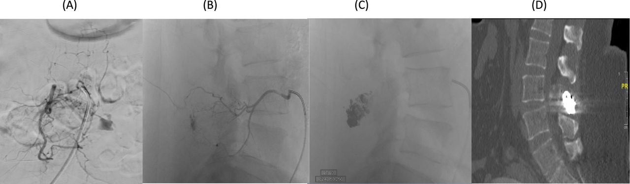
- Figure 3
(A) Right L3 segmental artery angiogram anteroposterior view demonstrating abnormal vascular blush and contrast pooling consistent with hemangioma. (B) Lateral view angiogram. (C) Fluoroscopic spot image obtained during spinal angiogram demonstrated embolization material (Onyx 18) in the spinous process lesion. (D) Spine computed tomography (CT) sagittal view showing embolization material (Onyx 18) seen in the spinous process of the L3 vertebra (postembolization).
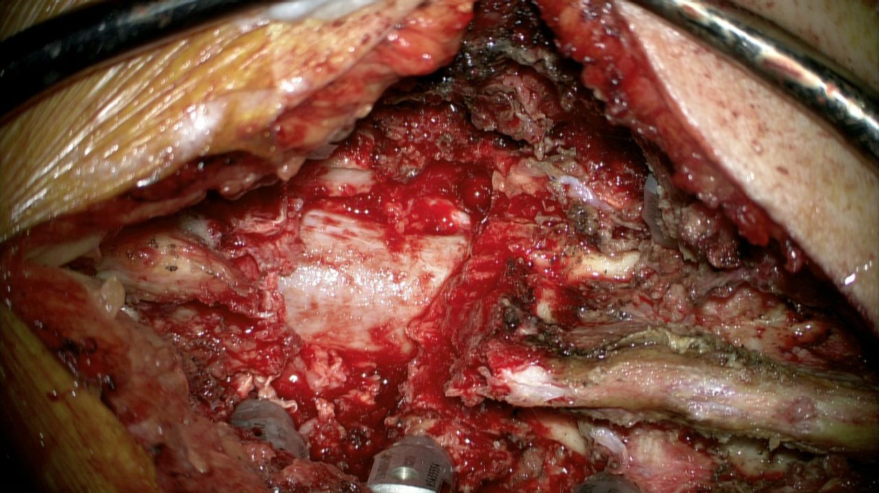
- Figure 4
Intraoperative image after decompression and excision of the tumor.
- Figure 5
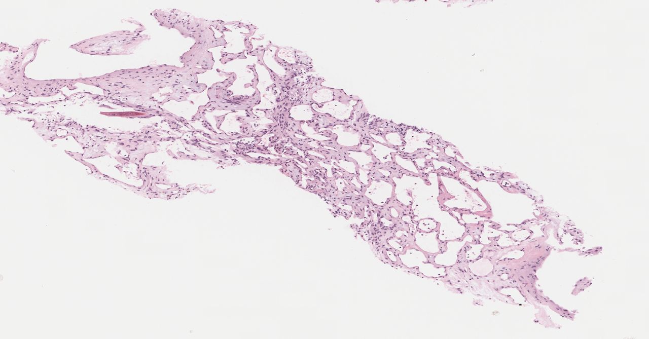
Histological examination reveals variable sizes of cystically dilated, thin-walled blood vessels that are lined by bland endothelial cells. No marked atypia, mitosis, or necrosis seen. Diagnosis: bland vascular proliferation consistent with hemangioma.
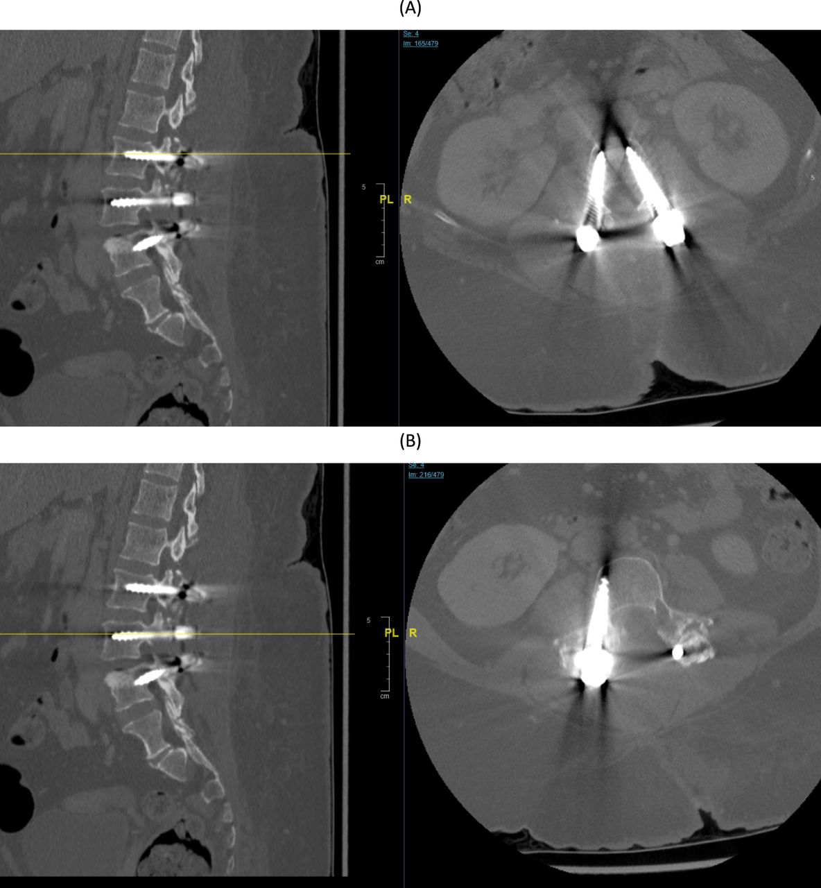
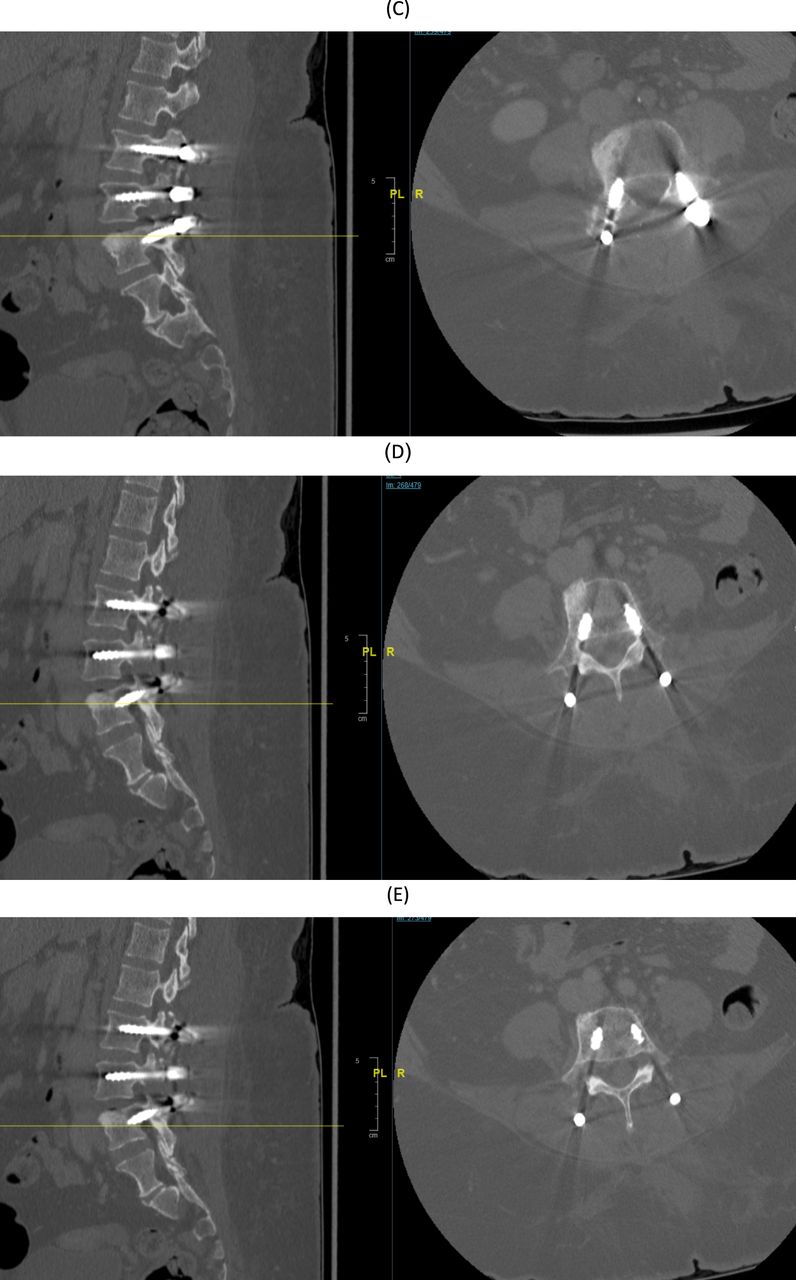
- Figure 6
(A) Postoperative sagittal and axial computed tomography (CT) spine at L2 pedicle. (B) Postoperative sagittal and axial CT spine at L3 pedicle. (C–E) Postoperative sagittal and axial CT spine at L4 pedicle. Axial image showing the amount of tumor that was resected including the medial wall of the left L3 pedicle.
- Figure 6
Continued.
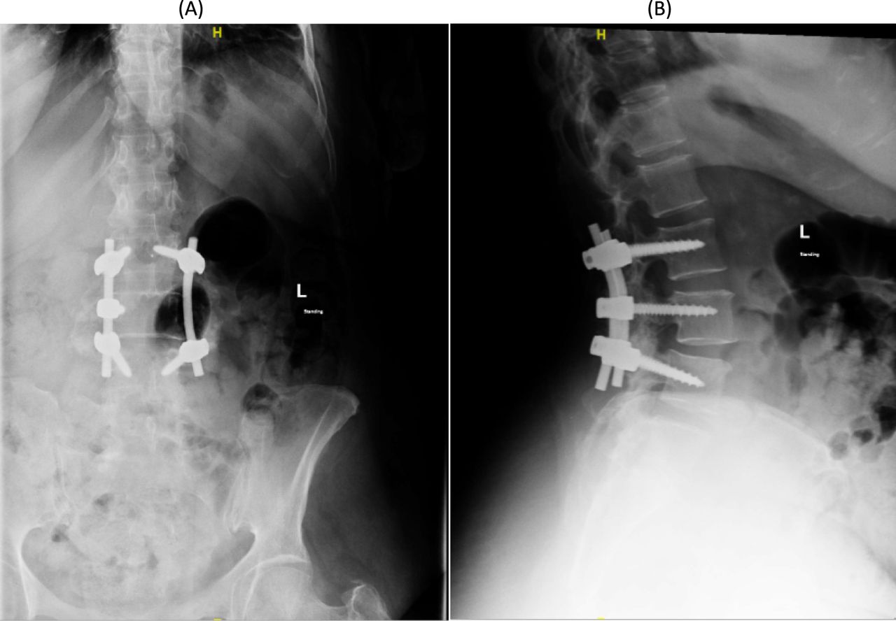
- Figure 7
(A, B) Anteroposterior and lateral postoperative x-rays, respectively, showing 2 rods and transpedicular screws seen transfixing the vertebral body from L2 to L4. No evidence of looseness or alignment deformity. There is no pedicle screw on the left L3 vertebra.
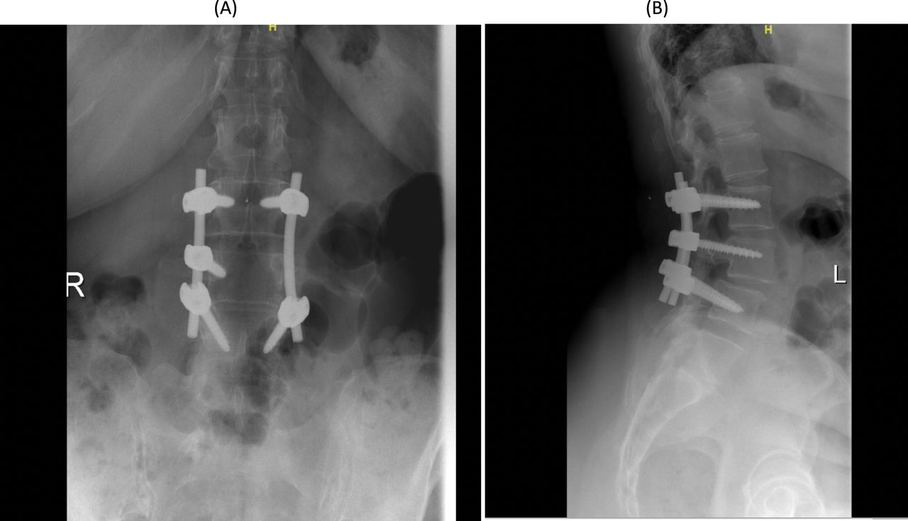
- Figure 8
(A, B) Anteroposterior and lateral views of follow-up x-rays, respectively, 2 years postoperatively showing 22 rods and transpedicular screws seen transfixing the vertebral body from L2 to L4. No evidence of looseness or alignment deformity. There is no pedicle screw on the left L3 vertebra.


Tables
In this issue


Jump to section
Related Articles
Cited By...
- No citing articles found.