
Main menu

  • Home
  • Content
    • Current Issue
    • Advance Online Publication
    • Archive
  • About Us
    • About ISASS
    • About the Journal
    • Author Instructions
    • Editorial Board
    • Reviewer Guidelines & Publication Criteria
  • More
    • Advertise
    • Subscribe
    • Alerts
    • Feedback
  • Join Us
  • Reprints & Permissions
  • Sponsored Content
  • Other Publications
    • ijss

User menu

  • My alerts

Search

  • Advanced search
International Journal of Spine Surgery
  • My alerts
International Journal of Spine Surgery

Advanced Search

  • Home
  • Content
    • Current Issue
    • Advance Online Publication
    • Archive
  • About Us
    • About ISASS
    • About the Journal
    • Author Instructions
    • Editorial Board
    • Reviewer Guidelines & Publication Criteria
  • More
    • Advertise
    • Subscribe
    • Alerts
    • Feedback
  • Join Us
  • Reprints & Permissions
  • Sponsored Content
  • Follow ijss on Twitter
  • Visit ijss on Facebook
Research ArticleCervical Spine

Dome Laminotomies at Adjacent Segments in Cervical Laminoplasty

Jeffrey H. Zimering and Konstantinos Margetis
International Journal of Spine Surgery October 2021, 15 (5) 871-878; DOI: https://doi.org/10.14444/8112
Jeffrey H. Zimering
Department of Neurosurgery, Mount Sinai Health System, New York, New York
MD
  • Find this author on Google Scholar
  • Find this author on PubMed
  • Search for this author on this site
Konstantinos Margetis
Department of Neurosurgery, Mount Sinai Health System, New York, New York
MD, PhD
  • Find this author on Google Scholar
  • Find this author on PubMed
  • Search for this author on this site
  • Article
  • Figures & Data
  • Info & Metrics
  • PDF
Loading

Article Figures & Data

Figures

  • Tables
  • Figure 1
    • Download figure
    • Open in new tab
    • Download powerpoint
    Figure 1

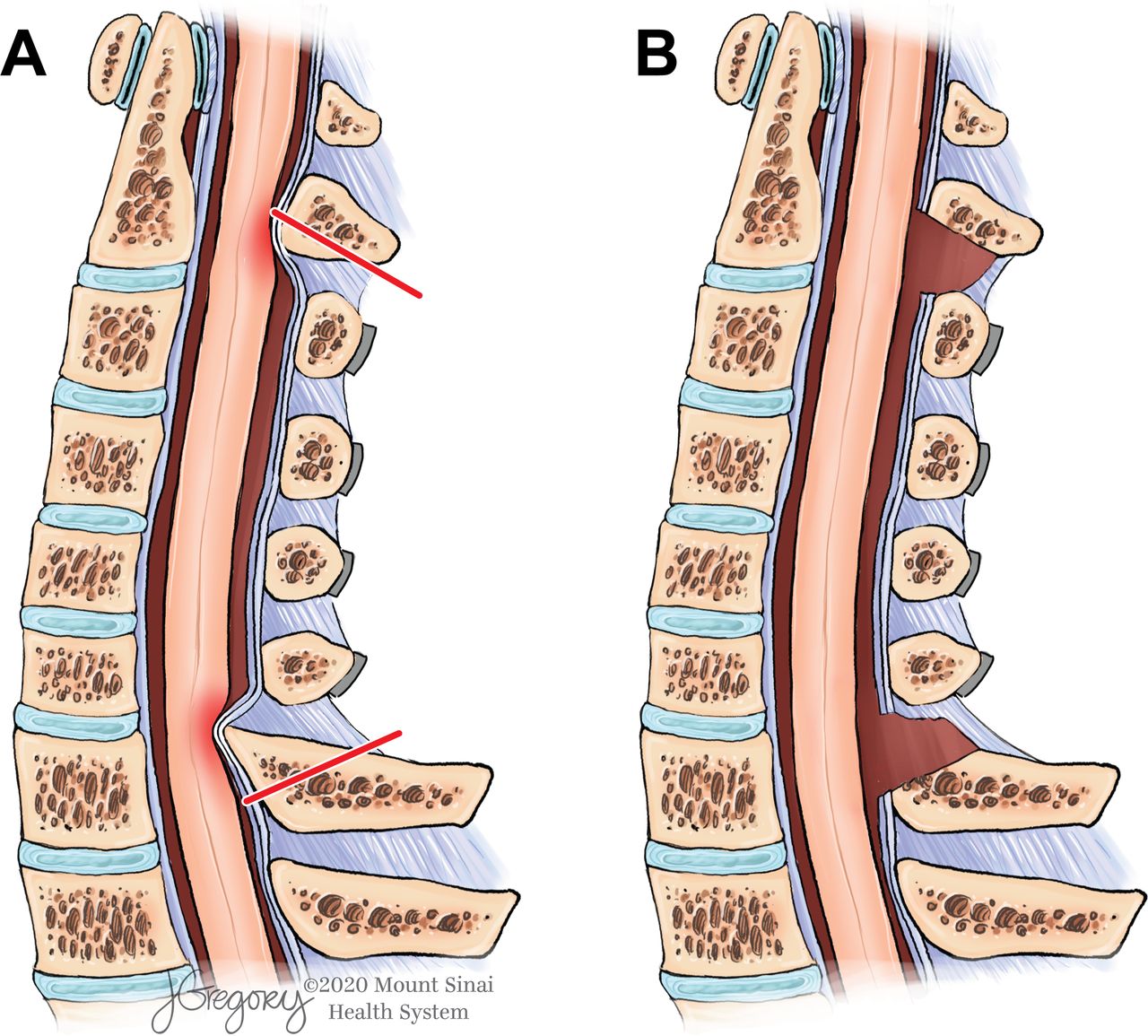
    (A) A sagittal, midline view of the cervical spine following a C3–C6 laminoplasty without dome laminotomies. The dorsal migration of the spinal cord following the decompressive effect of the laminoplasty at the C3–C6 levels leads to kinking and compression (red-shaded areas) of the spinal cord by the caudal edge of the C2 lamina and the cranial edge of the C7 lamina. The kinking can be static and/or dynamic. Adding C2 and C7 laminotomies, defined by the red lines, leads to an improved decompression, which is depicted in panel B. (B) The additional decompression afforded by the dome laminotomies prevents any spinal cord kinking. Illustration by Jill K. Gregory, used with permission of ©Mount Sinai Health System.

  • Figure 2
    • Download figure
    • Open in new tab
    • Download powerpoint
    Figure 2

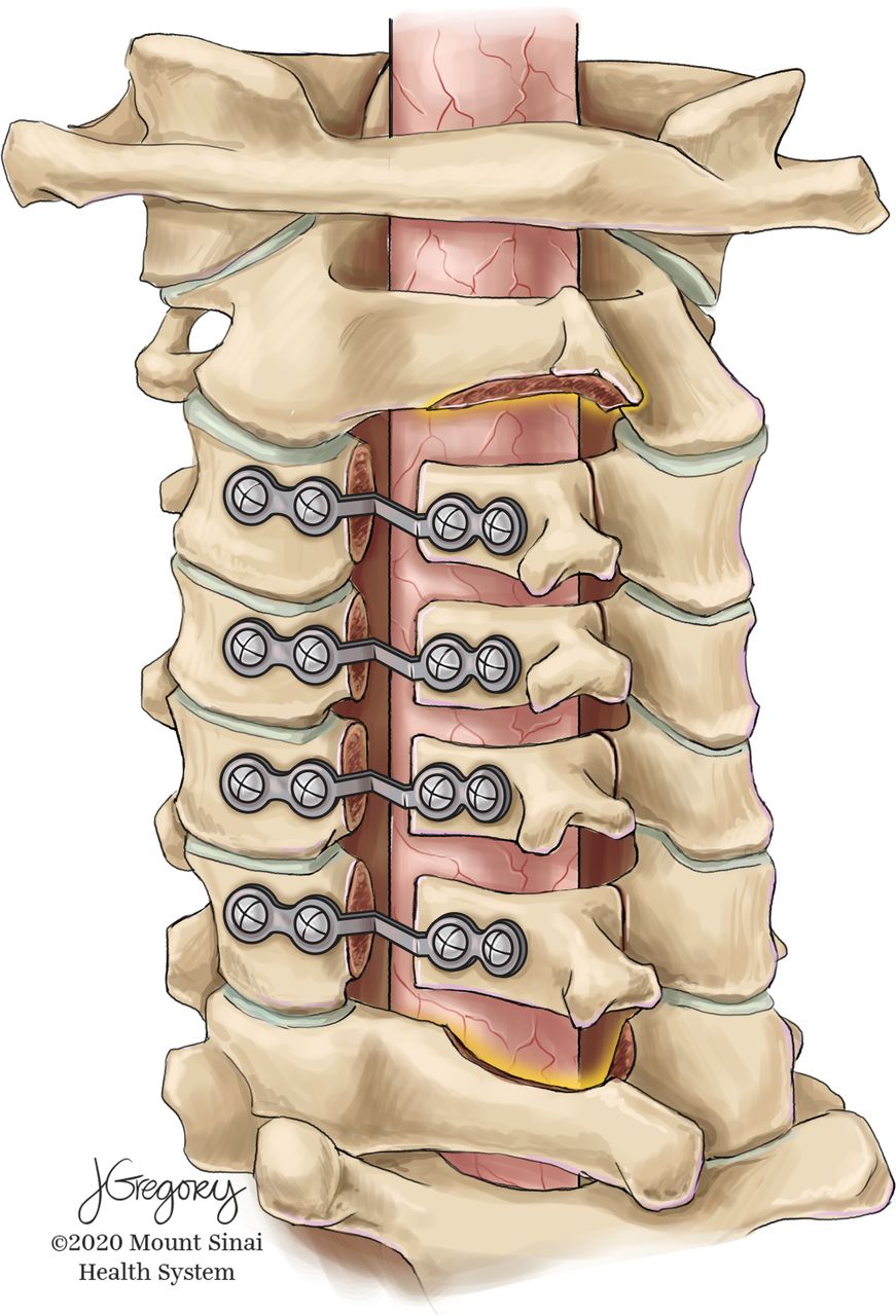
    A dorsolateral view of the osseous structures at the completion of C3–C6 laminoplasty and C2/C7 dome laminotomies. Illustration by Jill K. Gregory, used with permission of ©Mount Sinai Health System.

  • Figure 3
    • Download figure
    • Open in new tab
    • Download powerpoint
    Figure 3

    Axial pain before and after C3–C6 laminoplasty with dome laminotomy. Results are proportions of patients with no, mild, moderate, or severe pain. Baseline and early postoperative visit (n = 21): mean, 2.9 months; late postoperative visit (n = 16): mean, 14.9 months.

  • Figure 4
    • Download figure
    • Open in new tab
    • Download powerpoint
    Figure 4

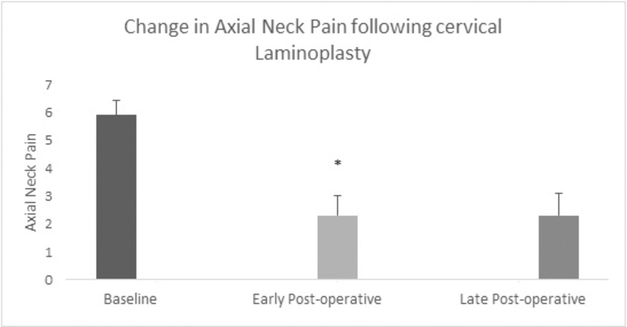
    Change in mean axial pain score at early and late postoperative time points. There was a significant reduction in the mean pain score at both the early and late postoperative examinations compared with mean baseline pain score. *P < .05 compared to baseline mean axial pain.

  • Figure 5
    • Download figure
    • Open in new tab
    • Download powerpoint
    Figure 5

    Recovery of neurologic function following C3–C6 laminoplasty with dome laminotomy. There was a significant improvement in hand function at the early and late examinations, and balance impairment improved significantly at the late examination. *P = .0001; +P = .001; ++P < .05 compared to baseline proportion.

  • Figure 6
    • Download figure
    • Open in new tab
    • Download powerpoint
    Figure 6

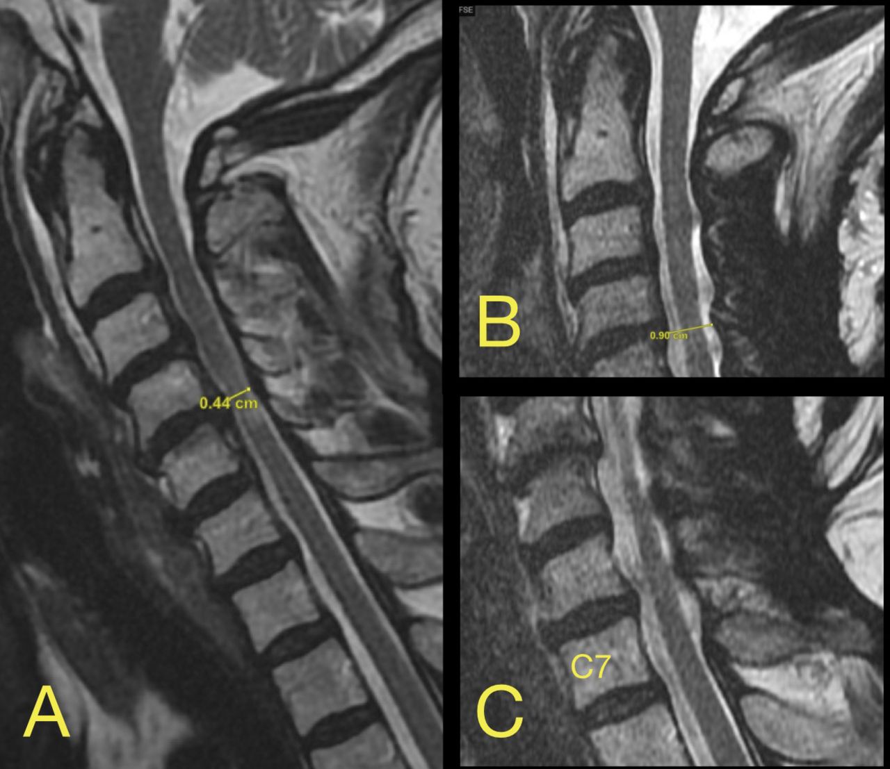
    (A) Preoperative sagittal T2 magnetic resonance image which revealed degenerative, superimposed on congenital stenosis with anterior-posterior diameter as low as 4.4 mm. (B) Decompressed C2–C3 level. (C) Decompressed C6–C7 level.

  • Figure 7
    • Download figure
    • Open in new tab
    • Download powerpoint
    Figure 7

    (A) Preoperative stenosis at the C6–C7 level with an AP diameter of the spinal canal of only 6 mm. (B) Image was obtained after a fall experienced 10 months after surgery which was associated with subjective weakness. It shows an increase in AP diameter of the spinal canal to 10.3 mm with adequate decompression of the spinal cord at the C6–C7 level. Of note, the patient had a stable mild spondylolisthesis at C7–T1 that remained unaltered postoperatively.

  • Figure 8
    • Download figure
    • Open in new tab
    • Download powerpoint
    Figure 8

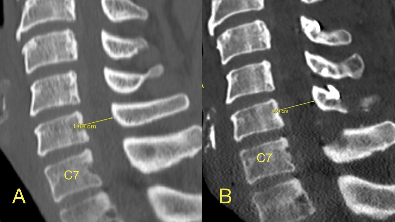
    (A) Preoperative sagittal CT. At the C6 level the AP diameter of the osseous spinal canal is 10 mm. (B) The dome laminotomy at C7 as well as the enlargement of the AP diameter of the osseous spinal canal to 14 mm. This is a case relatively early in this case series. The amount of bony resection was relatively more extensive in patients operated on later in the series.

Tables

  • Figures
  • Table 1
  • Table 2
PreviousNext
Back to top

In this issue

International Journal of Spine Surgery
Vol. 15, Issue 5
1 Oct 2021
  • Table of Contents
  • Index by author

Print
Download PDF
Article Alerts
Sign In to Email Alerts with your Email Address
Email Article

Thank you for your interest in spreading the word on International Journal of Spine Surgery.

NOTE: We only request your email address so that the person you are recommending the page to knows that you wanted them to see it, and that it is not junk mail. We do not capture any email address.

Enter multiple addresses on separate lines or separate them with commas.
Dome Laminotomies at Adjacent Segments in Cervical Laminoplasty
(Your Name) has sent you a message from International Journal of Spine Surgery
(Your Name) thought you would like to see the International Journal of Spine Surgery web site.
CAPTCHA
This question is for testing whether or not you are a human visitor and to prevent automated spam submissions.
Citation Tools
Dome Laminotomies at Adjacent Segments in Cervical Laminoplasty
Jeffrey H. Zimering, Konstantinos Margetis
International Journal of Spine Surgery Oct 2021, 15 (5) 871-878; DOI: 10.14444/8112

Citation Manager Formats

  • BibTeX
  • Bookends
  • EasyBib
  • EndNote (tagged)
  • EndNote 8 (xml)
  • Medlars
  • Mendeley
  • Papers
  • RefWorks Tagged
  • Ref Manager
  • RIS
  • Zotero
Share
Dome Laminotomies at Adjacent Segments in Cervical Laminoplasty
Jeffrey H. Zimering, Konstantinos Margetis
International Journal of Spine Surgery Oct 2021, 15 (5) 871-878; DOI: 10.14444/8112
Twitter logo Facebook logo Mendeley logo
  • Tweet Widget
  • Facebook Like
  • Google Plus One

Jump to section

  • Article
    • ABSTRACT
    • INTRODUCTION
    • METHODS
    • RESULTS
    • DISCUSSION
    • CONCLUSIONS
    • Footnotes
    • REFERENCES
  • Figures & Data
  • Info & Metrics
  • PDF

Related Articles

  • No related articles found.
  • PubMed
  • Google Scholar

Cited By...

  • No citing articles found.
  • Google Scholar

More in this TOC Section

  • Innovation and Adversity in Spine Surgery: A Retrospective
  • Cervical Myelopathy Secondary to Bilateral Atlantoaxial Pseudoarticulations in Rheumatoid Arthritis: A Case Report
  • Incidence of Traumatic Spinal Injury Following Public Policy Update on Moped Usage in South Carolina
Show more Cervical Spine

Similar Articles

Keywords

  • dome laminotomy
  • cervical laminoplasty
  • pain

Content

  • Current Issue
  • Latest Content
  • Archive

More Information

  • About IJSS
  • About ISASS
  • Privacy Policy

More

  • Subscribe
  • Alerts
  • Feedback

Other Services

  • Author Instructions
  • Join ISASS
  • Reprints & Permissions

© 2025 International Journal of Spine Surgery

International Journal of Spine Surgery Online ISSN: 2211-4599

Powered by HighWire