


Article Figures & Data
Figures
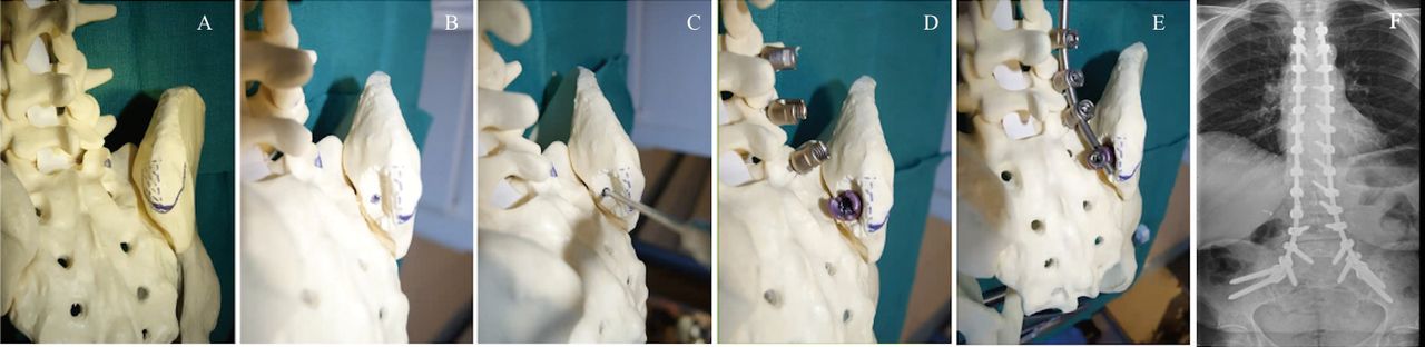
- Figure 1
Sawbones model illustrating right distal ventral iliac pathway (DVIP) recess creation and instrumentation. (A)–(E) Iliac screws are placed in line with S1 pedicle screws, and the vertical construct is completed with a single rod. (F) The DVIP recess is sufficient for placement of multiple iliac screws bilaterally.
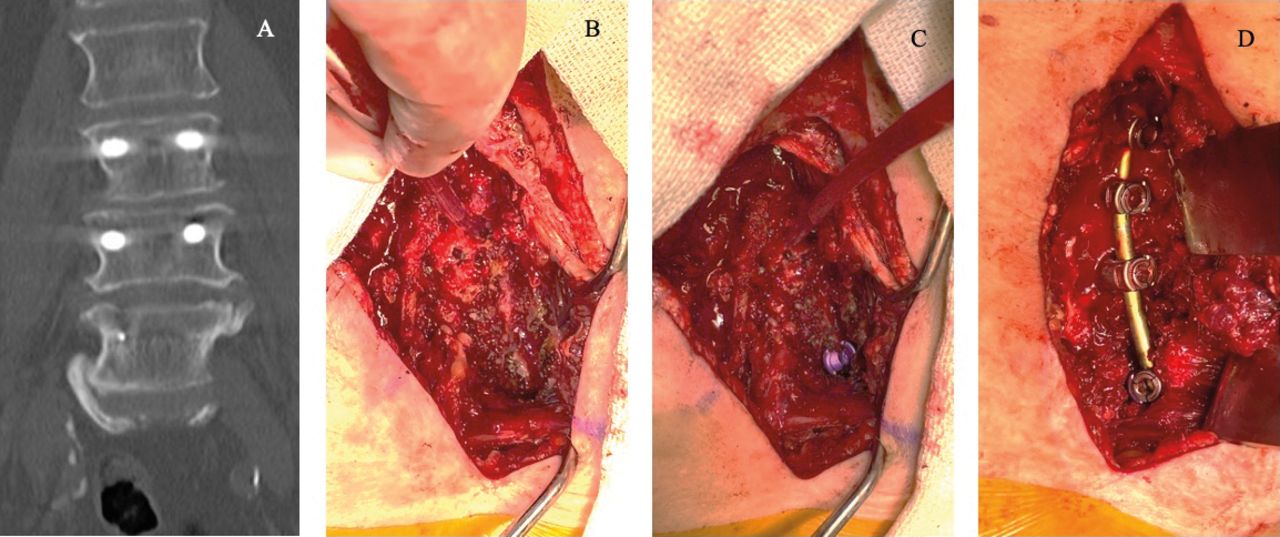
- Figure 2
Patient presenting with radicular pain was found to have distal junctional failure of L3–5 posterior instrumentation demonstrating kyphotic collapse postoperatively. Imaging reveals kyphotic collapse associated with vertebral body fracture and fixation failure at L5. Intraoperative photos of lumbosacral deformity treated with revision L3–L5 fusion and extension to the ilium with distal ventral iliac pathway screws.
- Figure 3
Preoperative and postoperative imaging of lumbosacral deformity treated with L1-ilium fusion with distal ventral iliac pathway screws. (A) and (B) Preoperative sagittal and coronal computed tomography scan of patient with vertebrae-anal-trachea-esophagus-renal syndrome and sagittal imbalance demonstrating autofusion of L3–5 with associated stenosis and listhesis as well as sacral dysgenesis. (C) and (D) Postoperative sagittal and anterior-posterior x-rays at 29 months follow up demonstrating solid fusion with absence of hardware failure.
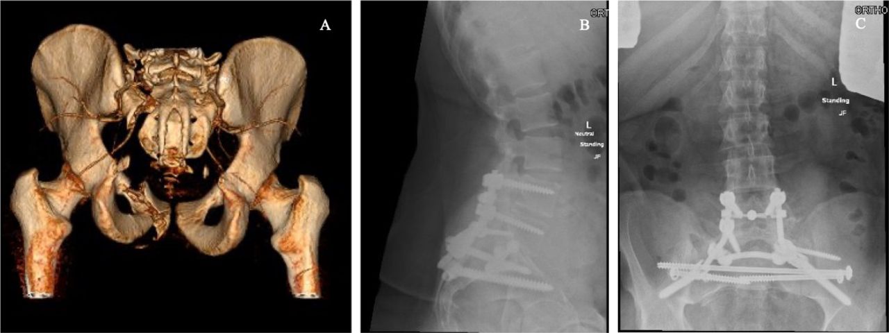
- Figure 4
Preoperative and postoperative imaging of spinopelvic trauma treated with L4-ilium fusion with distal ventral iliac pathway screws. (A) Three-dimensional reconstruction of preoperative computed tomography scan demonstrating left lateral compression 3 pelvic ring injury with a left complete displaced comminuted zone 2 sacral fracture, right sacroiliac joint dislocation, left segmental superior and inferior ramus fractures, L1–L5 left transverse process fractures, left L5 pedicle fracture, and right S1 pedicle fracture, constituting a form of spinopelvic dissociation. (B) and (C) Postoperative sagittal and anterior-posterior x-rays at 24 months follow up demonstrating stable fusion without hardware failure.
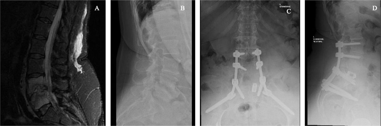
- Figure 5
Preoperative and postoperative imaging demonstrating osteomyelitis or discitis treated with L3-ilium fusion using distal ventral iliac pathway screws. Preoperative sagittal magnetic resonance imaging demonstrating osteomyelitis or discitis with infectious destruction of L4–5, epidural enhancement, kyphotic collapse, and central canal stenosis. Preoperative lateral x-ray of the lumbar spine demonstrating pathologic kyphotic collapse. Postoperative anterior-posterior and lateral x-rays at 24 months follow up demonstrating solid fusion without hardware failure.


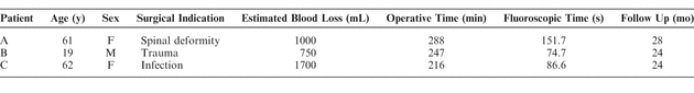
Tables
In this issue


Jump to section
Related Articles
Cited By...
- No citing articles found.