
Main menu

  • Home
  • Content
    • Current Issue
    • Advance Online Publication
    • Archive
  • About Us
    • About ISASS
    • About the Journal
    • Author Instructions
    • Editorial Board
    • Reviewer Guidelines & Publication Criteria
  • More
    • Advertise
    • Subscribe
    • Alerts
    • Feedback
  • Join Us
  • Reprints & Permissions
  • Sponsored Content
  • Other Publications
    • ijss

User menu

  • My alerts

Search

  • Advanced search
International Journal of Spine Surgery
  • My alerts
International Journal of Spine Surgery

Advanced Search

  • Home
  • Content
    • Current Issue
    • Advance Online Publication
    • Archive
  • About Us
    • About ISASS
    • About the Journal
    • Author Instructions
    • Editorial Board
    • Reviewer Guidelines & Publication Criteria
  • More
    • Advertise
    • Subscribe
    • Alerts
    • Feedback
  • Join Us
  • Reprints & Permissions
  • Sponsored Content
  • Follow ijss on Twitter
  • Visit ijss on Facebook
Research ArticleBiomechanics

Ligamentum Flavum Buckling vs Hypertrophy in Cervical Myelopathy Using Dynamic Imaging: Are Instability and Loss of Disc Height the Culprits?

Kalyan Kumar Varma Kalidindi, Jeevan Kumar Sharma, Gayatri Vishwakarma, Srinath Duggi and Harvinder Singh Chhabra
International Journal of Spine Surgery April 2022, 16 (2) 300-308; DOI: https://doi.org/10.14444/8221
Kalyan Kumar Varma Kalidindi
1 Department of Spine Service, Indian Spinal Injuries Center, Vasant Kunj, New Delhi, Delhi 110070, India
MS, DNB, FNB
  • Find this author on Google Scholar
  • Find this author on PubMed
  • Search for this author on this site
Jeevan Kumar Sharma
1 Department of Spine Service, Indian Spinal Injuries Center, Vasant Kunj, New Delhi, Delhi 110070, India
MS
  • Find this author on Google Scholar
  • Find this author on PubMed
  • Search for this author on this site
Gayatri Vishwakarma
2 Department of Biostatistics, Indian Spinal Injuries Center, Vasant Kunj, New Delhi, Delhi 110070, India
PhD
  • Find this author on Google Scholar
  • Find this author on PubMed
  • Search for this author on this site
Srinath Duggi
3 Department of Orthopaedics, Chalmeda AnandRao Institute of Medical Sciences, Karimnagar, Telangana 505001, India
MS
  • Find this author on Google Scholar
  • Find this author on PubMed
  • Search for this author on this site
Harvinder Singh Chhabra
1 Department of Spine Service, Indian Spinal Injuries Center, Vasant Kunj, New Delhi, Delhi 110070, India
MS
  • Find this author on Google Scholar
  • Find this author on PubMed
  • Search for this author on this site
  • Article
  • Figures & Data
  • Info & Metrics
  • PDF
Loading

Article Figures & Data

Figures

  • Tables
  • Figure 1
    • Download figure
    • Open in new tab
    • Download powerpoint
    Figure 1

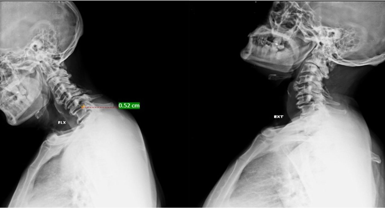
    Representative adequate dynamic x-ray images of a patient with cervical myelopathy depicting measurement of sagittal translational instability at C6-C7 level. Lines were drawn along the posterior border of the superior and inferior vertebrae, and the horizontal distance between the lines was measured. A difference of more than 3.5-mm translation or 20% was considered as sagittal translational instability.

  • Figure 2
    • Download figure
    • Open in new tab
    • Download powerpoint
    Figure 2

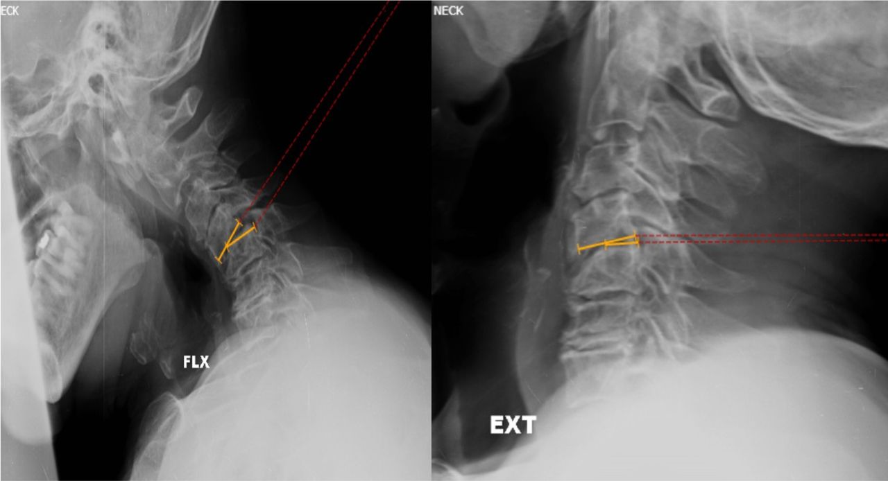
    Representative dynamic x-ray images of a patient with cervical myelopathy depicting measurement of sagittal rotational instability at C4-C5 level. Lines were drawn along the inferior end plate of the superior vertebra and the superior end plate of the inferior vertebra, and the angle between these lines was measured. The difference in angles formed between these lines in flexion and extension views was measured. A difference of more than 12° was considered as sagittal rotational instability.

  • Figure 3
    • Download figure
    • Open in new tab
    • Download powerpoint
    Figure 3

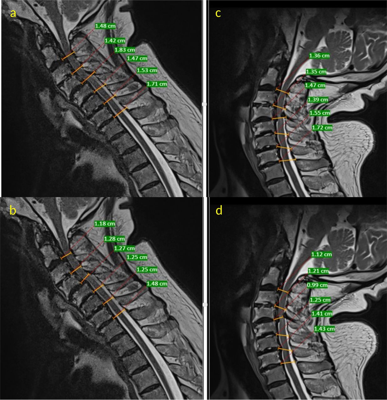
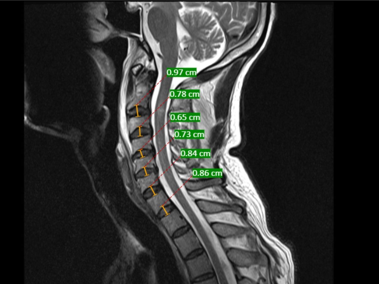
    Representative neutral T2-weighted sagittal magnetic resonance imaging image of the cervical spine depicting the measurement of disc height. Disc height was measured from the center of the inferior end plate of superior vertebra to the center of the superior end plate of the inferior vertebra.

  • Figure 4
    • Download figure
    • Open in new tab
    • Download powerpoint
    Figure 4

    Representative neutral T2-weighted sagittal magnetic resonance imaging (MRI) images of the cervical spine depicting measurements for ligamentum flavum (LF) thickness and buckling. At the midlevel of the intervertebral disc, 2 linear lines were drawn. One line measured the distance from the posterior margin of the disc to the base of spinous process (A) and another line measured the distance from the posterior margin of the disc to the posterior border of the cerebrospinal fluid (b) in a flexion MRI. The difference in value obtained (A, B) represents the LF thickness in flexion. (C) and (D) represent similar measurements performed on an extension MRI. The LF thickness in extension is represented by value (c-d). The difference in LF thickness in flexion and extension (C, D-[A, B]) is considered as buckling.

  • Figure 5
    • Download figure
    • Open in new tab
    • Download powerpoint
    Figure 5

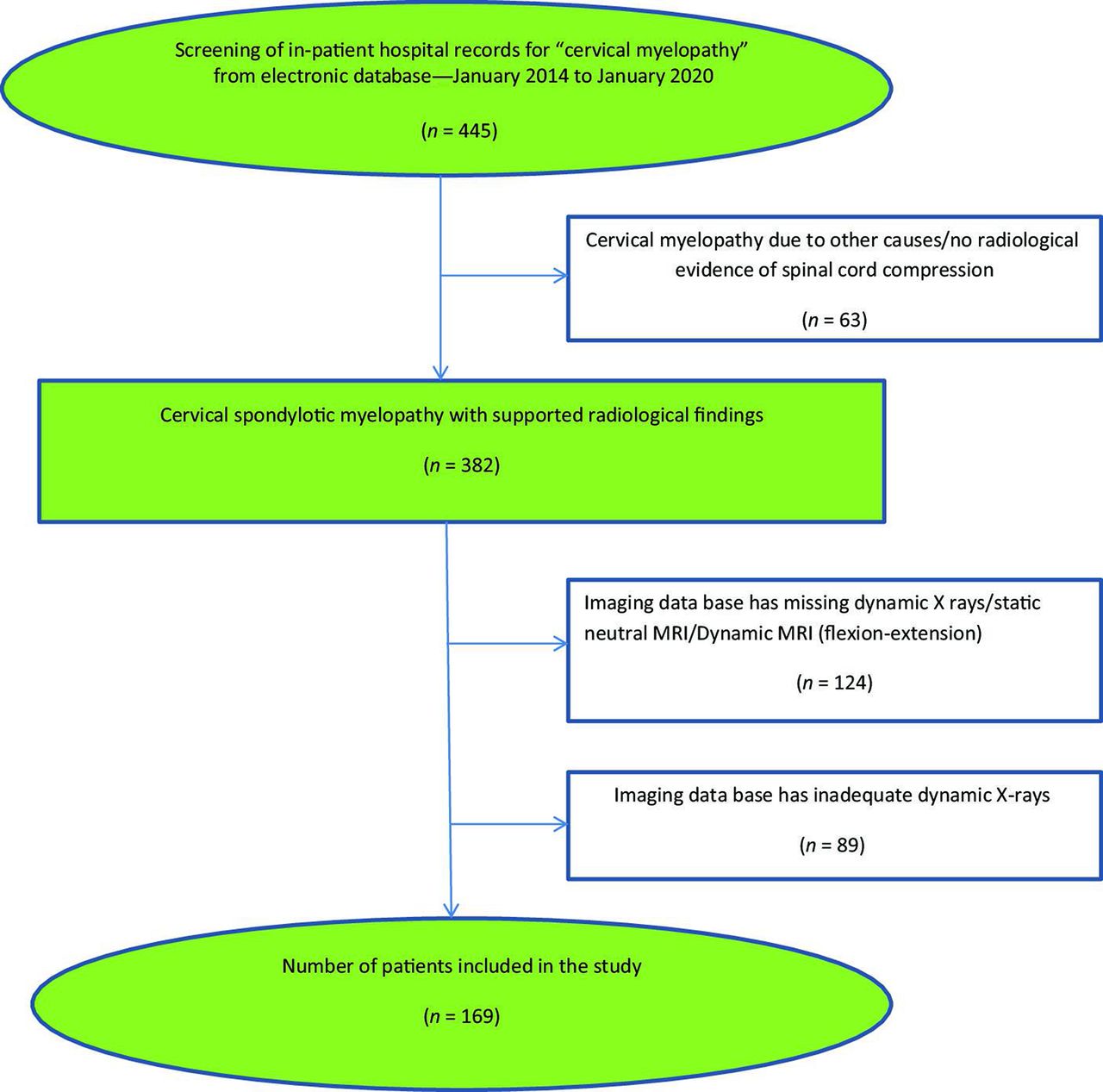
    A flowchart depicting the selection of study samples utilized for the study.

  • Figure 6
    • Download figure
    • Open in new tab
    • Download powerpoint
    Figure 6

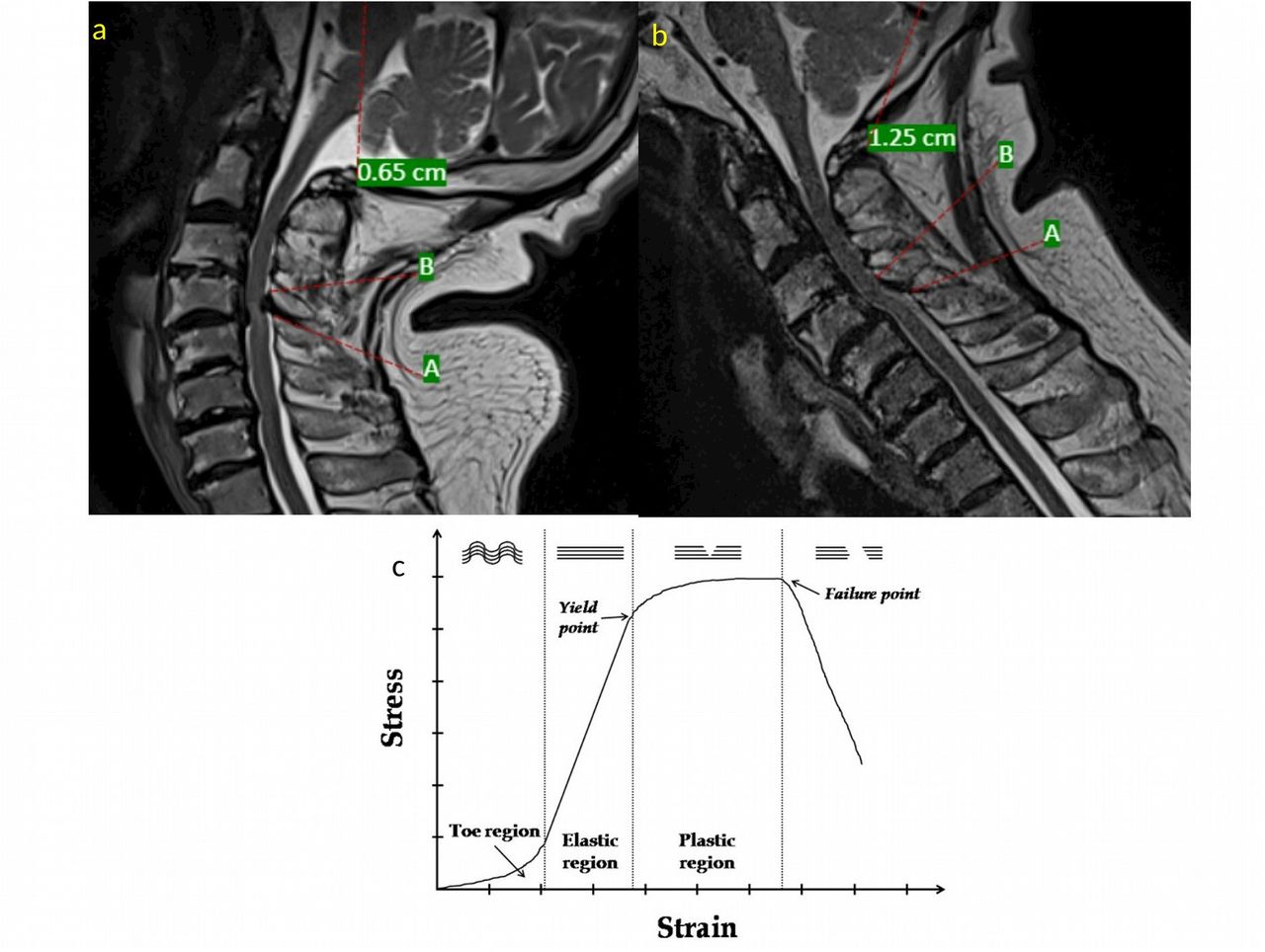
    (A, B) T2-weighted magnetic resonance images showing the changes in ligamentum flavum (LF) with neck motion. As the neck goes into flexion (B) from extension (A), the distance between the origin (marked A) and insertion (marked B) increases and puts strain on the LF. (C) Representation of a typical stress-strain curve of a ligament. The x-axis represents the strain on the ligament, and y-axis represents the stress applied to the ligament. Within the elastic region, repeated increases or decreases in tension load will not change the ligament length. The elastic region represents the tension loads that occur within the physiological range of motion of the neck. Beyond a point, the curve tends to flatten, indicating that the ligament deforms more per given increase in tension load (ie, the ligament undergoes permanent deformation [plastic deformation]). This is called the yield point. After the plastic region, the sudden failure of the ligament occurs and stress disappears. This is called the failure point. With permanent deformation or lengthening of the ligament, the LF tends to buckle into the spinal canal in extension because of the closer contact between the laminae (points A and B in Figure 6A and B).

Tables

  • Figures
    • View popup
    Table 1

    Descriptive statistics (mean ± standard deviation) according to the level. Analysis of variance (ANOVA) was used to compare the means of variables such as ligamentum flavum thickness/buckling across different cervical levels.

    CharacteristicC2-C3C3-C4C4-C5C5-C6C6-C7C7-T1P Valuea
    Thickness0.26 ± 0.060.28 ± 0.070.29 ± 0.110.28 ± 0.090.27 ± 0.080.31 ± 0.08<0.001
    Buckling0.03 ± 0.030.05 ± 0.050.10 ± 0.080.08 ± 0.090.05 ± 0.070.03 ± 0.04<0.001
    Disc height0.71 ± 0.130.71 ± 0.210.72 ± 0.140.75 ± 0.150.72 ± 0.160.72 ± 0.200.331
    Translation0.26 ± 0.450.27 ± 0.350.27 ± 0.280.48 ± 0.630.30 ± 0.410.26 ± 0.25<0.001
    Rotation3.03 ± 0.873.32 ± 1.534.02 ± 3.613.73 ± 1.773.27 ± 1.632.56 ± 1.22<0.001
    • aRepeated measures ANOVA (Greenhouse-Geisser).

    • View popup
    Table 2

    Distribution of the 2 groups of buckling (groups A and B) by cervical level.

    LevelNo. of Levels (%)
    Group A, Buckling≤0.1 mm (n = 798)Group B, Buckling>0.1 mm (n = 216)
    C2-C3156 (19.5)13 (6.0)
    C3-C4136(17.0)33 (15.3)
    C4-C597 (12.2)72 (33.3)
    C5-C6112 (14.0)57 (26.4)
    C6-C7148 (18.6)21 (9.7)
    C7-T1149 (18.7)20 (9.3)
    • View popup
    Table 3

    Correlation analysis of buckling with age at different levels. Only C7-T1 level had correlation of ligamentum flavum buckling with age.

    AgeC2-C3C3-C4C4-C5C5-C6C6-C7C7-T1
    Pearson correlation0.0010.0540.041−0.129−0.0790.187a
    P value0.9860.4870.5940.0950.3100.015
    • aCorrelation is significant at the 0.05 level (2 tailed).

    • View popup
    Table 4

    Disc height at different levels between the 2 groups of buckling.

    LevelGroup A, Buckling≤0.1 mm (n = 798)Group B, Buckling>0.1 mm (n = 216)P Value
    n Mean (SD)n Mean (SD)
    C2-C31560.71 (0.14)130.74 (0.09)0.435
    C3-C41360.68 (0.13)330.68 (0.13)<0.001
    C4-C5970.72 (0.71)720.71 (0.11)0.545
    C5-C61120.76 (0.17)570.72 (0.10)0.068
    C6-C71480.70 (0.17)210.83 (0.02)<0.001
    C7-T11490.72 (0.19)200.71 (0.28)0.901
PreviousNext
Back to top

In this issue

International Journal of Spine Surgery
Vol. 16, Issue 2
1 Apr 2022
  • Table of Contents
  • Index by author

Print
Download PDF
Article Alerts
Sign In to Email Alerts with your Email Address
Email Article

Thank you for your interest in spreading the word on International Journal of Spine Surgery.

NOTE: We only request your email address so that the person you are recommending the page to knows that you wanted them to see it, and that it is not junk mail. We do not capture any email address.

Enter multiple addresses on separate lines or separate them with commas.
Ligamentum Flavum Buckling vs Hypertrophy in Cervical Myelopathy Using Dynamic Imaging: Are Instability and Loss of Disc Height the Culprits?
(Your Name) has sent you a message from International Journal of Spine Surgery
(Your Name) thought you would like to see the International Journal of Spine Surgery web site.
CAPTCHA
This question is for testing whether or not you are a human visitor and to prevent automated spam submissions.
Citation Tools
Ligamentum Flavum Buckling vs Hypertrophy in Cervical Myelopathy Using Dynamic Imaging: Are Instability and Loss of Disc Height the Culprits?
Kalyan Kumar Varma Kalidindi, Jeevan Kumar Sharma, Gayatri Vishwakarma, Srinath Duggi, Harvinder Singh Chhabra
International Journal of Spine Surgery Apr 2022, 16 (2) 300-308; DOI: 10.14444/8221

Citation Manager Formats

  • BibTeX
  • Bookends
  • EasyBib
  • EndNote (tagged)
  • EndNote 8 (xml)
  • Medlars
  • Mendeley
  • Papers
  • RefWorks Tagged
  • Ref Manager
  • RIS
  • Zotero
Share
Ligamentum Flavum Buckling vs Hypertrophy in Cervical Myelopathy Using Dynamic Imaging: Are Instability and Loss of Disc Height the Culprits?
Kalyan Kumar Varma Kalidindi, Jeevan Kumar Sharma, Gayatri Vishwakarma, Srinath Duggi, Harvinder Singh Chhabra
International Journal of Spine Surgery Apr 2022, 16 (2) 300-308; DOI: 10.14444/8221
Twitter logo Facebook logo Mendeley logo
  • Tweet Widget
  • Facebook Like
  • Google Plus One

Jump to section

  • Article
    • Abstract
    • INTRODUCTION
    • MATERIALS AND METHODS
    • RESULTS
    • DISCUSSION
    • CONCLUSIONS
    • Footnotes
    • References
  • Figures & Data
  • Info & Metrics
  • PDF

Related Articles

  • No related articles found.
  • PubMed
  • Google Scholar

Cited By...

  • No citing articles found.
  • Google Scholar

More in this TOC Section

  • Sequential Anterior Longitudinal Ligament Release With Expandable Spacers for Lordosis Correction in Anterior-to-Psoas Lumbar Interbody Fusion: A Radiographic and Biomechanical Study
  • Comparative Biomechanical Analysis of Anterior Lumbar Interbody Fusion and Bilateral Expandable Transforaminal Lumbar Interbody Fusion Cages: A Finite Element Analysis Study
  • Impact of Different Operative Techniques for Patients With Adolescent Idiopathic Scoliosis on Frontal Curve Correction and Sagittal Balance
Show more Biomechanics

Similar Articles

Keywords

  • ligamentum flavum
  • buckling
  • hypertrophy
  • instability
  • disc height

Content

  • Current Issue
  • Latest Content
  • Archive

More Information

  • About IJSS
  • About ISASS
  • Privacy Policy

More

  • Subscribe
  • Alerts
  • Feedback

Other Services

  • Author Instructions
  • Join ISASS
  • Reprints & Permissions

© 2025 International Journal of Spine Surgery

International Journal of Spine Surgery Online ISSN: 2211-4599

Powered by HighWire