
Main menu

  • Home
  • Content
    • Current Issue
    • Advance Online Publication
    • Archive
  • About Us
    • About ISASS
    • About the Journal
    • Author Instructions
    • Editorial Board
    • Reviewer Guidelines & Publication Criteria
  • More
    • Advertise
    • Subscribe
    • Alerts
    • Feedback
  • Join Us
  • Reprints & Permissions
  • Sponsored Content
  • Other Publications
    • ijss

User menu

  • My alerts

Search

  • Advanced search
International Journal of Spine Surgery
  • My alerts
International Journal of Spine Surgery

Advanced Search

  • Home
  • Content
    • Current Issue
    • Advance Online Publication
    • Archive
  • About Us
    • About ISASS
    • About the Journal
    • Author Instructions
    • Editorial Board
    • Reviewer Guidelines & Publication Criteria
  • More
    • Advertise
    • Subscribe
    • Alerts
    • Feedback
  • Join Us
  • Reprints & Permissions
  • Sponsored Content
  • Follow ijss on Twitter
  • Visit ijss on Facebook
Research ArticleEndoscopic Minimally Invasive Surgery

Magnetic Resonance Imaging Documentation of Approach Trauma With Lumbar Endoscopic Interlaminar, Translaminar, Compared to Open Microsurgical Discectomy

Stefan Hellinger, Albert E. Telfeian and Kai-Uwe Lewandrowski
International Journal of Spine Surgery April 2022, 16 (2) 343-352; DOI: https://doi.org/10.14444/8226
Stefan Hellinger
1 Department of Orthopedic Surgery, Arabella Klinik, Munich, Germany
MD
  • Find this author on Google Scholar
  • Find this author on PubMed
  • Search for this author on this site
Albert E. Telfeian
2 Department of Neurosurgery, Rhode Island Hospital, Providence, RI, USA
3 Department of Neurosurgery, The Warren Alpert Medical School of Brown University, Providence, RI, USA
MD, PhD
  • Find this author on Google Scholar
  • Find this author on PubMed
  • Search for this author on this site
Kai-Uwe Lewandrowski
4 Center for Advanced Spine Care of Southern Arizona and Surgical Institute of Tucson, Tucson, AZ, USA
5 Department of Orthopaedics, Fundación Universitaria Sanitas, Bogotá, DC, Colombia
6 Department of Orthopaedic Surgery, UNIRIO, Rio de Janeiro, Brazil
MD
  • Find this author on Google Scholar
  • Find this author on PubMed
  • Search for this author on this site
  • Article
  • Figures & Data
  • Info & Metrics
  • PDF
Loading

Article Figures & Data

Figures

  • Tables
  • Figure 1
    • Download figure
    • Open in new tab
    • Download powerpoint
    Figure 1

    T2-weighted postoperative axial magnetic resonance images (MRIs) are shown to illustrate the extent of postoperative edema and fatty muscle degeneration following a full endoscopic interlaminar directly visualized videoendoscopic discectomy decompression (A, B) vs a traditional translaminar minimally invasive microsurgical decompression employing the operating microscope and a tubular retractor system (C, D). MRIs of study patients with symptomatic herniated disc were obtained at 4 days (A, C) and 1 year postoperatively (B, D). These exemplary postoperative images corroborate this study’s objective cross-sectional area measurements following both procedures by illustrating more edema immediately postoperatively with the standard microsurgical dissection (A vs C) and more atrophy of the paraspinal muscle not just on the approach side but also on the opposite nonsurgical side due to denervation-induced atrophy at 1 year postoperatively (B vs D) when compared with the endoscopic decompression. The observed atrophy and fatty degeneration on the nonsurgical side suggest functional segmental multifidus cross-innervation between both sides.
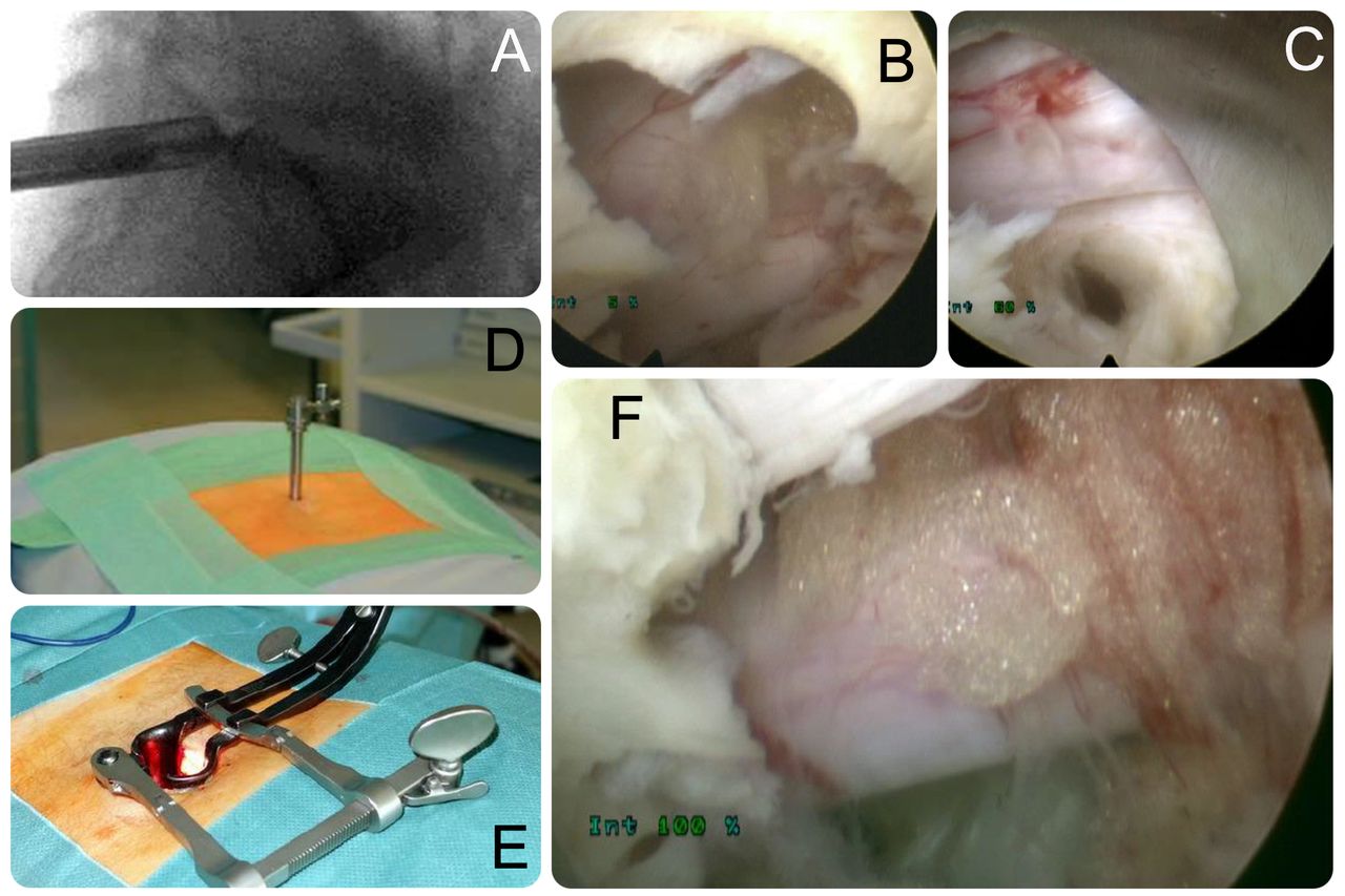
  • Figure 2
    • Download figure
    • Open in new tab
    • Download powerpoint
    Figure 2

    Clinical example of interlaminar endoscopic approach to the L5-S1 motion segment for a symptomatic herniated disc (A, D). First, the ligamentum flavum is removed (B). Then, the traversing nerve root is retracted with the beveled tip of the tubular working cannula before, and annulotomy is made (C). The alternative posterior translaminar microsurgical dissection and decompression are performed with a tubular retractor system (E). The discectomy is performed following medial facetectomy (F).
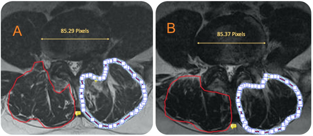
  • Figure 3
    • Download figure
    • Open in new tab
    • Download powerpoint
    Figure 3

    -weighted axial magnetic resonance imaging (MRI) image sequences obtained from patients with symptomatic lumbar disc herniations who underwent either microsurgical discectomy (A-C) or interlaminar endoscopic discectomy decompression (D–F). The MRI panels show the defect zone created by the 2 different surgical techniques. Preoperative measurements (A, D) were compared with measurements done on postoperative MRI images obtained at 4 days (B, E) and 1 year postoperatively (C, F). These area measurements summarized in Table suggested a significantly smaller defect size with the endoscopic vs the microsurgical technique (P < 0.001).

  • Figure 4
    • Download figure
    • Open in new tab
    • Download powerpoint
    Figure 4

    T2-weighted axial magnetic resonance image (MRI) sequences obtained from a 74-year-old patient with symptomatic left L4/5 extraforaminal lumbar disc herniations who underwent transforaminal endoscopic discectomy decompression. The MRI panels show the cross-sectional measurements of the paraspinal muscle zone following the transforaminal discectomy. Preoperative measurements (A) were compared to measurements done on postoperative MRI images obtained within one year from surgery (B). These area measurements summarized in Table 1 suggested no significant atrophy of the paraspinal muscles with the endoscopic transforaminal technique.
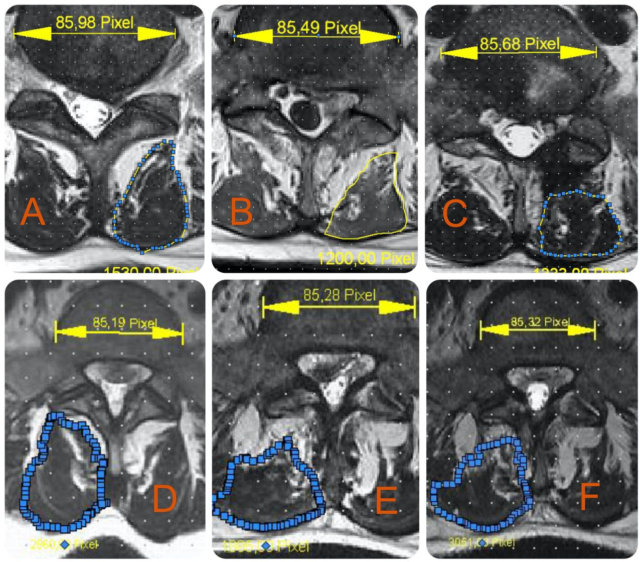
  • Figure 5
    • Download figure
    • Open in new tab
    • Download powerpoint
    Figure 5

    T2-weighted axial magnetic resonance image (MRI) sequences obtained from patients with symptomatic lumbar disc herniations who underwent either microsurgical discectomy (A–C) or interlaminar endoscopic discectomy decompression (D–F). The MRI panels show the cross-sectional measurements of the paraspinal muscle zone following discectomy by the 2 different surgical techniques. Preoperative measurements (A, D) were compared with measurements done on postoperative MRI images obtained at 4 days (B, E) and 1 year postoperatively (C, F). These area measurements summarized in Table suggested significantly less atrophy of the paraspinal muscles with the endoscopic vs the microsurgical technique (P < 0.001). Moreover, there was more paraspinal muscle atrophy with the microsurgical technique not only on the approach side but also opposite from it, implying disruption of cross-innervation between the bilateral paraspinal muscle groups, including the multifidus muscles.

Tables

  • Figures
    • View popup
    Table

    Changes in surgical defect zone and paraspinal muscle zone data obtained at 4 d and 1 y postoperatively for interlaminar endoscopy and microsurgical patients.

    Surgical TechniqueArea MeasurementMean ± SD
    ΔrCSA Immediately PostoperativelyΔrCSA Within 1-y Postoperatively
    MicrosurgeryDefect zone41.2% ± 11.8%62.9% ± 18.3%
     Muscle degeneration22.6% ± 10.3%23.6% ± 11.8%
    Interlaminar endoscopyDefect zone17.6% ± 9.4%6.4 ± 3.2%
     Muscle degeneration20.3% ± 8.6%2.1% ± 1.9%
    Transforaminal endoscopyDefect zone5.5% ± 3.7%6.3% ± 4.5%
     Muscle degeneration6.9% ± 2.8%7.8% ± 5.1%
    • Δ rCSA, relative cross-sectional area.

PreviousNext
Back to top

In this issue

International Journal of Spine Surgery
Vol. 16, Issue 2
1 Apr 2022
  • Table of Contents
  • Index by author

Print
Download PDF
Article Alerts
Sign In to Email Alerts with your Email Address
Email Article

Thank you for your interest in spreading the word on International Journal of Spine Surgery.

NOTE: We only request your email address so that the person you are recommending the page to knows that you wanted them to see it, and that it is not junk mail. We do not capture any email address.

Enter multiple addresses on separate lines or separate them with commas.
Magnetic Resonance Imaging Documentation of Approach Trauma With Lumbar Endoscopic Interlaminar, Translaminar, Compared to Open Microsurgical Discectomy
(Your Name) has sent you a message from International Journal of Spine Surgery
(Your Name) thought you would like to see the International Journal of Spine Surgery web site.
CAPTCHA
This question is for testing whether or not you are a human visitor and to prevent automated spam submissions.
Citation Tools
Magnetic Resonance Imaging Documentation of Approach Trauma With Lumbar Endoscopic Interlaminar, Translaminar, Compared to Open Microsurgical Discectomy
Stefan Hellinger, Albert E. Telfeian, Kai-Uwe Lewandrowski
International Journal of Spine Surgery Apr 2022, 16 (2) 343-352; DOI: 10.14444/8226

Citation Manager Formats

  • BibTeX
  • Bookends
  • EasyBib
  • EndNote (tagged)
  • EndNote 8 (xml)
  • Medlars
  • Mendeley
  • Papers
  • RefWorks Tagged
  • Ref Manager
  • RIS
  • Zotero
Share
Magnetic Resonance Imaging Documentation of Approach Trauma With Lumbar Endoscopic Interlaminar, Translaminar, Compared to Open Microsurgical Discectomy
Stefan Hellinger, Albert E. Telfeian, Kai-Uwe Lewandrowski
International Journal of Spine Surgery Apr 2022, 16 (2) 343-352; DOI: 10.14444/8226
Twitter logo Facebook logo Mendeley logo
  • Tweet Widget
  • Facebook Like
  • Google Plus One

Jump to section

  • Article
    • Abstract
    • INTRODUCTION
    • MATERIALS AND METHODS
    • RESULTS
    • DISCUSSION
    • CONCLUSIONS
    • Footnotes
    • References
  • Figures & Data
  • Info & Metrics
  • PDF

Related Articles

  • No related articles found.
  • PubMed
  • Google Scholar

Cited By...

  • No citing articles found.
  • Google Scholar

More in this TOC Section

  • Patient Perspectives on Awake Transforaminal Endoscopic Decompression Surgery Outcomes
  • Endoscopic Spine Surgery: A French National Survey on Practices, Motivations, and Challenges
  • Effective Biportal Endoscopic Spine Surgery Technique With Better Facet Joint Preserving for Lumbar Lateral Recess Stenosis
Show more Endoscopic Minimally Invasive Surgery

Similar Articles

Keywords

  • tissue damage
  • endoscopic discectomy

Content

  • Current Issue
  • Latest Content
  • Archive

More Information

  • About IJSS
  • About ISASS
  • Privacy Policy

More

  • Subscribe
  • Alerts
  • Feedback

Other Services

  • Author Instructions
  • Join ISASS
  • Reprints & Permissions

© 2025 International Journal of Spine Surgery

International Journal of Spine Surgery Online ISSN: 2211-4599

Powered by HighWire