


Article Figures & Data
Figures
- Figure 1
(A) Measurement of functional cross-sectional area of erector spinae outlined with red line and multifidus outlined with blue line on T1 axial magnetic resonance imaging images. Fatty infiltration scale: (B) grade 0 ( 10%); (C) grade 1 (10%–50%); and (D) grade 2 (>50%).
- Figure 2
Box plot with interquartile range and median value showing for the single-level lumbar endoscopic unilateral laminotomy with bilateral decompression preoperative and 6-month follow-up changes in functional cross-sectional area of (A) ipsilateral multifidus (MF), (B) contralateral MF, (C) ipsilateral erector spinae (ES), and (D) contralateral ES.
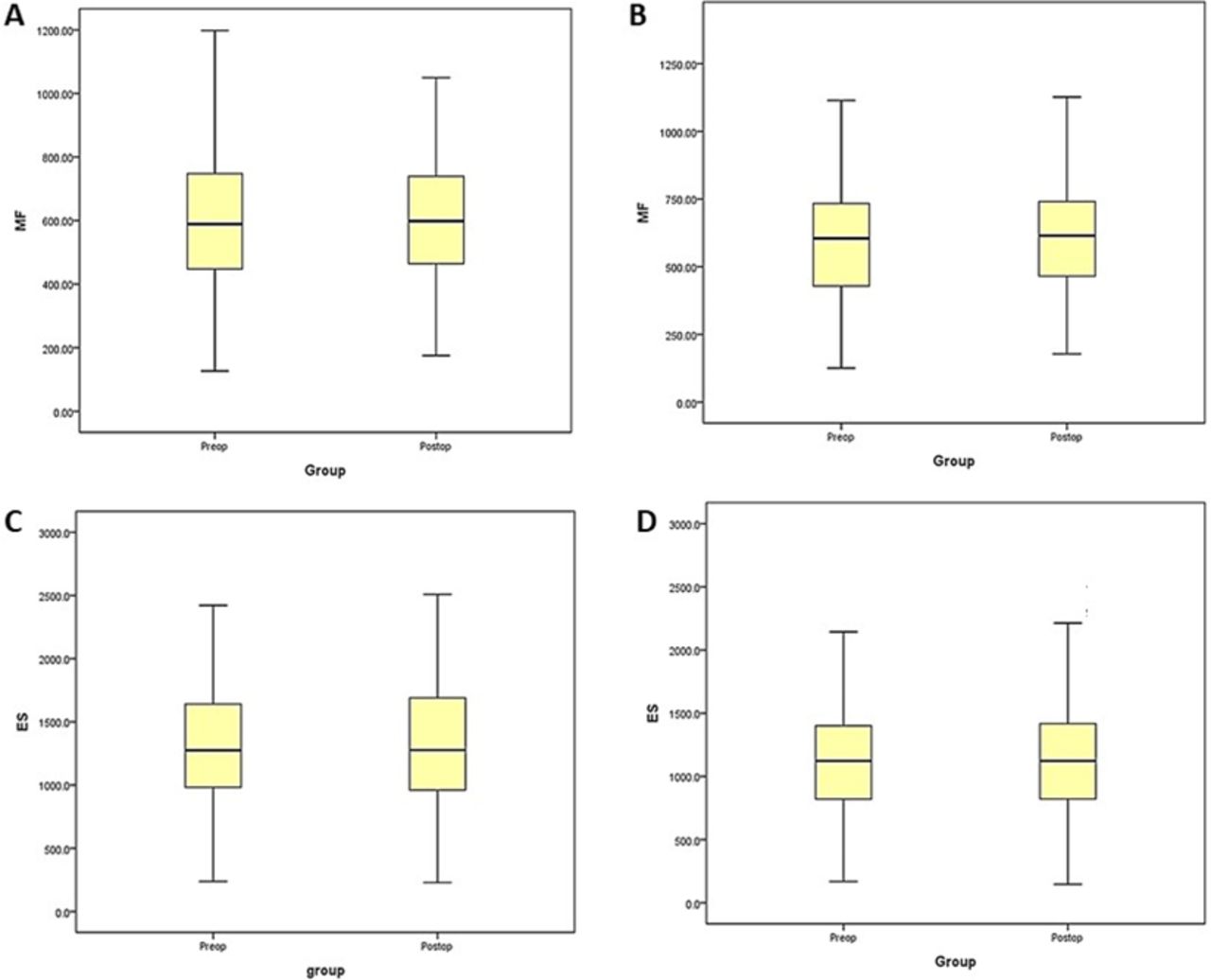
- Figure 3
Box plot with interquartile range and median value showing for the multilevel lumbar endoscopic unilateral laminotomy with bilateral decompression preoperative and 6-month follow-up changes in functional cross-sectional area of (A) ipsilateral multifidus (MF), (B) contralateral MF, (C) ipsilateral erector spinae (ES), and (D) contralateral ES.
- Figure 4
Saggital and axial T2-weighted images of a 56-year-old female patient operated for single-level lumbar endoscopic unilateral laminotomy with bilateral decompression L4-5 showing significant increase and maintenance of dural sac cross-sectional area for long-term follow-up without post-operative paraspinal muscle atrophy.


Tables
Single-Level ESLD Group Multilevel ESLD Group P Value Patients 120 39 Age, y, mean 60.5 67.28 Gender (M:F) 60:60 (50%) 18:21 (46.15%) Levels of ESLD 120 83 L1-2 1 2 L2-3 9 15 L3-4 15 26 L4-5 82 28 L5-S1 13 12 VAS score, mean ± SD Preoperative 7.83 ± 1.37 7.84 ± 1.38 Immediate postoperative 3.15 ± 0.67 3.50 ± 0.60 <0.001* 6-mo follow-up 2.19 ± 0.88 2.44 ± 0.79 <0.001* ODI, mean ± SD Preoperative 74.09 ± 7.18 74.1 ± 7.72 Immediate postoperative 27.88 ± 4.40 31.30 ± 4.46 <0.001* 6-mo follow-up 23.88 ± 4.56 24.90 ± 4.75 <0.001* Statistically significant values are marked with an asterisk (*).
ESLD, end-stage liver disease; ODI, Oswestry Disability Index; VAS, visual analog scale.
Outcome Measure Preoperative Six-mo Follow-Up P Value Single-level ESLD MF FCSA (in mm2) Ipsilateral 594.47 ± 208.24 604.56 ± 199.99 0.112 Contralateral 597.96 ± 227.05 613.79 ± 219.18 0.66 ES FCSA (in mm2) Ipsilateral 1101.26 ± 429.1 1177.88 ± 636.6 0.123 Contralateral 1138.26 ± 465.4 1144.65 ± 463.2 0.621 Fatty infiltration scale, n (%) 0 47 (39.83) 62 (52.54) 0.221 1 51 (43.22) 42 (35.59) 0.358 2 22 (16.95) 14 (11.86) 0.119 DS CSA (in mm2) 79.23 ± 28.54 163.97 ± 37.86 <0.001* Multilevel ESLD MF FCSA (in mm2) Ipsilateral 513.81 ± 198.56 520.92 ± 189.22 0.458 Contralateral 507.48 ± 218.49 516.23 ± 206.64 0.344 ES FCSA (in mm2) Ipsilateral 1316.89 ± 523.7 1332.92 ± 541.68 0.432 Contralateral 1296.36 ± 563.25 1316.27 ± 563.43 0.311 Fatty infiltration scale, n (%) 0 37 (97.37) 38 (100) 0.865 1 33 (86.84) 33 (86.84) 0.697 2 13 (28.95) 10 (26.32) 0.771 DS CSA (in mm2) 69.83 ± 19.94 157.81 ± 23.6 <0.001* Note: Data presented as mean ± SD unless otherwise noted. Statistically significant values are marked with an asterisk (*).
CSA, cross-sectional area; DS, dural sac; ES, erector spinae; ESLD, end-stage liver disease; FCSA, functional cross-sectional area; MF, multifidus.
In this issue


Jump to section
Related Articles
Cited By...
- No citing articles found.