
Main menu

  • Home
  • Content
    • Current Issue
    • Advance Online Publication
    • Archive
  • About Us
    • About ISASS
    • About the Journal
    • Author Instructions
    • Editorial Board
    • Reviewer Guidelines & Publication Criteria
  • More
    • Advertise
    • Subscribe
    • Alerts
    • Feedback
  • Join Us
  • Reprints & Permissions
  • Sponsored Content
  • Other Publications
    • ijss

User menu

  • My alerts

Search

  • Advanced search
International Journal of Spine Surgery
  • My alerts
International Journal of Spine Surgery

Advanced Search

  • Home
  • Content
    • Current Issue
    • Advance Online Publication
    • Archive
  • About Us
    • About ISASS
    • About the Journal
    • Author Instructions
    • Editorial Board
    • Reviewer Guidelines & Publication Criteria
  • More
    • Advertise
    • Subscribe
    • Alerts
    • Feedback
  • Join Us
  • Reprints & Permissions
  • Sponsored Content
  • Follow ijss on Twitter
  • Visit ijss on Facebook
Research ArticleEndoscopic Minimally Invasive Surgery

Full Endoscopic Lumbar Stenosis Decompression: A Future Gold Standard in Managing Degenerative Lumbar Canal Stenosis

Asrafi Rizki Gatam, Luthfi Gatam, Phedy, Harmantya Mahadhipta, Ajiantoro and Dina Aprilya
International Journal of Spine Surgery October 2022, 16 (5) 821-830; DOI: https://doi.org/10.14444/8338
Asrafi Rizki Gatam
1 Orthopaedic spine surgeon, Fatmawati General Hospital, Jakarta, Indonesia
2 Orthopaedic and spine center, Gatam Institute, Eka Hospital BSD, Banten, Indonesia
3 Orthopaedic spine surgeon, Premier Bintaro Hospital, Banten, Indonesia
MD
  • Find this author on Google Scholar
  • Find this author on PubMed
  • Search for this author on this site
Luthfi Gatam
1 Orthopaedic spine surgeon, Fatmawati General Hospital, Jakarta, Indonesia
2 Orthopaedic and spine center, Gatam Institute, Eka Hospital BSD, Banten, Indonesia
3 Orthopaedic spine surgeon, Premier Bintaro Hospital, Banten, Indonesia
MD, PhD
  • Find this author on Google Scholar
  • Find this author on PubMed
  • Search for this author on this site
Phedy
1 Orthopaedic spine surgeon, Fatmawati General Hospital, Jakarta, Indonesia
2 Orthopaedic and spine center, Gatam Institute, Eka Hospital BSD, Banten, Indonesia
MD
  • Find this author on Google Scholar
  • Find this author on PubMed
  • Search for this author on this site
Harmantya Mahadhipta
2 Orthopaedic and spine center, Gatam Institute, Eka Hospital BSD, Banten, Indonesia
4 Orthopaedic spine surgeon, Tangerang General Hospital, Banten, Indonesia
MD
  • Find this author on Google Scholar
  • Find this author on PubMed
  • Search for this author on this site
Ajiantoro
3 Orthopaedic spine surgeon, Premier Bintaro Hospital, Banten, Indonesia
MD
  • Find this author on Google Scholar
  • Find this author on PubMed
  • Search for this author on this site
Dina Aprilya
2 Orthopaedic and spine center, Gatam Institute, Eka Hospital BSD, Banten, Indonesia
MD
  • Find this author on Google Scholar
  • Find this author on PubMed
  • Search for this author on this site
  • Article
  • Figures & Data
  • Info & Metrics
  • PDF
Loading

Article Figures & Data

Figures

  • Tables
  • Supplementary Materials
  • Figure 1
    • Download figure
    • Open in new tab
    • Download powerpoint
    Figure 1

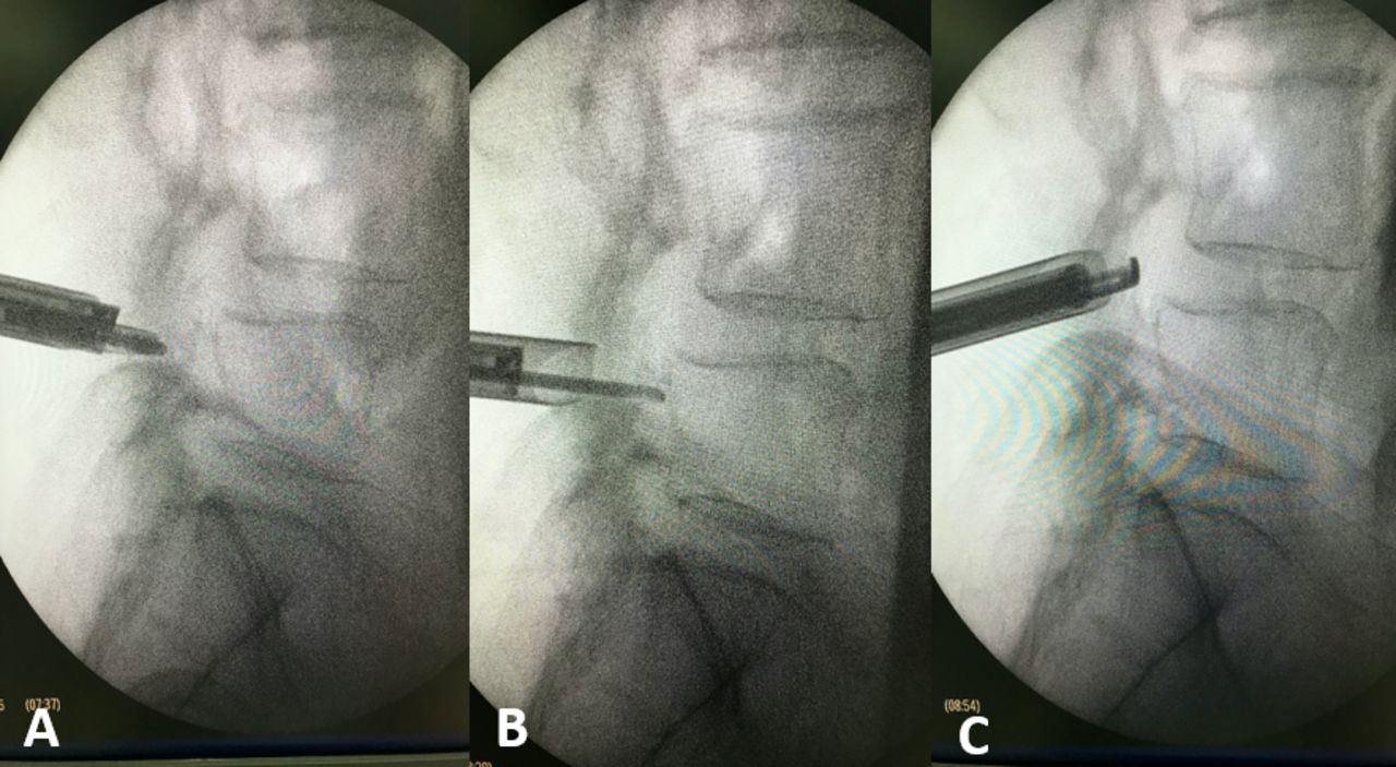
    Drilling medial facet started from the midpedicle (A–B) until the tip of the superior articular process (C) to make sure complete decompression.

  • Figure 2
    • Download figure
    • Open in new tab
    • Download powerpoint
    Figure 2

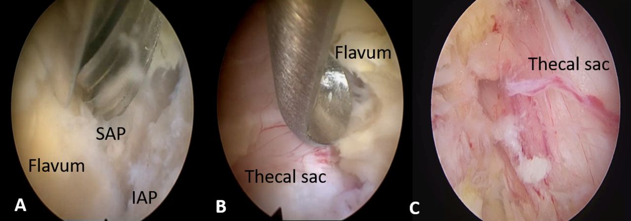
    (A) Opening the lamina in the medial side. (B) Removing flavum ligament using Kerrison punch. (C) Free thecal sac after endoscopic decompression. SAP, superior articular process; IAP, inferior articular process

  • Figure 3
    • Download figure
    • Open in new tab
    • Download powerpoint
    Figure 3

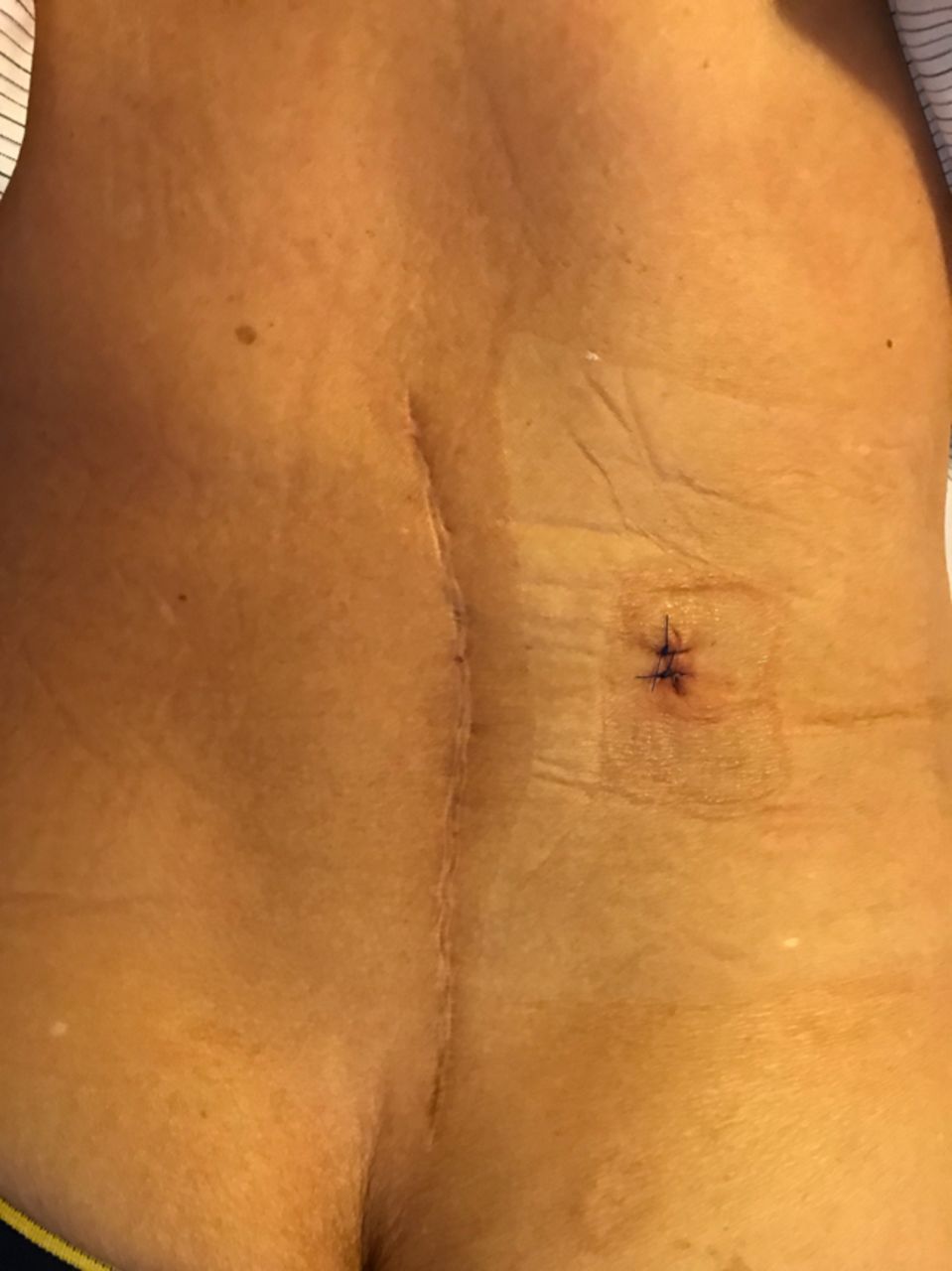
    Location of incision for paracentral endoscopic decompressive foraminotomy.

  • Figure 4
    • Download figure
    • Open in new tab
    • Download powerpoint
    Figure 4

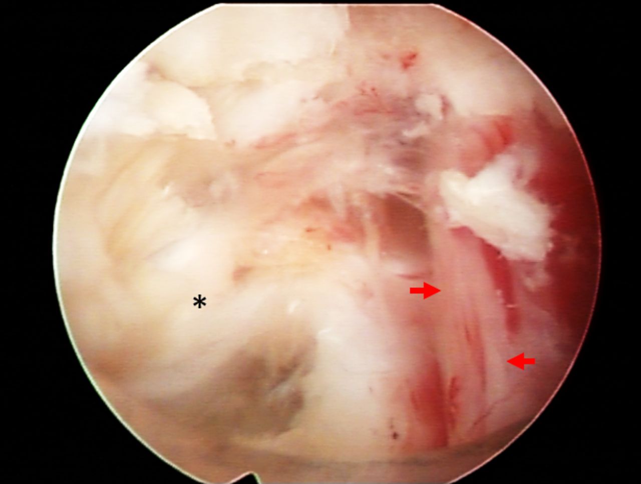
    Free exiting nerve root and disc material that was removed. Asterisk, disc material; blue dot, disc space; red arrows, exiting root.
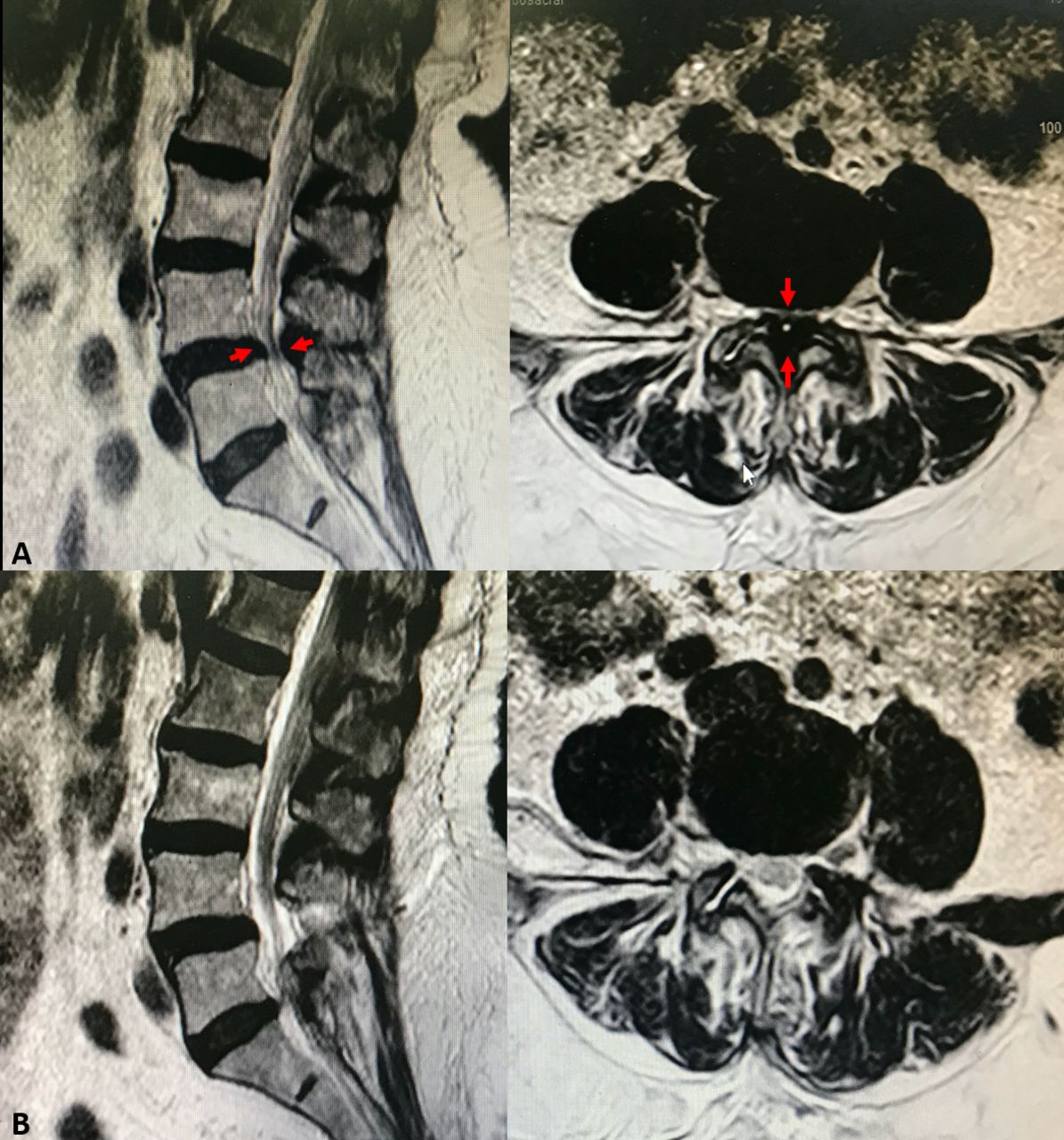
  • Figure 5
    • Download figure
    • Open in new tab
    • Download powerpoint
    Figure 5

    Magnetic resonance imaging (MRI) of a patient with central lumbar canal stenosis. (A) Preoperative MRI: the central canal was compressed anteriorly by the disk and posteriorly by the thick flavum (arrows). (B) Postendoscopic decompression.
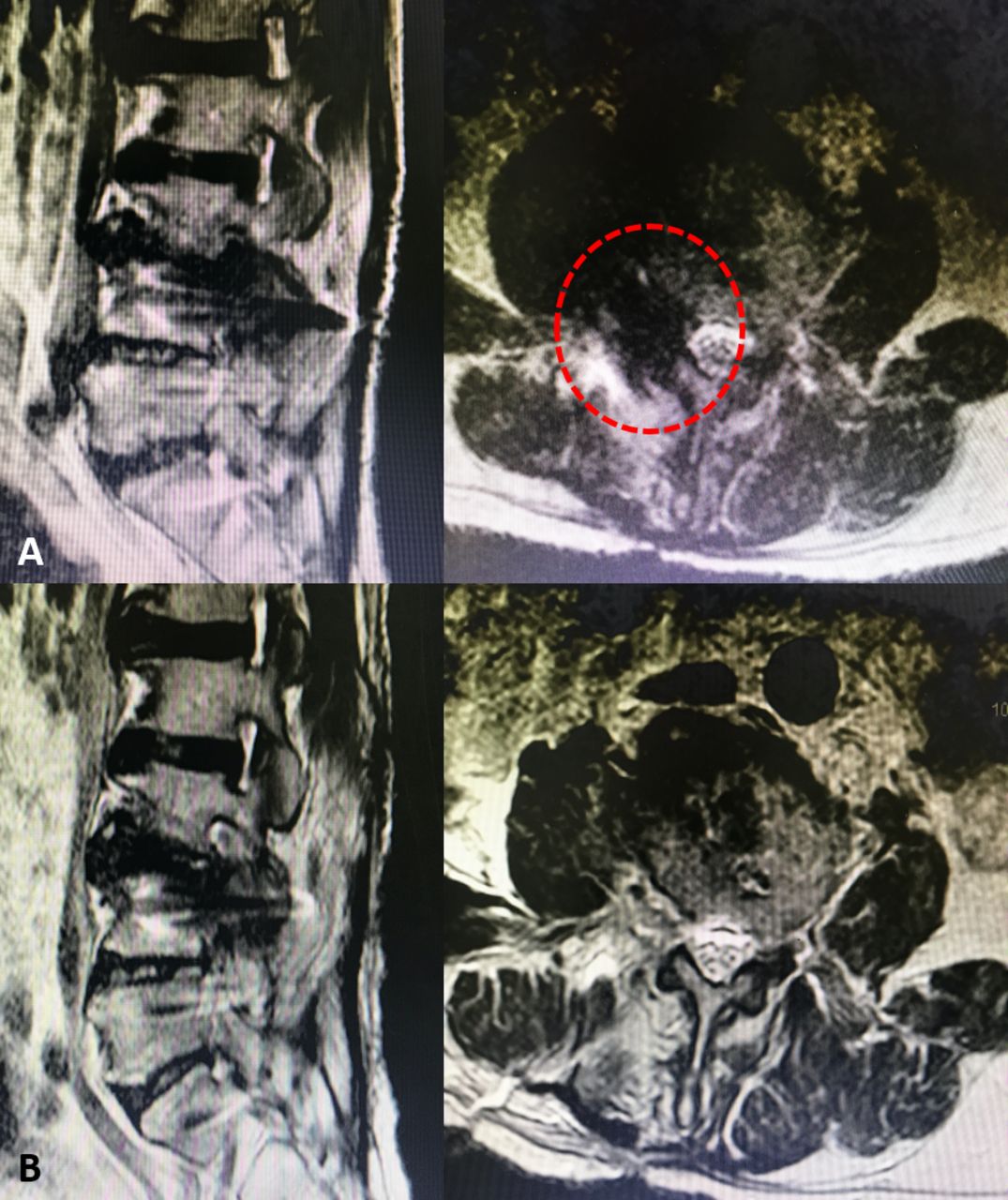
  • Figure 6
    • Download figure
    • Open in new tab
    • Download powerpoint
    Figure 6

    Magnetic resonance imaging of a patient with L3-L4 foraminal stenosis. (A) Preoperative: The right paracentral compression (red dots). (B) After endoscopic foraminotomy.

  • Figure 7
    • Download figure
    • Open in new tab
    • Download powerpoint
    Figure 7

    Image of dural tear during endoscopic procedure (red circle).

Tables

  • Figures
  • Supplementary Materials
    • View popup
    Table 1

    Clinical and demographic characteristics of degenerative lumbar canal stenosis patients managed with conventional decompression and full endoscopic decompression.

    Patient CharacteristicsConventional DecompressionFull Endoscopic DecompressionMean Difference (CI 95%) P Value
    N = 132 N = 163
    Age, y, mean (SD)57.6 (6.13)53.1 (11.31)4.5 (−1.9 to 10.9)0.160
    Sex, n (%)0.130
     Male72 (54.7)102 (62.5)
     Female60 (45.2)61 (36.8)
    Level of decompression, n (%)0.686
     L3-L419 (14.2)16 (10.0)
     L4-L575 (57.1)106 (65.0)
     L5-S138 (28.5)41 (25.0)
    Intraoperative bleeding, mL, median (IQR)84 (50–150)30 (10–50)0.001
    Operation duration, min, median (IQR)45 (30–75)65 (50–110)0.032
    Duration of hospital stay, d, median (IQR)3.4 (3–4)1.52 (1–3)0.034
    Mobilization time, h, median (IQR)14.3 (10–18)4.2 (3–5)0.001
    • Abbreviations: IQR, interquartile range.

    • View popup
    Table 2

    VAS of patients who underwent conventional decompression and full endoscopic decompression.

    VASConventional DecompressionFull Endoscopic DecompressionMean Difference
    (95% CI)
    P Value
    N = 132 N = 163
    Back
     Preoperation1 (0–2)1.1 (0–2)0 (−1 to 1)0.708
     Postoperation
      0 mo4 (3–5.3)2 (1.5–3)0 (−1 to 1)0.033
      3 mo2 (1–3)1 (0–2)0 (−1 to 1)0.112
      6 mo1 (0–2)1 (0–2)0 (0–1)0.134
      12 mo1(0–2)0(0)0.111
    Leg
     Preoperation6 (5.8–7)6 (6–7)0 (−1 to 1)0.909
      Postoperation
      0 mo3 (1–5)2 (0–3)0 (0–1)0.05
      3 mo1 (0–2)1 (0–2)1 (0–1)0.071
      6 mo1 (0–1)1 (0–1)1 (0–2)0.075
      12 mo1 (0–1)1(0–1)1(0–2)0.080
    • Abbreviations: VAS, visual analog scale.

    • Note: Decompression data presented as median (interquartile range).

    • View popup
    Table 3

    Oswestry Disability Index of patients who underwent conventional decompression and full endoscopic decompression.

    Oswestry Disability IndexConventional DecompressionFull Endoscopic DecompressionMedian Difference
    (95% CI)
    P Value
    N = 132 N = 163
    Preoperation62 (56.5–70.5)58 (52–63.5)6 (−2 to 14)0.103
    Postoperation
     0 mo12 (5.5–14.5)16 (8–32.5)−4 (−12 to 2)0.232
     3 mo8 (4–12.5)12 (4–19)−2 (−8 to 4)0.483
     6 mo5.5 (1.5–10)10 (3.5–17.5)−3 (−10 to 2)0.184
     12 mo5.7 (1.5–9.2)9.5 (3.6–15.6)−3 (−9 to 2)0.188
    • Note: Decompression data presented as median (interquartile range).

Supplementary Materials

  • Figures
  • Tables
  • Online Supplementary Video 1.

    [8338supp001.mp4]

  • Online Supplementary Video 2.

    [8338supp002.mp4]

PreviousNext
Back to top

In this issue

International Journal of Spine Surgery
Vol. 16, Issue 5
1 Oct 2022
  • Table of Contents
  • Index by author

Print
Download PDF
Article Alerts
Sign In to Email Alerts with your Email Address
Email Article

Thank you for your interest in spreading the word on International Journal of Spine Surgery.

NOTE: We only request your email address so that the person you are recommending the page to knows that you wanted them to see it, and that it is not junk mail. We do not capture any email address.

Enter multiple addresses on separate lines or separate them with commas.
Full Endoscopic Lumbar Stenosis Decompression: A Future Gold Standard in Managing Degenerative Lumbar Canal Stenosis
(Your Name) has sent you a message from International Journal of Spine Surgery
(Your Name) thought you would like to see the International Journal of Spine Surgery web site.
CAPTCHA
This question is for testing whether or not you are a human visitor and to prevent automated spam submissions.
Citation Tools
Full Endoscopic Lumbar Stenosis Decompression: A Future Gold Standard in Managing Degenerative Lumbar Canal Stenosis
Asrafi Rizki Gatam, Luthfi Gatam, Phedy, Harmantya Mahadhipta, Ajiantoro, Dina Aprilya
International Journal of Spine Surgery Oct 2022, 16 (5) 821-830; DOI: 10.14444/8338

Citation Manager Formats

  • BibTeX
  • Bookends
  • EasyBib
  • EndNote (tagged)
  • EndNote 8 (xml)
  • Medlars
  • Mendeley
  • Papers
  • RefWorks Tagged
  • Ref Manager
  • RIS
  • Zotero
Share
Full Endoscopic Lumbar Stenosis Decompression: A Future Gold Standard in Managing Degenerative Lumbar Canal Stenosis
Asrafi Rizki Gatam, Luthfi Gatam, Phedy, Harmantya Mahadhipta, Ajiantoro, Dina Aprilya
International Journal of Spine Surgery Oct 2022, 16 (5) 821-830; DOI: 10.14444/8338
Twitter logo Facebook logo Mendeley logo
  • Tweet Widget
  • Facebook Like
  • Google Plus One

Jump to section

  • Article
    • Abstract
    • INTRODUCTION
    • MATERIALS AND METHODS
    • SURGICAL TECHNIQUES
    • RESULTS
    • DISCUSSION
    • CONCLUSION
    • Footnotes
    • References
  • Figures & Data
  • Info & Metrics
  • PDF

Related Articles

  • No related articles found.
  • PubMed
  • Google Scholar

Cited By...

  • No citing articles found.
  • Google Scholar

More in this TOC Section

  • Patient Perspectives on Awake Transforaminal Endoscopic Decompression Surgery Outcomes
  • Endoscopic Spine Surgery: A French National Survey on Practices, Motivations, and Challenges
  • Effective Biportal Endoscopic Spine Surgery Technique With Better Facet Joint Preserving for Lumbar Lateral Recess Stenosis
Show more Endoscopic Minimally Invasive Surgery

Similar Articles

Keywords

  • Degenerative lumbar spine
  • spinal stenosis
  • minimally invasive spine surgery
  • full endoscopic spine surgery

Content

  • Current Issue
  • Latest Content
  • Archive

More Information

  • About IJSS
  • About ISASS
  • Privacy Policy

More

  • Subscribe
  • Alerts
  • Feedback

Other Services

  • Author Instructions
  • Join ISASS
  • Reprints & Permissions

© 2025 International Journal of Spine Surgery

International Journal of Spine Surgery Online ISSN: 2211-4599

Powered by HighWire