
Main menu

  • Home
  • Content
    • Current Issue
    • Advance Online Publication
    • Archive
  • About Us
    • About ISASS
    • About the Journal
    • Author Instructions
    • Editorial Board
    • Reviewer Guidelines & Publication Criteria
  • More
    • Advertise
    • Subscribe
    • Alerts
    • Feedback
  • Join Us
  • Reprints & Permissions
  • Sponsored Content
  • Other Publications
    • ijss

User menu

  • My alerts

Search

  • Advanced search
International Journal of Spine Surgery
  • My alerts
International Journal of Spine Surgery

Advanced Search

  • Home
  • Content
    • Current Issue
    • Advance Online Publication
    • Archive
  • About Us
    • About ISASS
    • About the Journal
    • Author Instructions
    • Editorial Board
    • Reviewer Guidelines & Publication Criteria
  • More
    • Advertise
    • Subscribe
    • Alerts
    • Feedback
  • Join Us
  • Reprints & Permissions
  • Sponsored Content
  • Follow ijss on Twitter
  • Visit ijss on Facebook
Research ArticleLumbar Spine

Single-Position Fluoroscopy-Guided Lateral Lumbar Interbody Fusion With Intraoperative Computed Tomography–Navigated Posterior Pedicle Screw Fixation: Technical Report and Literature Review

Jacob L. Goldberg, Lynn B. McGrath, Sertac Kirnaz, Fabian Sommer, Joseph A. Carnevale, Branden Medary and Roger Härtl
International Journal of Spine Surgery April 2022, 16 (S1) S9-S16; DOI: https://doi.org/10.14444/8231
Jacob L. Goldberg
1 Department of Neurosurgery, New York Presbyterian Hospital/Weill Cornell Medicine 1 New York, NY, USA
MD
  • Find this author on Google Scholar
  • Find this author on PubMed
  • Search for this author on this site
Lynn B. McGrath Jr
1 Department of Neurosurgery, New York Presbyterian Hospital/Weill Cornell Medicine 1 New York, NY, USA
MD
  • Find this author on Google Scholar
  • Find this author on PubMed
  • Search for this author on this site
Sertac Kirnaz
1 Department of Neurosurgery, New York Presbyterian Hospital/Weill Cornell Medicine 1 New York, NY, USA
MD
  • Find this author on Google Scholar
  • Find this author on PubMed
  • Search for this author on this site
Fabian Sommer
1 Department of Neurosurgery, New York Presbyterian Hospital/Weill Cornell Medicine 1 New York, NY, USA
MD
  • Find this author on Google Scholar
  • Find this author on PubMed
  • Search for this author on this site
Joseph A. Carnevale
1 Department of Neurosurgery, New York Presbyterian Hospital/Weill Cornell Medicine 1 New York, NY, USA
MD
  • Find this author on Google Scholar
  • Find this author on PubMed
  • Search for this author on this site
Branden Medary
1 Department of Neurosurgery, New York Presbyterian Hospital/Weill Cornell Medicine 1 New York, NY, USA
BS
  • Find this author on Google Scholar
  • Find this author on PubMed
  • Search for this author on this site
Roger Härtl
1 Department of Neurosurgery, New York Presbyterian Hospital/Weill Cornell Medicine 1 New York, NY, USA
MD
  • Find this author on Google Scholar
  • Find this author on PubMed
  • Search for this author on this site
  • Article
  • Figures & Data
  • Info & Metrics
  • PDF
Loading

Article Figures & Data

Figures

  • Tables
  • Figure 1
    • Download figure
    • Open in new tab
    • Download powerpoint
    Figure 1

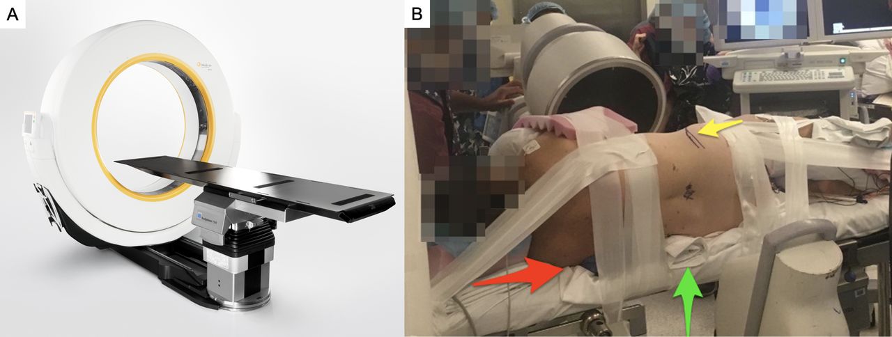
    (A) Intraoperative computed tomography (CT) with integrated flat operative bed (AIRO, Brainlab. Photograph used with permission from Brainlab). (B) Patient positioning. Patient is on a flat, integrated CT table and is secured with silk tape and padded. Both lateral and posterior operative sites are accessible. The iliac crest is marked (yellow arrow). The patient is as close to the edge of the operative table as safely possible (red arrow). A sheet is rolled to create lateral flexion and to minimize patient movement during the procedure (green arrow).

  • Figure 2
    • Download figure
    • Open in new tab
    • Download powerpoint
    Figure 2

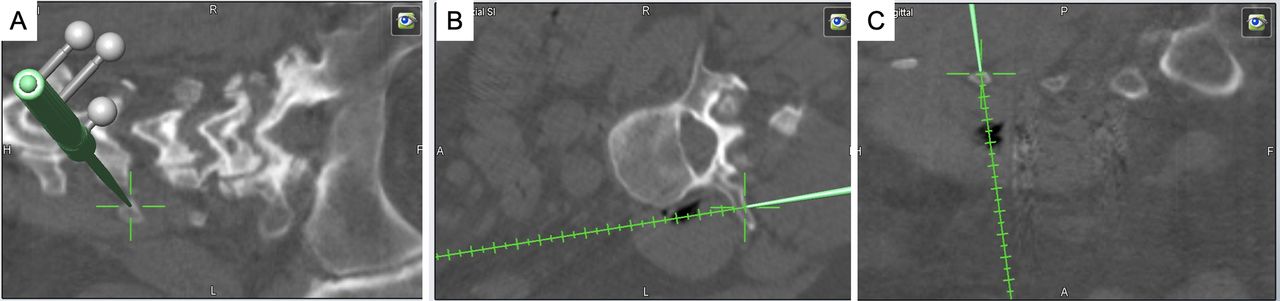
    Accuracy of intraoperative navigation is verified at the transverse process of the most cranial level (furthest from iliac crest reference array). (A) Coronal, (B) axial, and (C) sagittal.

  • Figure 3
    • Download figure
    • Open in new tab
    • Download powerpoint
    Figure 3

    Pedicle screws of various sizes and dimensions can be virtually sized (red). (A) Axial and (B) sagittal.

  • Figure 4
    • Download figure
    • Open in new tab
    • Download powerpoint
    Figure 4

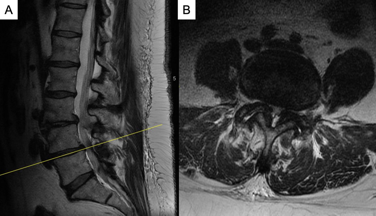
    Magnetic resonance imaging (MRI) lumbar spine. (A) Sagittal T2 MRI lumbar spine most notable for loss of disc height at L3/4 and L4/5. Severe stenosis is evident at L3/4 and L4/5. (B) Axial T2 MRI at L4/5 demonstrating severe central stenosis.

  • Figure 5
    • Download figure
    • Open in new tab
    • Download powerpoint
    Figure 5

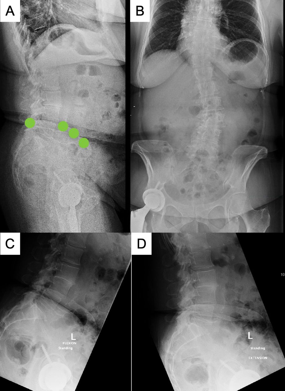
    Plain film x-ray images. (A) Standing lateral x-ray image. Significant disc height loss is evident in L3/4 and L4/5. Note level of iliac crest (green dots) allowing access to L4/5 disc space laterally. (B) Standing anteroposterior x-ray image without major coronal imbalance. Levoscoliosis with apex at L3 measures 24° and minor curve between T7 and T12 measures 18°. (C) Flexion x-ray images and (D) extension view demonstrate instability with pathologic movement most notable at L4/5.

  • Figure 6
    • Download figure
    • Open in new tab
    • Download powerpoint
    Figure 6

    Intraoperative fluoroscopy (lateral view) demonstrating lordotic interbody cages placed at L3/4 and L4/5.

  • Figure 7
    • Download figure
    • Open in new tab
    • Download powerpoint
    Figure 7

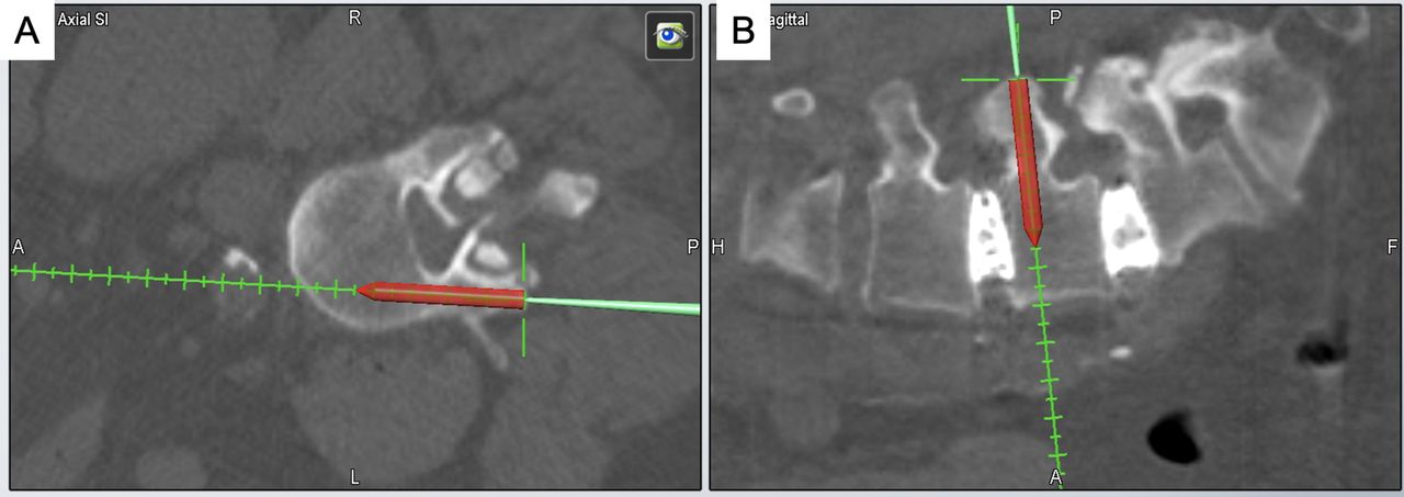
    Percutaneous pedicle screws with integrated stylet tip (red) are used. The stylet tip is placed at the entry point for the pedicle screw, and the trajectory is optimized. (A) Axial and (B) sagittal.

  • Figure 8
    • Download figure
    • Open in new tab
    • Download powerpoint
    Figure 8

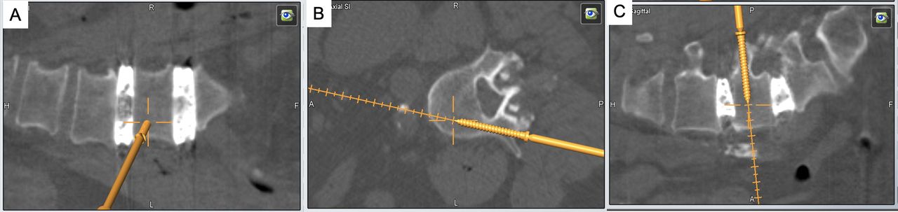
    As the pedicle screw is advanced, the trajectory is continuously monitored via navigation. Additionally, after the screw enters the vertebral body, the stylet tip is retracted (red tip no longer visible). (A) Coronal, (B) axial, and (C) sagittal.

  • Figure 9
    • Download figure
    • Open in new tab
    • Download powerpoint
    Figure 9

    Postoperative x-ray images demonstrating intact hardware and increased disc space height at L3/4 and L4/5 with improved lumbar lordosis. (A) Lateral and (B) anteroposterior.

Tables

  • Figures
    • View popup
    Table

    Single-stage lateral lumbar interbody fusion publications.

    AuthorsNumber of PatientsDesign of StudySimultaneous Lateral and Posterior Instrumentation (Parallel) or Sequential (Series)Intraoperative Imaging for Pedicle Screw PlacementTotal Operative Time (Average Minutes)Pedicle Breach RateRate of Screw Misplacement Requiring RevisionReturn to OR for Screw Revision (n)
    Drazin et al13 10 (SP), 10 (DP)RetrospectiveSeriesFluoroscopy131 (SP), 190 (DP)NRNR1(SP), 0 (DP)
    Blizzard et al14 72 (SP), 0 (DP)RetrospectiveSeriesFluoroscopy87.95.1%2.8% 2
    Ziino et al19 42 (SP), 24 (DP)RetrospectiveSeriesFluoroscopy149 (SP), 226 (DP)NRNR2 (SP), 0 (DP)
    Sellin et al15 4 (SP), 0 (DP)RetrospectiveParallelCone beam iCT138 (SP)14%14%1 (SP)
    Huntsman et al20 55 (SP), 0 (DP)RetrospectiveSeriesRobotic guidance: preop CT (14 pts), fan or cone beam iCT (41 pts)155 (SP)000
    Hiyama et al16 19 (SP), 26 (DP)RetrospectiveSeriesFluoroscopy98 (SP), 130 (DP)NRNRNR
    Ouchida et al17 51 (SP), 51 (DP)RetrospectiveParallelFan beam iCT93 (SP), 121 (DP)9.5% (SP), 12.5% (DP)00
    Buckland et al18 237 (SP), 153 (DP)RetrospectiveSeriesFlouroscopy, iCT, iCT with robotic guidance103 (SP), 306 (DP)NR1.56% (SP), 2.34% (DP)NR
    • CT, computed tomography; DP, dual position; iCT, intraoperative computed tomography; NR, not reported; SP, single position.

PreviousNext
Back to top

In this issue

International Journal of Spine Surgery: 16 (S1)
International Journal of Spine Surgery
Vol. 16, Issue S1
1 Apr 2022
  • Table of Contents
  • Index by author

Print
Download PDF
Article Alerts
Sign In to Email Alerts with your Email Address
Email Article

Thank you for your interest in spreading the word on International Journal of Spine Surgery.

NOTE: We only request your email address so that the person you are recommending the page to knows that you wanted them to see it, and that it is not junk mail. We do not capture any email address.

Enter multiple addresses on separate lines or separate them with commas.
Single-Position Fluoroscopy-Guided Lateral Lumbar Interbody Fusion With Intraoperative Computed Tomography–Navigated Posterior Pedicle Screw Fixation: Technical Report and Literature Review
(Your Name) has sent you a message from International Journal of Spine Surgery
(Your Name) thought you would like to see the International Journal of Spine Surgery web site.
CAPTCHA
This question is for testing whether or not you are a human visitor and to prevent automated spam submissions.
Citation Tools
Single-Position Fluoroscopy-Guided Lateral Lumbar Interbody Fusion With Intraoperative Computed Tomography–Navigated Posterior Pedicle Screw Fixation: Technical Report and Literature Review
Jacob L. Goldberg, Lynn B. McGrath, Sertac Kirnaz, Fabian Sommer, Joseph A. Carnevale, Branden Medary, Roger Härtl
International Journal of Spine Surgery Apr 2022, 16 (S1) S9-S16; DOI: 10.14444/8231

Citation Manager Formats

  • BibTeX
  • Bookends
  • EasyBib
  • EndNote (tagged)
  • EndNote 8 (xml)
  • Medlars
  • Mendeley
  • Papers
  • RefWorks Tagged
  • Ref Manager
  • RIS
  • Zotero
Share
Single-Position Fluoroscopy-Guided Lateral Lumbar Interbody Fusion With Intraoperative Computed Tomography–Navigated Posterior Pedicle Screw Fixation: Technical Report and Literature Review
Jacob L. Goldberg, Lynn B. McGrath, Sertac Kirnaz, Fabian Sommer, Joseph A. Carnevale, Branden Medary, Roger Härtl
International Journal of Spine Surgery Apr 2022, 16 (S1) S9-S16; DOI: 10.14444/8231
Twitter logo Facebook logo Mendeley logo
  • Tweet Widget
  • Facebook Like
  • Google Plus One

Jump to section

  • Article
    • Abstract
    • Introduction
    • OPERATIVE TECHNIQUE
    • ILLUSTRATIVE CASE
    • DISCUSSION
    • Conclusions
    • Footnotes
    • References
  • Figures & Data
  • Info & Metrics
  • PDF

Related Articles

  • No related articles found.
  • PubMed
  • Google Scholar

Cited By...

  • No citing articles found.
  • Google Scholar

More in this TOC Section

  • Comparison of Stand-Alone Anterior Lumbar Interbody Fusion, 360° Anterior Lumbar Interbody Fusion, and Arthroplasty for Recurrent Lumbar Disc Herniation: Focus on Nerve Decompression and Painful Spinal Instability Resolution
  • Recovery Trajectories After Lumbar Fusion Stratified by Baseline Patient-Reported Outcomes Measurement Information System Physical Function Disability Levels
  • Association Between Nonsteroidal Anti-inflammatory Drugs Use and Surgical Outcomes Following Posterior Lumbar Fusion: A Medical Claims Database Analysis
Show more Lumbar Spine

Similar Articles

Keywords

  • single-position lateral lumbar interbody fusion
  • LLIF
  • intraoperative navigation

Content

  • Current Issue
  • Latest Content
  • Archive

More Information

  • About IJSS
  • About ISASS
  • Privacy Policy

More

  • Subscribe
  • Alerts
  • Feedback

Other Services

  • Author Instructions
  • Join ISASS
  • Reprints & Permissions

© 2025 International Journal of Spine Surgery

International Journal of Spine Surgery Online ISSN: 2211-4599

Powered by HighWire