
Main menu

  • Home
  • Content
    • Current Issue
    • Advance Online Publication
    • Archive
  • About Us
    • About ISASS
    • About the Journal
    • Author Instructions
    • Editorial Board
    • Reviewer Guidelines & Publication Criteria
  • More
    • Advertise
    • Subscribe
    • Alerts
    • Feedback
  • Join Us
  • Reprints & Permissions
  • Sponsored Content
  • Other Publications
    • ijss

User menu

  • My alerts

Search

  • Advanced search
International Journal of Spine Surgery
  • My alerts
International Journal of Spine Surgery

Advanced Search

  • Home
  • Content
    • Current Issue
    • Advance Online Publication
    • Archive
  • About Us
    • About ISASS
    • About the Journal
    • Author Instructions
    • Editorial Board
    • Reviewer Guidelines & Publication Criteria
  • More
    • Advertise
    • Subscribe
    • Alerts
    • Feedback
  • Join Us
  • Reprints & Permissions
  • Sponsored Content
  • Follow ijss on Twitter
  • Visit ijss on Facebook
Research ArticleCervical Spine

Evaluation of Haplo-Paraspinal-Muscle-Preserving Technique to Prevent Postoperative Axial Pain in Cervical Laminoplasty

XiaoLong Shen, Chen Xu, Ruizhe Wang, Zifan Zhang, Min Qi, Yizhi Zhang, Huiqiao Wu, Yang Liu, Huajiang Chen and Wen Yuan
International Journal of Spine Surgery April 2023, 17 (2) 281-291; DOI: https://doi.org/10.14444/8416
XiaoLong Shen
1 Spine Center, Department of Orthopedics, Shanghai Changzheng Hospital, Naval Medical University, Shanghai, China
PʜD
  • Find this author on Google Scholar
  • Find this author on PubMed
  • Search for this author on this site
Chen Xu
1 Spine Center, Department of Orthopedics, Shanghai Changzheng Hospital, Naval Medical University, Shanghai, China
PʜD
  • Find this author on Google Scholar
  • Find this author on PubMed
  • Search for this author on this site
Ruizhe Wang
1 Spine Center, Department of Orthopedics, Shanghai Changzheng Hospital, Naval Medical University, Shanghai, China
MD
  • Find this author on Google Scholar
  • Find this author on PubMed
  • Search for this author on this site
Zifan Zhang
1 Spine Center, Department of Orthopedics, Shanghai Changzheng Hospital, Naval Medical University, Shanghai, China
MD
  • Find this author on Google Scholar
  • Find this author on PubMed
  • Search for this author on this site
Min Qi
1 Spine Center, Department of Orthopedics, Shanghai Changzheng Hospital, Naval Medical University, Shanghai, China
PʜD
  • Find this author on Google Scholar
  • Find this author on PubMed
  • Search for this author on this site
Yizhi Zhang
1 Spine Center, Department of Orthopedics, Shanghai Changzheng Hospital, Naval Medical University, Shanghai, China
MD
  • Find this author on Google Scholar
  • Find this author on PubMed
  • Search for this author on this site
Huiqiao Wu
1 Spine Center, Department of Orthopedics, Shanghai Changzheng Hospital, Naval Medical University, Shanghai, China
PʜD
  • Find this author on Google Scholar
  • Find this author on PubMed
  • Search for this author on this site
Yang Liu
1 Spine Center, Department of Orthopedics, Shanghai Changzheng Hospital, Naval Medical University, Shanghai, China
PʜD
  • Find this author on Google Scholar
  • Find this author on PubMed
  • Search for this author on this site
Huajiang Chen
1 Spine Center, Department of Orthopedics, Shanghai Changzheng Hospital, Naval Medical University, Shanghai, China
PʜD
  • Find this author on Google Scholar
  • Find this author on PubMed
  • Search for this author on this site
Wen Yuan
1 Spine Center, Department of Orthopedics, Shanghai Changzheng Hospital, Naval Medical University, Shanghai, China
PʜD
  • Find this author on Google Scholar
  • Find this author on PubMed
  • Search for this author on this site
  • Article
  • Figures & Data
  • Info & Metrics
  • PDF
Loading

Article Figures & Data

Figures

  • Tables
  • Figure 1
    • Download figure
    • Open in new tab
    • Download powerpoint
    Figure 1

    Illustration showing the surgical steps of haplo-paraspinal-muscle-preserving (HMP) technique in laminoplasty. (A) Representative image showing the structure of the cervical vertebra and its posteriorly attached muscle. (B) The main difference between the HMP technique and traditional laminoplasty is that only half of the laminae that are to be opened during the laminoplasty procedure are exposed. The paraspinal muscle is only dissected on the left side of the spinous process, and the right side of the paraspinal muscle remains intact. (C) After exposure, the left sides of the laminae are opened and flipped using a small vertebral spreader. During the opening, the spreader slowly separates the laminae to prevent hinge side laminae fracture. (D) After opening the spinal canal, an ARCH plate is fixed to maintain the canal enlargement. PSM, paraspinal muscles; SL, spinous process–attached ligaments; SM, spinous process–attached muscles.

  • Figure 2
    • Download figure
    • Open in new tab
    • Download powerpoint
    Figure 2

    The surgical view, radiographic outcome, and intraoperative images showing the process of laminoplasty using the haplo-paraspinal-muscle-preserving (HMP) technique. (A) Only one side of the laminae is exposed and the paraspinal muscles, as well as spinous process–attached ligaments and muscles of the other side, were prevented from being dissected. (B) The laminae were cut to create an opening of the spinal canal with the assistance of a small vertebral spreader. (C) ARCH plates were used after the opening to finish the laminoplasty procedure. The radiological images show the preoperative magnetic resonance images (D), preoperative reconstructed computed tomography (E), and postoperative reconstructed computed tomography (F) of the same patient that received HMP laminoplasty. Note that the spinal canal is significantly enlarged after the surgery. LA, laminae; LM, dissected laminae–attached muscles; SP, spinous process.

  • Figure 3
    • Download figure
    • Open in new tab
    • Download powerpoint
    Figure 3

    Computed tomography (CT) images showing the typical characteristics of hinge side fractures in different laminoplasty techniques. Typical CT images of a hinge side fracture in the C4 level of an haplo-paraspinal-muscle-preserving laminoplasty–treated patient (A–C). Stenosis is observed preoperatively (A), and the spinal canal enlarged significantly after the surgery (B), with a fracture in the cortical bone of the ventral side of the hinge laminar while the dorsal side of the hinge remained intact. The fracture is healed 6 mo after the surgery, with no sign of cortical bone fracture of the hinge side (C). Typical CT images of hinge side fractures in the C3 (D) and C4 (E) levels of a traditional laminoplasty–treated patient. The C5 (F) level is intact. Both ventral and dorsal sides of the hinge side laminae were compromised when the fracture occurred (D–E); typically, the dorsal side of the hinge is thinned due to the removal of cortical bone to form a hinge (F).
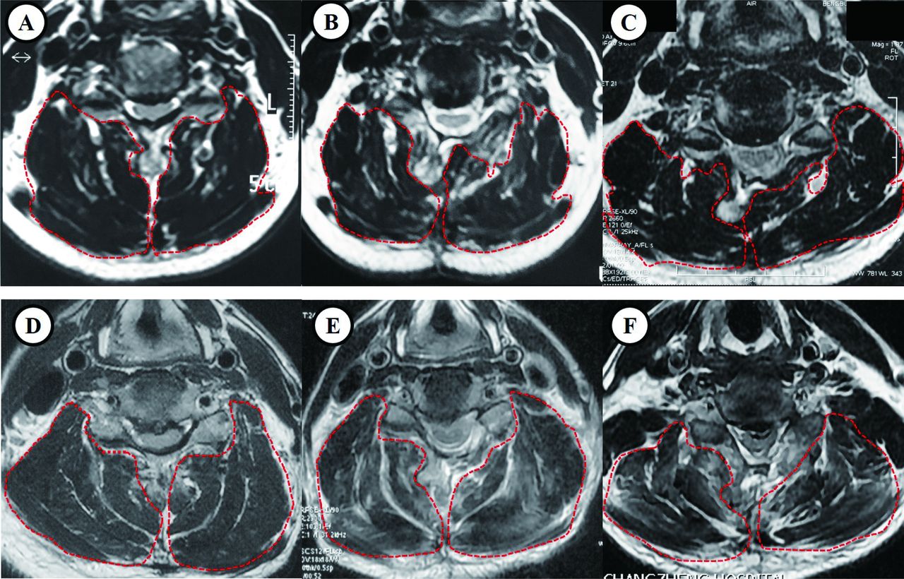
  • Figure 4
    • Download figure
    • Open in new tab
    • Download powerpoint
    Figure 4

    Magnetic resonance images (MRIs) showing the posterior cervical paravertebral muscle volumes between different laminoplasty groups. Case example of a 55-year-old female patient who received traditional laminoplasty, with the T2-weighted axial MRI showing the paravertebral muscle volumes (A) preoperation, (B) 3 days postoperation, and (C) 1 year postoperation. Case example of a 57-year-old male patient who received haplo-paraspinal-muscle-preserving laminoplasty, the T2-weighted axial MRI showing the paravertebral muscle volumes (D) preoperation, (E) 3 days postoperation, and (F) 1 year postoperation.

Tables

  • Figures
    • View popup
    Table 1

    Comparison of patient demographics by laminoplasty group.

    DemographicsMP GroupLP Group P Value
    Number2246–
    Age61.2 ± 6.460.9 ± 5.90.85
    Gender (male:female)(14:8)(29:17)0.96
    BMI26.6 ± 3.027.7 ± 2.70.08
    Disc herniation (patients)1939–
    OPLL (patients)820–
    Operation level0.74
     C3–C648–
     C3–C71629–
     C2–C614–
     C2–C715–
    Preoperative JOA9.1 ± 7.98.7 ± 7.20.84
    Preoperative VAS5.2 ± 2.55.4 ± 2.30.75
    Follow-up period (mo)15.8 ± 3.816.8 ± 3.20.55
    • Note: Data are shown as the mean ± SD; ‘-’ represents values were not compared for differences.

    • BMI, body mass index; JOA, Japanese Orthopaedic Association; LP Group, traditional open-door laminoplasty-treated patients; MP Group, haplo-paraspinal-muscle-preserving laminoplasty-treated patients; OPLL, ossification of posterior longitudinal ligament; VAS, visual analog scale.

    • View popup
    Table 2

    Comparison of surgical parameters between laminoplasty groups.

    Surgical ParametersMP GroupLP Group P Value
    Time of surgery, min50.5 ± 12.874.0 ± 17.8 0.01
    Blood loss volume, mL92.5 ± 49.3189.4 ± 62.7 <0.01
    Spinal canal expansion, mm3.9 ± 1.34.2 ± 1.20.35
    CSF leakagea 010.68
    • Note: Spinal cord expansion was measured and compared using preoperative and postoperative computed tomography. Data expressed as mean ± SD. Boldface indicates statistically significant findings.

    • ↵a The CSF leakage here represents leakage that was found during the surgery.

    • CSF, cerebrospinal fluid; LP Group, traditional open-door laminoplasty-treated patients; MP Group, haplo-paraspinal-muscle-preserving laminoplasty-treated patients.

    • View popup
    Table 3

    Postoperative clinical data comparison of the patients by laminoplasty group.

    Postoperative Clinical DataMP GroupLP Group P Value
    JOA score
     Before surgery9.1 ± 7.98.7 ± 7.20.84
     6 mo after surgery12.6 ± 2.212.7 ± 2.40.87
     12 mo after surgery13.8 ± 3.113.6 ± 2.80.64
     IR (%)59.5 ± 9.259.0 ± 8.90.23
    VAS
     Before surgery5.3 ± 2.65.2 ± 2.70.75
     6 mo after surgery2.5 ± 1.63.8 ± 1.6<0.01
     12 mo after surgery2.1 ± 1.52.7 ± 1.60.15
    C2–C7 Cobb angle, degree
     Before surgery10.2 ± 2.810.8 ± 2.60.65
     6 mo after surgery11.4 ± 2.810.6 ± 2.70.05
     12 mo after surgery10.7 ± 2.39.2 ± 2.4  <0.01
     Loss of lordosis (final follow-up)0.7 ± 1.11.4 ± 1.2  <0.01
    Postoperative complications, n (%)
     Persistent axial neck pain2 (9.1)7 (15.2)0.75
     C5 nerve root palsy0 (0.0)1 (2.2)0.68
     CSF leakage1 (4.5)4 (8.7)0.91
    • Note: Data presented as mean ± SD except where otherwise noted. Boldface indicates statistically significant findings.

    • CSF, cerebrospinal fluid; IR, improvement rate; JOA, Japanese Orthopaedic Association; LP Group, traditional open-door laminoplasty-treated patients; MP Group, haplo-paraspinal-muscle-preserving laminoplasty-treated patients; VAS, visual analog scale.

    • View popup
    Table 4

    Comparison of hinge side fractures between laminoplasty groups.

    Hinge Fracture TypeMP Group
    (107 levels)
    LP Group
    (227 levels)
    P Value
    Ventral cortex fracture67 (59.8%)57 (25.1%) <0.01
    Dorsal cortex fracture9 (8.4%)--
    Complete fracture9 (8.4%)57 (25.1%) <0.01
    Hinge side displacement0 (0.0%)3 (1.3%)0.31
    • Note: Data presented as number of levels (%). Boldface indicates statistically significant findings

    • LP Group, traditional open-door laminoplasty-treated patients; MP Group, haplo-paraspinal-muscle-preserving laminoplasty-treated patients.

    • View popup
    Table 5

    Comparison of the posterior cervical paravertebral muscle volumes.

    Muscle Volume (cm2)MP Group
    (n = 22)
    LP Group
    (n = 46)
    P Value
    Open-door side
     Preoperation76.84 ± 6.2377.69 ± 7.210.82
     Postoperation65.23 ± 5.2266.79 ± 4.670.42
     1-year follow-up72.61 ± 5.2473.23 ± 5.120.79
    Hinge side
     Preoperation76.62 ± 6.1177.42 ± 6.750.78
     Postoperation75.27 ± 5.8368.54 ± 5.48 <0.01
     1-year follow-up76.22 ± 4.9674.63 ± 6.74 <0.01
    • Note: Data are expressed as the mean ± SD. Boldface indicates statistically significant findings.

    • LP Group, traditional open-door laminoplasty-treated patients; MP Group, haplo-paraspinal-muscle-preserving laminoplasty-treated patients.

PreviousNext
Back to top

In this issue

International Journal of Spine Surgery
Vol. 17, Issue 2
1 Apr 2023
  • Table of Contents
  • Index by author

Print
Download PDF
Article Alerts
Sign In to Email Alerts with your Email Address
Email Article

Thank you for your interest in spreading the word on International Journal of Spine Surgery.

NOTE: We only request your email address so that the person you are recommending the page to knows that you wanted them to see it, and that it is not junk mail. We do not capture any email address.

Enter multiple addresses on separate lines or separate them with commas.
Evaluation of Haplo-Paraspinal-Muscle-Preserving Technique to Prevent Postoperative Axial Pain in Cervical Laminoplasty
(Your Name) has sent you a message from International Journal of Spine Surgery
(Your Name) thought you would like to see the International Journal of Spine Surgery web site.
CAPTCHA
This question is for testing whether or not you are a human visitor and to prevent automated spam submissions.
Citation Tools
Evaluation of Haplo-Paraspinal-Muscle-Preserving Technique to Prevent Postoperative Axial Pain in Cervical Laminoplasty
XiaoLong Shen, Chen Xu, Ruizhe Wang, Zifan Zhang, Min Qi, Yizhi Zhang, Huiqiao Wu, Yang Liu, Huajiang Chen, Wen Yuan
International Journal of Spine Surgery Apr 2023, 17 (2) 281-291; DOI: 10.14444/8416

Citation Manager Formats

  • BibTeX
  • Bookends
  • EasyBib
  • EndNote (tagged)
  • EndNote 8 (xml)
  • Medlars
  • Mendeley
  • Papers
  • RefWorks Tagged
  • Ref Manager
  • RIS
  • Zotero
Share
Evaluation of Haplo-Paraspinal-Muscle-Preserving Technique to Prevent Postoperative Axial Pain in Cervical Laminoplasty
XiaoLong Shen, Chen Xu, Ruizhe Wang, Zifan Zhang, Min Qi, Yizhi Zhang, Huiqiao Wu, Yang Liu, Huajiang Chen, Wen Yuan
International Journal of Spine Surgery Apr 2023, 17 (2) 281-291; DOI: 10.14444/8416
Twitter logo Facebook logo Mendeley logo
  • Tweet Widget
  • Facebook Like
  • Google Plus One

Jump to section

  • Article
    • Abstract
    • Introduction
    • Materials and Methods
    • Results
    • Discussion
    • Conclusions
    • Acknowledgments
    • Footnotes
    • References
  • Figures & Data
  • Info & Metrics
  • PDF

Related Articles

  • No related articles found.
  • PubMed
  • Google Scholar

Cited By...

  • No citing articles found.
  • Google Scholar

More in this TOC Section

  • Innovation and Adversity in Spine Surgery: A Retrospective
  • Cervical Myelopathy Secondary to Bilateral Atlantoaxial Pseudoarticulations in Rheumatoid Arthritis: A Case Report
  • Incidence of Traumatic Spinal Injury Following Public Policy Update on Moped Usage in South Carolina
Show more Cervical Spine

Similar Articles

Keywords

  • laminoplasty
  • paraspinal muscle
  • axial pain
  • cervical myelopathy

Content

  • Current Issue
  • Latest Content
  • Archive

More Information

  • About IJSS
  • About ISASS
  • Privacy Policy

More

  • Subscribe
  • Alerts
  • Feedback

Other Services

  • Author Instructions
  • Join ISASS
  • Reprints & Permissions

© 2025 International Journal of Spine Surgery

International Journal of Spine Surgery Online ISSN: 2211-4599

Powered by HighWire