
Main menu

  • Home
  • Content
    • Current Issue
    • Advance Online Publication
    • Archive
  • About Us
    • About ISASS
    • About the Journal
    • Author Instructions
    • Editorial Board
    • Reviewer Guidelines & Publication Criteria
  • More
    • Advertise
    • Subscribe
    • Alerts
    • Feedback
  • Join Us
  • Reprints & Permissions
  • Sponsored Content
  • Other Publications
    • ijss

User menu

  • My alerts

Search

  • Advanced search
International Journal of Spine Surgery
  • My alerts
International Journal of Spine Surgery

Advanced Search

  • Home
  • Content
    • Current Issue
    • Advance Online Publication
    • Archive
  • About Us
    • About ISASS
    • About the Journal
    • Author Instructions
    • Editorial Board
    • Reviewer Guidelines & Publication Criteria
  • More
    • Advertise
    • Subscribe
    • Alerts
    • Feedback
  • Join Us
  • Reprints & Permissions
  • Sponsored Content
  • Follow ijss on Twitter
  • Visit ijss on Facebook
Research ArticleEndoscopic Minimally Invasive Surgery

Clinical Outcomes of Interlaminar Percutaneous Endoscopic Uniportal Pars Decompression for Lumbar Spondylolysis

Pornpavit Sriphirom, Chaiyaporn Siramanakul, Manoch Sumritsopak, Parinya Chokviriyaprasert, Nantawat Uttamo and Kittinon Songchou
International Journal of Spine Surgery June 2023, 17 (3) 335-342; DOI: https://doi.org/10.14444/8413
Pornpavit Sriphirom
1 Department of Orthopaedic Surgery, Rajavithi Hospital, Rangsit University, Bangkok, Thailand
MD
  • Find this author on Google Scholar
  • Find this author on PubMed
  • Search for this author on this site
Chaiyaporn Siramanakul
2 Department of Orthopaedic Surgery, Paolo Phaholyothin Hospital, Bangkok, Thailand
MD
  • Find this author on Google Scholar
  • Find this author on PubMed
  • Search for this author on this site
  • For correspondence: chaisira@hotmail.com
Manoch Sumritsopak
2 Department of Orthopaedic Surgery, Paolo Phaholyothin Hospital, Bangkok, Thailand
MD
  • Find this author on Google Scholar
  • Find this author on PubMed
  • Search for this author on this site
Parinya Chokviriyaprasert
3 Department of Orthopaedic Surgery, Sakon Nakhon Hospital, Sakon Nakhon, Thailand
MD
  • Find this author on Google Scholar
  • Find this author on PubMed
  • Search for this author on this site
Nantawat Uttamo
4 Department of Orthopaedic Surgery, Phyathai 2 International Hospital, Bangkok, Thailand
MD
  • Find this author on Google Scholar
  • Find this author on PubMed
  • Search for this author on this site
Kittinon Songchou
5 Department of Orthopaedic Surgery, Maechan Hospital, Chiangrai, Thailand
MD
  • Find this author on Google Scholar
  • Find this author on PubMed
  • Search for this author on this site
  • Article
  • Figures & Data
  • Info & Metrics
  • PDF
Loading

Article Figures & Data

Figures

  • Tables
  • Figure 1
    • Download figure
    • Open in new tab
    • Download powerpoint
    Figure 1

    The entry point of endoscope for L4 spondylolysis (L4-L5 spondylolisthesis) is the L3-L4 interlaminar window.

  • Figure 2
    • Download figure
    • Open in new tab
    • Download powerpoint
    Figure 2

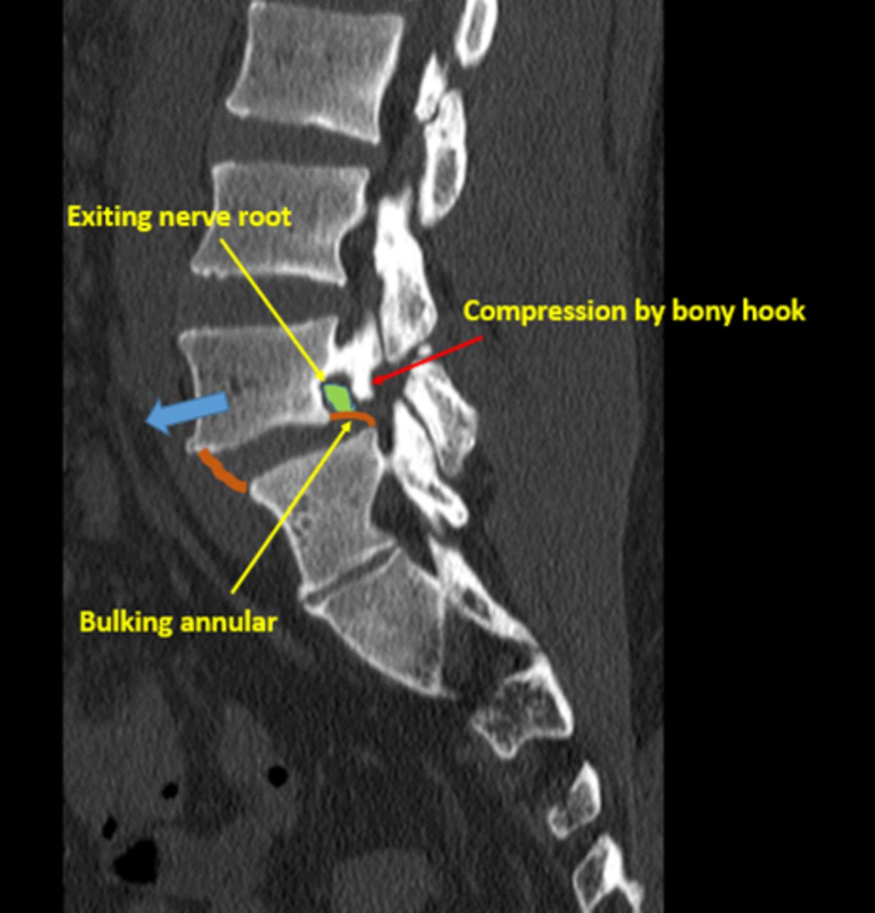
    The bony hook is the hook-like remnant of the proximal deficient lamina of the L5 lamina below the pars defect in L5-S1 spondylolisthesis, which compresses the L5 exiting nerve root (red arrow).

  • Figure 3
    • Download figure
    • Open in new tab
    • Download powerpoint
    Figure 3

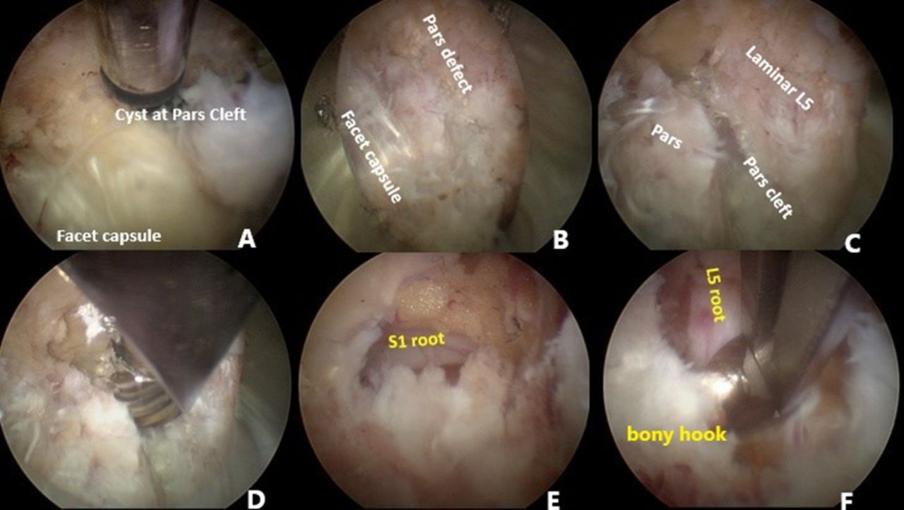
    Steps of percutaneous endoscopic pars decompression in L5 spondylolysis (L5-S1 spondylolisthesis). (A) The facet joint of L4-L5 is identified. (B, C) The endoscope cannula is moved to the caudal region adjacent to the facet joint to find the pars defect of L5. (D) The soft tissue and fibrocartilaginous mass in the gap are removed with a burr. (E) After the gap is clear, the S1 traversing nerve is identified. (F) The bony hook below the gap is identified and removed using a Kerrison rongeur until the L5 exiting nerve root is free.

  • Figure 4
    • Download figure
    • Open in new tab
    • Download powerpoint
    Figure 4

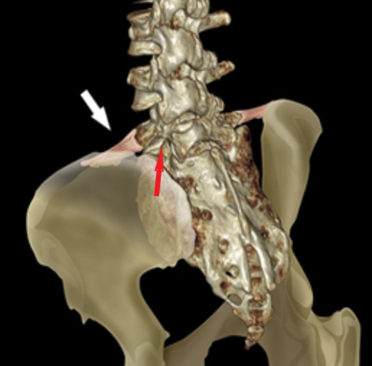
    The red arrow indicates L5 pars defect. The iliolumbar ligament binds between the L5 transverse process and the ilium (white arrow).

  • Figure 5
    • Download figure
    • Open in new tab
    • Download powerpoint
    Figure 5

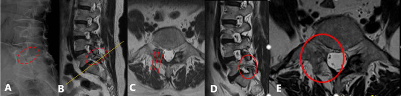
    Comparison of pre- and postoperative magnetic resonance images of L5 spondylolysis in a patient who underwent percutaneous endoscopic right L5 pars decompression. (A) The pars defect at L5 observed in a lateral radiograph. (B, C) The right L5 pars defect and L5 exiting nerve root compression in the L5-S1 intervertebral foramen. (D, E) The right L5 exiting nerve root is free after surgery.

Tables

  • Figures
    • View popup
    Table 1

    Demographics and clinical data of the patients.

    Demographics N = 11
    Gender, men:women6:5 (54.55%:45.45%)
    Age, y, mean ± SD62.45 ± 10.65
    Follow-up period, mo, mean ± SD22.64 ± 15.40
    Spondylolysis alone5 (45.45%)
     L3-L4 and L4-L5 spondylolysis1
     L5-S1 spondylolysis4
    Spondylolysis with spondylolisthesis6 (54.54%)
     Grade 1 spondylolisthesis L3-L41
     Grade 1 spondylolisthesis L4-L51
     Grade 1 spondylolisthesis L5-S13
     Grade 2 spondylolysis L5-S11
    • View popup
    Table 2

    Level of full endoscopic pars decompression.

    Level of Decompression n (%)
    L3-L41 (9%)
    L4-L51 (9%)
    L5-S17 (64%)
    L3-L4 and L4-L51 (9%)
    Bilateral L5-S11 (9%)
    • View popup
    Table 3

    Comparison of clinical outcomes and slippage percentage of interlaminar percutaneous endoscopic pars decompression between the preoperative and postoperative periods.

    Clinical OutcomePreoperativePostoperative P Value
    Visual analog scale for leg pain5.18 ± 3.340.82 ± 0.980.007
    Oswestry Disability Index41.72 ± 19.2417.78 ± 12.260.005
    Percent slippage8.03 ± 11.277.42 ± 9.361.000
    • Note: The variables were compared using the Wilcoxon signed-rank test. The P values were determined to be significant at the 0.05 level.

    • View popup
    Table 4

    Improvement rate for VAS and ODI scores after percutaneous endoscopic interlaminar pars decompression.

    Improvement RateVAS for Leg Pain, n (%)ODI, n (%)
    76%‒100%6 (54.55%)3 (27.28%)
    51%‒75%4 (36.36%)4 (36.36%)
    26%‒50%1 (9.09%)2 (18.18%)
    0%‒25%02 (18.18%)
    • Abbreviations: ODI, Oswestry Disability Index; VAS, visual analog scale.

    • View popup
    Table 5

    Comparison of VAS and ODI improvement between spondylolysis alone and spondylolysis with spondylolisthesis after percutaneous endoscopic pars decompression.

    Outcome MeasureSpondylolysis Alone (n = 5), Mean ± SDSpondylolysis With Spondylolisthesis (n = 6), Mean ± SD P Value
    VAS improvement87.00% ± 18.57%53.98% ± 43.91%0.154
    ODI improvement81.91% ± 16.97%27.57% ± 21.45%0.001
    • Abbreviations: ODI, Oswestry Disability Index; VAS, visual analog scale.

    • Note: An independent sample t test was used to compare the results. P values <0.05 were considered to be statistically significant.

PreviousNext
Back to top

In this issue

International Journal of Spine Surgery
Vol. 17, Issue 3
1 Jun 2023
  • Table of Contents
  • Index by author

Print
Download PDF
Article Alerts
Sign In to Email Alerts with your Email Address
Email Article

Thank you for your interest in spreading the word on International Journal of Spine Surgery.

NOTE: We only request your email address so that the person you are recommending the page to knows that you wanted them to see it, and that it is not junk mail. We do not capture any email address.

Enter multiple addresses on separate lines or separate them with commas.
Clinical Outcomes of Interlaminar Percutaneous Endoscopic Uniportal Pars Decompression for Lumbar Spondylolysis
(Your Name) has sent you a message from International Journal of Spine Surgery
(Your Name) thought you would like to see the International Journal of Spine Surgery web site.
CAPTCHA
This question is for testing whether or not you are a human visitor and to prevent automated spam submissions.
Citation Tools
Clinical Outcomes of Interlaminar Percutaneous Endoscopic Uniportal Pars Decompression for Lumbar Spondylolysis
Pornpavit Sriphirom, Chaiyaporn Siramanakul, Manoch Sumritsopak, Parinya Chokviriyaprasert, Nantawat Uttamo, Kittinon Songchou
International Journal of Spine Surgery Jun 2023, 17 (3) 335-342; DOI: 10.14444/8413

Citation Manager Formats

  • BibTeX
  • Bookends
  • EasyBib
  • EndNote (tagged)
  • EndNote 8 (xml)
  • Medlars
  • Mendeley
  • Papers
  • RefWorks Tagged
  • Ref Manager
  • RIS
  • Zotero
Share
Clinical Outcomes of Interlaminar Percutaneous Endoscopic Uniportal Pars Decompression for Lumbar Spondylolysis
Pornpavit Sriphirom, Chaiyaporn Siramanakul, Manoch Sumritsopak, Parinya Chokviriyaprasert, Nantawat Uttamo, Kittinon Songchou
International Journal of Spine Surgery Jun 2023, 17 (3) 335-342; DOI: 10.14444/8413
Twitter logo Facebook logo Mendeley logo
  • Tweet Widget
  • Facebook Like
  • Google Plus One

Jump to section

  • Article
    • Abstract
    • Introduction
    • Methods
    • Results
    • Discussion
    • Conclusions
    • Footnotes
    • References
  • Figures & Data
  • Info & Metrics
  • PDF

Related Articles

  • No related articles found.
  • PubMed
  • Google Scholar

Cited By...

  • Efficacy and Safety of Biportal Endoscopic Decompressive Laminectomy in Octogenarians With Severe Lumbar Spinal Stenosis
  • Google Scholar

More in this TOC Section

  • Effective Biportal Endoscopic Spine Surgery Technique With Better Facet Joint Preserving for Lumbar Lateral Recess Stenosis
  • Risk Analysis of Neurological Deterioration Associated With Fluid Insufflation in Uniportal Spine Endoscopy: A Case Series and Literature Review
  • Comparison of Pain and Functional Outcomes Among Geriatric and Nongeriatric Adults Following Full Endoscopic Spine Surgery for Degenerative Lumbar Pathology
Show more Endoscopic Minimally Invasive Surgery

Similar Articles

Keywords

  • spondylolysis
  • nerve root compression
  • interlaminar percutaneous endoscopic uniportal pars decompression

Content

  • Current Issue
  • Latest Content
  • Archive

More Information

  • About IJSS
  • About ISASS
  • Privacy Policy

More

  • Subscribe
  • Alerts
  • Feedback

Other Services

  • Author Instructions
  • Join ISASS
  • Reprints & Permissions

© 2025 International Journal of Spine Surgery

International Journal of Spine Surgery Online ISSN: 2211-4599

Powered by HighWire